Digestive System of Human
Share
Digestive System of Human
Animal Nutrition of Class 11
In humans digestion is performed by 8 to 10 m long tube called alimentary canal. This tube starts at buccal aperture or mouth and ends at anal aperture or anus. Different part of the digestive system are shown in the diagram given below.
Mouth
It harbors buccal cavity. Buccal cavity divides into two parts.
1. Vestibule
2. Proper
The space between lips and teeth is termed as vestibule. In vestibule labial glands are present which secrete mucus.
The space between lips and uvula is called cavity proper. The roof of the buccal cavity proper is formed by Palate while floor is formed by Tongue and lateral wall is formed by cheeks.
Palate: The roof of buccal cavity is palate that separates it from nasal chamber, consist of hard and soft palate. Mucus epithelium has thick transverse folds called palatine rugae (thick in case of carnivorous animals) and helps to hold the prey. Soft palate is soft and formed of muscles and soft tissues. Terminal part of soft palate hangs in the throat called uvula. Uvula is
surrounded by a ring of tonsils called Waldeyer’s ring. It is formed by Palantine, Lingual, Tubal and Pharyngeal tonsils. Enlarged Pharyngeal tonsils are called Adenoids.
Tongue
It is spoon like thick, musculo-sensory organ present in the buccal cavity and attached to hyoid bone with the help of glossengial muscles besdies it is also attached to the floor of buccal cavity with the help of ligament like structure called Frenelum linguae. It is covered by the mucous membrane of thick stratified squamous epithelium with papillae of 4 types.
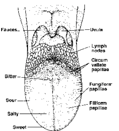
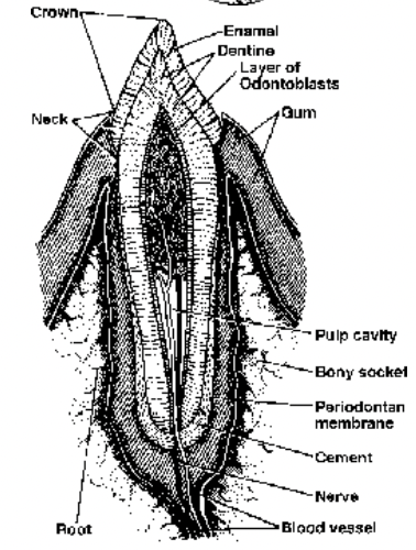
Fig. Upper surface of tongue showing papillae Fig. L.S. of a typical single-rooted tooth in situ
Filiform : Most abundant short filamentous (absent in Rabbit). Taste buds are absent and papillae are used to increase the surface area.
Fungiform : Small mushroom shaped on the upper part with taste buds.
Foliate : Small leaf-like folds on the lateral sides.
Circumvallate : Largest cup like, on the posterior part.
Teeth
It originatee from ectoderm and mesoderm, tooth has three parts:
Crown: Outer part covered with enamel (hardest substance) secreted by Amoeloblast cells.
Neck : Joint between crown and root covered with gum or gingivae.
Root: Part within the socket of jaw bone. It has a cavity called pulp cavity which is lined by odontoblast cells, these cells secrete Dentine or Ivory. Pulp cavity is made up of soft connective tissue called pulp along with blood vessels, lymphatic vessels and nerve fibres. A peridontal membrane is present between the root and the socket. Dentine constitutes main part of the tooth with a pulp cavity which contains
connective tissue, blood vessels and nerves.
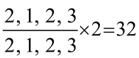
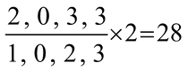
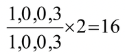
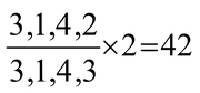
Dental formula =
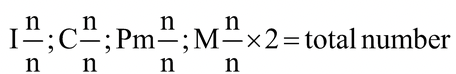
n = half number of respective teeth in upper and lower jaws.
Man
 Rabbit
Rat
 Dog
Dog
Mole/Horse/Pig
 Cat
Cat
Elephant
 Monkey
Squirrel
 Cow, sheep and goat
Opossum
 Kangaroo
Lemur
Type : Attachment of teeth
1. Acrodont : Teeth flat surface of jaw bone e.g., Amphibia (frog), lizards.
2. Pleurodont : Teeth attached by their sides to the rim of jaw or in groves as in certain lizards.
3. Thecodont : Teeth attached within socket of jaw bone e.g., in mammals and crocodiles.
Succestion of teeth :
1. Monophyodont : Strenians toothless whole, eg. Platypus, marsupials, moles, (some mammals)
2. Diphyodont : eg. Most mammals
3. Polyphyodont : ef lower vertebrates frogs
Different shape of teeth :
(C) 1. Homodont : Similar in shape & size eg. Fishes, Amphibians, Reptiles.
2. Hetrodont : Different types of teeth e.g.. incisor, canine, premolar, and molar as in mammals.
(D) Kinds of teeth :
1. Incisors : Adapted for seizing, culting and biting. In rodents and lagomorpha they grow throughout lofe due to open drooped.
Elephant tusks are modified upper incisors with open roots
They are totally absent in sloths and lacking in upper jaw of OX.
2. Canines : There are generally elongated, single rooted and with conical sharp monocuspid crown. Meant for piercing, tearing offence & defense.
They are present only in upper jaw in male musk deer.
Upper canines forms turks in walrus for digging mollusks and for locomotion on ice.
Absent in ungulates, lagomorphs, rodents (diastema).
3. Cheek teeth : Pm & m for crushing, grinding and chewing.
Carnassial teeth : Last premolar in upper jaw and first molar in lower jaw in carnivores have very sharp curps for cracking bones and shearing tendons.
Cheek teeth are of various types depending on the no; shape and arrangement of curps.
(a) Triconodont : 3 cones are arranged in a straight lone of fossils Mesozoic mammals.
(b) Trituber culate : 3 cones or tubercles are arranged in the form of a triangle of fossile mammals.
(c) bunodont : their crown bear small, separate, blunt and rounded tubercles for crushing of man, monkey and pig.
(d) Secondong : Teeth have sharp cutting edges for tearing and cutting flesh of carmiores.
(e) selenodont : Grinding herbivores grazingammals with shorth root called brachydong (eg. Squirrer) high crown & low roots elongated teeth in horse & cattle (hyprodont)
(f) Lophodont : eg. Elephant
Salivary Glands (3 pairs in man, 4 pairs in rabbit, 5 pairs in cat).
These are compound tubulo-alveolar type.
Parotid gland: Largest gland, at the base of pinna. Its duct, Stenson’s duct opens behind the upper second molar.
Submandibular or submaxillary glands: Situated at the angles of lower and upper jaws; its duct Wharton’s duct opens behind the lower central incisors.
Sub-lingual glands: Situated below the tongue, with many short ducts of Rivinus, opening below the tip of tongue. The duct systems are called ducts of Rivinus.
Infra-orbital (or zygomatic) glands: Situated below the eye orbit, its duct opens near the upper molars; absent in human (but present in rabbit)
Salivary glands secrete Saliva which contain Mucin that lubricates the food passage ; Lysozyme-antibacterial enzyme and ptyalin that digests starch. Food in mouth, its smell, taste etc. stimulate the secretions of saliva. It is largely controlled by parasympathetic system.
Pharynx
Uvula divides the pharynx into postero-dorsal nasopharynx and antero-ventral oropharynx. Epiglottis (elastic cartilage plate) covers the glottis at the time of swallowing of food.
Histology of Gut
In all vertebrates the wall of alimentary canal is basically made on same plan and consists of 4 layers :
Layers Tissue Origin
1. Serosa/peritoneum (outermost layer) Squamous epithelium Mesodermal
2. Muscular layer Smooth muscles –do–
(a) outer longitudinal
(b) inner circular
3. Submucosa (lamina propria) Areolar connective tissue –do–
4. Mucosa (innermost layer) Simple columnar epithelium Endodermal
In vertebrates all the four layers are similar except the fact that in mammals all structures are of higher degree. In different functional parts these layers are modified :
Serosa is also followed by subserosa, a fibrous connective tissue layer for its support.
Mouth and Pharynx
It is Lined with stratified squamous epithelium of ectodermal origin, hence represent stomodeal part. With possesses striated muscle layer, hence the movement of food here is: voluntary.
Oesophagus
All four layers are typical as it serves only as the passage. Lamina propria, Muscularis externa are abscent. The part of oesphagus which is present outside abdomianal cavity is lined by a
fibrous coat Adventitia externa or it’s thoracic part is without serosa layer which shifts to form the mediastinal wall.
Stomach
Stomach is sickle shaped pouch like structure present below diaphragm towards left hand side. It has thick and highly distensible wall, when empty it forms inner longitudinal folds, rugae. In ruminants (e.g. cattle, sheep, goat, camel and deer) have stomach is compound and divides into following four chambers:
(i) Rumen - first and the largest part for storage of food and digestion of cellulose by symbiotic bacteria living here, (Rumenococcus)
(ii) Reticulum (honey comb)- next chamber also the site for cellulose digestion.
(iii) Omasum (psalterium) - concentrates the food by absorbing H2O and HCO3 (absent in camel and deer)
(iv) Abomasum or true stomach - last part as secretion of gastric juice and the digestion takes place here only.
Camel’s rumen and reticulum have many diverticula (water pockets) but these do not store water. Camel can live without water for two weeks. Its hump is store of fats. For every 100 gms of fats metabolized it produces 120 ml of H2O.
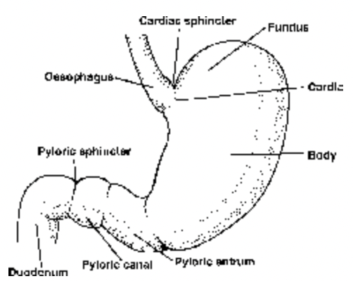
Fig. Parts of human stomach
In humans stomach divides into three parts,
1. Fundus
2. Corpus
3. Pylorus
In fundus part a small cardiac area is present which has mucus producing cardiac glands are present. Fundus usually remains filled with gases. In Corpus and Pylorus gastric glands are present these are of two
types
1. Chief Glands or Oxyntic glands
2. Pyloric glands
Chief glands are the main gastric glands and present throughout Corpus and Pylorus while Pyloric glands are usually present in Pylorus only.
In humans muscular layer is thickest of all parts with third and innermost oblique muscle layer. Submucosa has no speciality.
Mucosa forms glands by invaginating into submucosa and has four types of cells :
(i) Mucous (Goblet) cells secrete mucin
(ii) Oxyntic (Parietal) cells secrete HC1
(iii) Chief cells (Peptic or zymogenic) cells secrete enzymes.
(iv) Argentaffin cells
The glands of pylorus has two types of cells only
1. Mucus producing cells
2. G cells which secrete Gastrin, which is a polypeptide hormone and induce the secretion of gastric glands.
Small Intestine
Small intestine is 6.25 m long tube starts from Pyloric sphicter and continue till Ileocoecal valve. It divides into three parts,
1. Dueodenum
2. Jejunum
3. Ileum
Dueodenum is 25 cm long while Jejunum is 2.5 m long and Ileum is 3.5 m long. Dueodenum is U shaped usually remains lined by thick alkaline mucus due to Brunner’s gland present in the submucosa of it which also secrete secretin and cholecystokinin. Jejunum and Ileum is highly folded and convoluted due to presense of Submucosal and mucosal folds which form inner transverse finger-like folds to increase the surface area of absorption. In mammals, the primary folds are called as folds of Kerkering (or valvulae conniventes or plica circularis), which bears true villi as secondary folds. In small intestine 4 to 5 million villi are present. The size and shape of villi varies from Dueodenum to Ileum. Muscle layer is typical (no speciality). Submucosa is most developed and specialised of all parts with following structures:
(i) Highly organised system of blood capillaries up to villi for the transportation of absorbed food.
(ii) Fine lymphatic channels (lacteals) are extended upto villi for absorption and transportation of lipids.
(iii) Muscle layer (muscularis mucosa) is present just behind mucosa for movement of villi.
(iv) Two nerve plexuses of autonomic nervous system (ANS) are :
(a) Auerbachs plexuses - controls movement of food in gut.
(b) Meissner’s plexuses - controls movement of villi.
(v) The lymph nodes, Peyer’s patches are present which act as biological filters.
In submucosa Folds of Kerkering increase surface area of absorption by 3 times, villi increase by 10 times and microvilli of mucosal cells increase by 20 times, thus total increase is by 600 times that comes to about 250 mtr 2 in man. Mucosa is the main functional layer for digestion and absorption with brush border cells both secretory and absorptive. Mucosa forms glands embedded in submucosa are :
(a) Brunner’s gland (duodenal gland) : Present mainly in duodenum; secretes mucous ; absent in frog.
(b) Crypts of Lieberkuhn : Small flasked-shaped gland in the crypts between two villi. They have four different kind of cells
1. Cells of Paneth in it secrete digestive enzymes
2. Undifferentiated cells
3. Mucus cells
4. Argentaffin cells secrete intestinal hormones
In small intestine approximately 33 different hormones are produced, some hormones of importance are
Dueocrinin : Activates Brunner’s glands
Villicrinin: Activates glands of small intestine
Enterocrinin: Activates the glands of small intestine
Enterogastrone: Inhibits the glands of stomach
Gastric Inhibitory Peptide (GIP): Blocks the glands of stomach
Secretin: Activates pancreatic duct system to release large amount of NaHCO3 rich secretin also blocks the secretion of gastric glnads
Cholecystokinin (CCK): It is most important gland of intestine and performs many functions, it stimulates the secretion of Gall Bladder and pancreas, it relaxes the sphincter of Boyden and Oddi and allow the entry bile into intestine. All the secretions of small intestine are collectively called Succus Entericus.
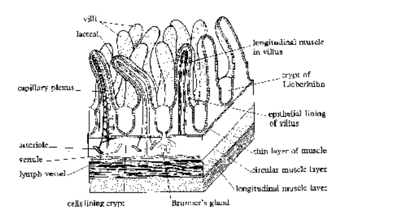
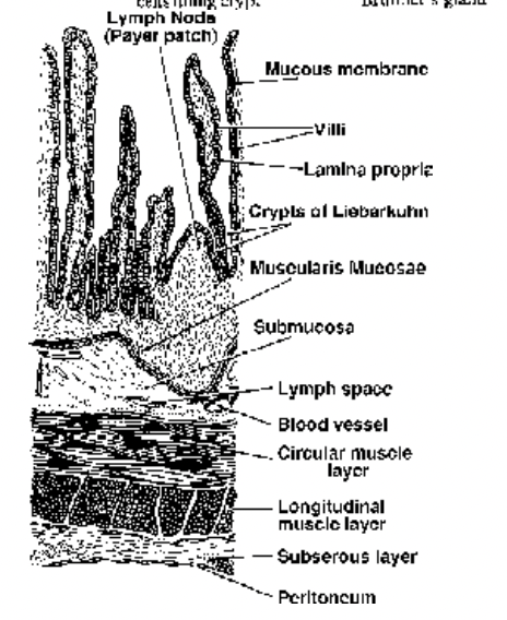
Fig. Part of intestinal wall to show villi Fig. T.S. of ileum of rabbit
Large Intestine
Large intestine starts from Ileocaecal valve and ends at anal sphincter. It divides into three parts
1. Caecum
2. Colon
3. Rectum
Caecum
It is smallest part of large intestine approx. 6 cm in length, in the distal part a 5 to 9 cm long finger like projection is present called
Vermiform appendix. Vermiform appendix is a vestigial organ. Its infection causes appendicitis.
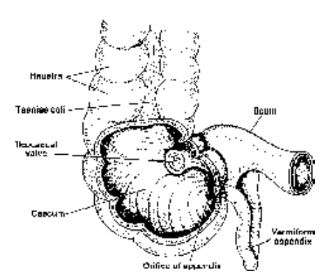
Fig. Relationships of colon, caecum and appendix
Colon
Colon (2 mtr long) takes a characteristic form and course. From the caecum on right side it moves upwards in the abdominal cavity as ascending colon (shortest), then turns at right angle
(horizontally) towards left side as transverse colon, then moves downwards on the left side as descending colon and lastly terminates as pelvic (or sigmoid) colon. It is the longest and
largest part of the large intestine. It has no specialized glands only mucus producing goblet cells are there besides mucus bicarbonate secretions are also released. There are three inner
longitudinal folds called as taeniae - specialized structures for absorption of water. The transverse membranous folds between two taeniae form series of pockets, haustra, highly
developed in desert or dryland animals like rabbit, goat, camel etc. but, less developed in man.
Rectum
It is the lower most part of the large intestine approx. 16-20 c.m. long. It has six or more folds called rectal papillae for absorption of water. Goblet cells secrete mucous to facilitate defecation.
Sphincters
Thickened circular muscle layer, acts as valve at the opening of one part into another, like :
(a) Pyloric sphincter - between pylorus and duodenum.
(b) Anal sphincter - at the opening of anus outside.
Ileocaecal (Ileocolic or Ileocaecolic) valve
Present in sacculus rotundus and operates between ileum and caecum to direct the food in between ileum, caecum and colon.