
Female Reproductive System
Human Reproduction of Class 12
Consists of a pair of ovaries, a pair of oviduct (fallopian tubes), a sac-like uterus (paired in some mammals), a vagina and the external genitalia like vulva and genital glands.
Ovary
Almond shaped structures in the upper part of pelvis attached to dorsal abdominal wall through mesovarium near kidney. In women each measures about 3 cm × 2 cm × 1 cm surrounded by the fold of peritoneum.
Blood vessels and nerves enter through a narrow connecting part, hilus having continuity with mesovarium.

Fig.Female reproductive organs
Fallopian tubes (oviducts)
A pair of muscular tubes (10 cm) extend laterally from the uterus on either side of the wall of pelvis. It is divided into 3 parts : isthmus, ampulla, and infundibulum. Ampulla is thin walled, longest part lined with ciliated epithelium and is the site of fertilization. Infundibulum has fimbriated (or oviducal) funnel with many filger-like projections, fimbrae, around the opening called ostium through which egg enters. Tubectomy (or Tubal ligation), the cutting or ligation of fallopian tube prevents the entry of ovum into it. It is a permanent method of contraception (sterilization) Mullerian ducts of embryo develop into fallopian tubes.
Uterus (Metra or Hystera or Womb)
For viviparous development in mammals it makes the house of embryo. Depending upon the degree of fusion of paired gonoduct behind fallopian tube, it is of various types based upon joining of two tubes: as duplex (bats, marsupials and rodents); bicornuate (elephant); bipartite (carnivores) and simplex (higher primates including human). In women it is sac-like, thick walled, pear shaped, muscular and glandular part of 8 × 5 × 2 cm size; distinct in three parts: (i) upper fundus (ii) middle corpus (body) and (iii) lower narrow cervix. Its wall consists of myometrium, the smooth muscle layer, followed by perimetrium or submucosa as areolar connective tissue layer with blood vessels, nerves etc.and endometrium.
Endometrium (or mucosa) consists of inner epithelial layer and underlying connective tissue layer (lamina propria) with many coiled tubular glands and screw-like blood vessels. This undergoes cyclic changes during menstruation. Cervix has two openings, os internalis towards corpus and os externalis towards vagina. After implantation and pregnancy placenta is formed by the contribution of uterine wall. Hysterectomy is the surgical removal of uterus. Vagina (8 to 10 cm) .It extends from the cervix to the outside of body.
It receives penis during copulation and also serves as birth canal. Lined with non-keratinized stratified squamous epithelium with numerous transverse folds called rugae undergoing cyclic changes during
ovarian cycle. Initially its opening remains covered with a membranous hymen which ruptures either during sexual intercourse or during other physical activity. Presence or absence of hymen is not a sign of virginity of female.

Fig.Female reproductive system
External genitalia (vulva)
The external part around the vaginal pore is vulva with upper pad-like part, mons pubis (or mons veneris) with hairs. The middle cut, vestibule, is laterally bound with two pairs of skin fold, outer labia majora and inner labia minora. Labia majora are two large thick folds of skin which form the boundary of vulva. These are homologous to scrotum of male. Labia minora are two smaller folds of skin which lie between labia majora. Posterorly the labia minora are fused together to forms fourchette. These contain numerous sebaceous glands. In the vestibule the urethral opening is present on upper side (below clitoris) and vaginal orifice at the lower side. Clitoris is highly sensitive and erectile projected part homologous to penis.
Vestibular glands
Vestibular glands are of two types i.e. lesser vestibular glands and greater vestibular glands. The lesser vestibular glands (or Paraurethral glands or glands of Skene) are numerous minute glands present around urethral orifice. These are homologous to prostate gland of male & secrete mucus. The greater vestibular glands (or Bartholin’s glands) are paired glands situated one on each side of uaginal orifice. These are homologous to bulbourethral (cowper’s) glands of male and secrete viscid fluid that helps in lubrication during sexual intercourse.
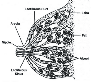
Mammary glands or Breasts
Modified sweat glands over the pectoral muscles. In male mammary glands are rudimentary. In females these begin to develop under the influence of oestrogen and progesterone during puberty. Externally, the breast is covered with skin and has a nipple surrounded by a pigmented area, the areola.
The mammary glands consists of the glandular tissue, the fibrous tissue (connective tissue) and the fatty or adipose tissue.
- The glandular tissue comprises about 20 lobes in each breast. Each lobe is made up of a number of lobules. Each louble consists of a group of glandular alveoli which open into small ducts which unite to form large ducts, the lactiferous ducts. Just before the nipple, the lactiferous ducts become expanded to form lactiferous sinuses which act as reservoirs for milk during lactation. Narrow ducts lead from these sinuses and open on the surface at the nipple.
- The fibrous tissue (connective tissue) consists of fibrous strands. It supports the glandular tissue and the ducts.
- The fatty or adipose tissue is found between the lobes and covers the surface of the gland. The amount of the adipose tissue determines thesize of the breasts.
Human milk
Composed of about 80% water, 7% lactose, 4% fat and 1% protein (casein and lactalbumin), Mineral salts (Na, Ca, P, K) vitamins, antibodies (IgA). Milk is poor in iron.
About 5 lakhs primary follicles are formed only once when female foetus is only 3 months old in her mother’s womb. Follicles remain arrested in diplotene stage of meiotic prophase-I (called Dictyate oocytes) and become active only after 10-12 years of age for maturation influenced by FSH.
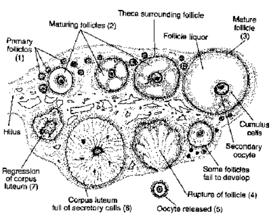
Fig.Structure of human ovary
Maturing follicles secrete estrogen that maintains the sex organs and secondary sexual characters. In human only one follicle releases ovum each month and only 400 to 450 follicles attain maturity in the entire reproductive age of a female, except during pregnancy. Majority of follicles degenerate by a genetically programed process called follicular atresia.
Graafian (or mature) follicle
By accumulating antral fluid and mitosis of follicle cells a mature follicle grows upto 2.5 cm in diameter; becomes invested with ovarian connective tissue (cells with fibrous layer), theca externa and theca interna followed by the layer of follicular cells membrana granulosa. Antrum (or follicular cavity) formed in the midst of granulosa cells keep increasing and pushing the oocyte and its surrounding granulosa cells to one side. The fluid, liquor folliculi, in the antrum secreted by these cells contains nutrients and estrogens (secreted by granulosa cells). Ultimately the ovum along the follicular wall occupies a place on little hill of granulosa cells called cumulus ovaricus or discus proligerous or cumulus oophorus or germ hill. Jelly-like covering around the oocyte is zona pellucida, a primary membrane secreted by the egg itself. The layer of granulosa cells around the oocyte is called corona radiata for having radiating process to draw nutrients from liquor folliculi.
Estrogens
Secreted by the granulosa cells it is group of steroids which include estradiol, estrone and estriol, of which first is the major. In adult human ovary, at one time, there may be several follicles with antrum of different sizes but, only one reaches complete maturity taking about 2 weeks time. In some cases more than one ovum may be ovulated, the common cause of multiple pregnancies. Rupture of follicle wall with ovarian wall causes the release of ovum (= ovulation) along with zona pellucida and cumulus, into the coelomic cavity. Released oocyte is actually secondary oocyte.
Corpus luteum
The ruptured Graafian follicle after ovulation forms glandular structure called as corpus luteum (yellow body). It has lutein protein & carotene pigment. Macula lutea is yellow spot in retina of eye. It secretes a little amount of estrogen and large amount of progesterone hormone that maintains pregnancy. It develops extensively if pregnancy occurs, otherwise, degenerates to form corpus albicans (white body) after nearly 10 days of its formation.
Reproductive cycle
Ovulation and all related phenomenon of ovary and uterus occur in cyclic manner. Period of cycle varies in different mammals, like, in rat every 5 - 6 days; rabbit 8 - 10 days; cow 15 days ; human 28 days.
It is two types :
- Estrous cycle and
- Menstrual cycle.
Estrous cycle :
It occurs in non-primate mammals (cow, rat, rabbit etc.) ‘Estrous’ means ‘heat’ which denotes the ovulation time with very high level of estrogen in the female body. The entire period is divided into four phases :
- Proestrous phase – Vaginal epithelium proliferates, follicles are near maturing stage, the estrogen level starts increasing.
- Estrous phase – The ‘heat phase’, when female develops strong desire for sex; the estrogen level is at its peak ; vaginal epithelium thickens and becomes keratinized; ovulation takes place. In some animals like cat, rabbit the ovulation does not occur without copulation hence, they are induced ovulators.
- Metestrous phase – Corpus luteum starts developing, the keratinized layer of vaginal epithelium breaks off. In monoestrous animals (e.g. bitch) hypertrophic mucosa of uterus breaks down and is discharged.
- Diestrous phase – The quiescent period, without any activity in the ovary and other parts. In monoestrous animals the period may be as long as the next breeding season (after one year) hence called as anestrous phase. In polyestrous animals it is the gap between two cycles like rat.
Menstrual cycle :
(mensem = month) In primates (human, ape, monkey) the cycle of 28 days is marked by the vaginal bleeding called menstruation or “weeping of uterus” as major event. Depending upon the changes in ovary, uterus i.e., preparation for conception and pregnancy, and associated hormonal changes, the cycle is divided into four phases:
- Menstrual phase (destructive phase) – If fertilization/pregnancy doesn’t take place the breakdown of overgrown uterine tissue with blood vessels begins. Discharge of blood and ruptured endometrium through vagina mark the whole phase that lasts for 3 - 5 days. Estrogen level is low, LH and progesterone also come down. Average blood loss equals 50 to 150 ml.
- Follicular phase (resting phase) – Bleeding stops; follicles start growing; level of FSH, estrogen starts rising; reformation and growth (proliferation) of uterine wall begins and thus preparation for next pregnancy sets in; also called as proliferative phase; lasts for 7 - 8 days.
- Ovulatory phase – By the end of 13th day one of the mature follicles undergoes ovulation. The estrogen level is highest hence corresponds to ‘estrus’ phase of estrous cycle.
High level of LH from pituitary induces ovulation, FSH level comes down :

Fig. Graphic representation of menstrual cycle
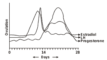
- Luteal phase (or Secretory phase): After ovulation till the onset (or 1st day) of next menstruation it is the longest phase of about 13 days. Ruptured follicle under the influence of LH, develops as corpus luteum to secrete progesterone. After a fall for 2-3 days the estrogen level rises again. High level of estrogen and progesterone during this period inhibits FSH, hence, maturation of follicle stops, but the growth of uterine wall increases multifold. Menstrual cycle remain suspended during pregnancy.
- Menopause (or female climacteric) – The menstruation stops at 45 - 50 years of age, women loses reproductive ability. Urine of a menopausal women contain large amount of FSH because estrogen production by ovaries stops and pituitary gets relieved of inhibitory effect of estrogen.Menarche – The first menstruation which marks the onset of puberty.
- Dysmenorrhoea – Abnormal or irregular menstruation with cramps and profuse bleeding due to locally produced prostaglandins. Amenorrhoea – Menstruation does not occur due to many physiological/hormonal or mental defects this is the sign of sterility.
Luteal phase (or Secretory phase):
After ovulation till the onset (or 1st day) of next menstruation it is the longest phase of about 13 days. Ruptured follicle under the influence of LH, develops as corpus luteum to secrete progesterone. After a fall for 2-3 days the estrogen level rises again. High level of estrogen and progesterone during this period inhibits FSH, hence, maturation of follicle stops, but the growth of uterine wall increases multifold. Menstrual cycle remain suspended during pregnancy.
- Menopause (or female climacteric) – The menstruation stops at 45 - 50 years of age, women loses reproductive ability. Urine of a menopausal women contain large amount of FSH because estrogen production by ovaries stops and pituitary gets relieved of inhibitory effect of estrogen.
- Menarche – The first menstruation which marks the onset of puberty.
- Dysmenorrhoea – Abnormal or irregular menstruation with cramps and profuse bleeding due to locally produced prostaglandins.
- Amenorrhoea – Menstruation does not occur due to many physiological/hormonal or mental defects this is the sign of sterility.
