
Joints
Movement and Locomotion of Class 11
JOINTS
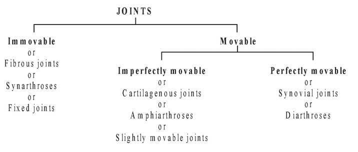
Fibrous Joints
No synovial cavity. Two bones remain held together by thin layer of fibrous tissue or dense fibrous tissue or cement or sutures.
Sutures (Synostoses—Suture during infancy but fusion afterwards as in Frontal bone). Bones are thin and plate like, held together by interdigitations. e.g., skull bones.
Gomphosis One bone remains embedded in the socket of other attached through fibres or cement layer. e.g., Thecodont teeth of mammals.
Shindylases One bone fits into slit of other. e.g., ethmoid bone into vomer.
Syndesmosis Two bones are united by dense fibrous tissue. e.g., joint between skull bones and bones of upper jaw, distal ends of tibia and fibula.
Cartilaginous joints
No synovial cavity, articulating bones are united by cartilage.
Synchondrosis Connecting material is hyaline cartilage. e.g., temporary joint between diaphysis and epiphysis of a long bone and permanent joint between true ribs and sternum.
Symphysis Connecting material is broad flat disc of fibrocartilage. e.g., Intervertebral disc and symphysis pubis.
Synovial Joints
Most perfect, freely movable, most common type of joints. Allows free movement in one or more directions. There is no direct contact between two bones, end is covered with hyaline cartilage cap (articular cartilage) and the whole structure is covered with ligament. The synovial cavity between two bones is lined with synovial membrane and filled with synovial fluid secreted by this membrane. Synovial fluid acts as lubricant and shock absorber and also provides nourishment to articular cartilage. In old age stiffness of joints occur due to decrease in synovial fluid and erosion of cartilaginious part. Synovial membrane is composed of loose connective tissue with elastic fibres and a variable amount of adipose tissue. Synovial fluid also contains phagocytic cells and removes microbes and debris resulting from wear and tear of joints. It also contains Hyaluronic acid and interstitial fluid formed from blood plasma and is similar in appearance, consistency to uncooked egg white. When there is no movement, the fluid is quite viscous, but as movement increases, fluid becomes less viscous. Amount of synovial fluid varies in different joints of body, ranging from a thin viscous layer to about 3.5 ml of free fluid in large joint such as knee. It also removes metabolic wastes from the joint. Arthroscopy is examination of interior of a joint, usually knee by an arthroscope. One interesting feature of some synovial joints is their ability to produce a cracking sound when pulled apart. Articular discs (menisci) These are pads of fibrocartilage that lie between articular surfaces of some bones. These allow 2 bones of different shapes to fit tightly, these modify the shape of joint surfaces of the articulating bones. Articular discs help to maintain the stability of joints and direct the flow of synovial fluid to areas of greatest friction. Bursae Sac like structures situated between tendons and bones, muscles and bones, ligaments and bones, skin and bones. Their wall has connective tissue lined by synovial membrane, they are also filled with a fluid similar to synovial fluid. Inflammation of Bursa is called Bursitis.
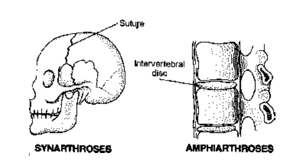
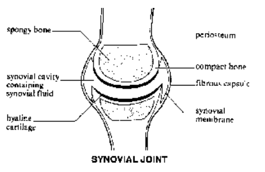
Fig. Types of joints
Types of Synovial Joints
Ball & Socket joint Articulate end of one bone is like a ball whereas other bone end is like a cup shaped socket. It permits triaxial movements, i.e., movement in three planes. e.g., Acetabulum of pelvic girdles and head of femur, glenoid cavity of pectoral girdles and head of humerus.
Angular/Condyloid/Ellipsoid joint Half ball and socket type joint with one biaxial movement in two planes, back and forth and side to side. Oval condyle of one bone fits into elliptical cavity of other bone like
joints of phalanges with metacarpals, radius and carpals. (radiocarpal joint)
Gliding/Sliding joint or Arthrodia Articulating surfaces of bones are usually flat. Only side to side and back and forth movements are permitted. Twisting and rotation are inhibited because ligaments or
adjacent bones restrict the range of movement. Since gliding joints do not move around an axis, they are referred to as Non-axial. e.g., joints between carpal bones, tarsal bones, sternum and clavicle, scapula and clavicle, pre and post zygapophysis of vertebrae.
Hinge joint or Ginglymus Convex surface of one bone fits into concave surface of another bone movement is primarily in a single plane and the joint is known as monoaxial/uniaxial. Motion is similar to that of a hinged door. e.g., elbow, ankle, interphalangeal joint.
Saddle/Sellaris joint Articular surface of one bone is saddle shaped and that of other bone is shaped like a rider sitting in a saddle. It is a modified ellipsoidal joint in which movement is somewhat freer (biaxial).
e.g., joint between trapezium of carpus and metacarpals of thumb (carpometacarpal joint).
Pivot joint/Rotatoria/Trochoid A rounded, pointed or conical surface of one bone articulates within a ring formed partly by another bone and partly by ligament. Primary movement permitted is rotation and the
joint is Monoaxial. e.g., joint between atlas, and axis, and between proximal ends of radius and ulna (Atlantoaxial and Radioulnar joint).
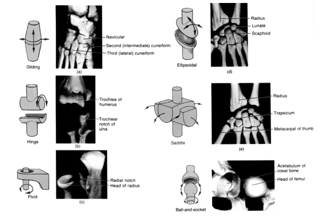
Fig. Fig. Different types of joints
