
Nucleus
Structure of Cell of Class 11
Reported by Robert Brown (1831) in orchid cells.
Strasburger (1882) proved that nucleus arises from pre-existing nucleus by division.
Hertwig and Van Beneden showed the role of nucleus in fertilization.
Hammerling (1953) by his grafting experiments on Acetabularia (largest unicellular green, marine alga) proved the role of nucleus in heredity, growth and morphology.
Found in all eukaryotic cells, except mature phloem sieve tube elements and mature red blood cells of mammals.
A cell may be uni-or multinucleate.If a multinucleated condition arises due to fusion of cells, it is called syncytium e.g. plasmodium, body of slime moulds, young xylem vessels and if due to repeated nuclear
divisions without cytokinesis, it is called coenocytic e.g. Vaucheria, Rhizopus.
1/10th of volume of cell is occupied by nucleus. In a cell, there is a definite nucleo-cytoplasmic ratio.
Nucleus has 80% proteins (65% non-histone, acidic, rich in tyrosine and tryptophan) 15% proteins are basic, histone proteins, rich in lysine and arginine]
Structure :
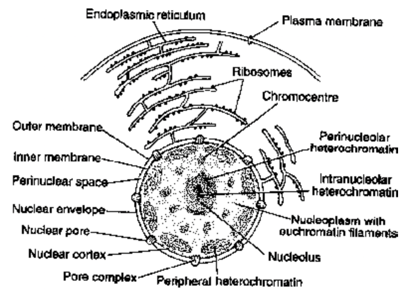
Fig. Diagrammatic Structure of nucleus
1. Nuclear envelope :
Composed of two membranes. The outer membrane is continuous with the endoplasmic reticulum (ER) and it may be covered with ribosomes carrying out protein synthesis.
Perforated by nuclear pores which allow exchange of substances between the nucleus and the cytoplasm. The pore has a definite structure formed by fusion of the outer and inner membranes of the envelope.
The pores are octagonal orifices about 80 nm in diameter which are plugged by a cylinder of proteins materials.
The inner membrane of the nuclear membrane is attached to a layer of fibrous proteins 50 to 80 nm thick that is known as the nuclear lamina. Terminal ends of chromatin fibres or telomeres are embedded in nuclear or fibrous lamina.
2. Nucleoplasm / Nuclear sap / Karyolymph - Strasburger : proteinaceous, jelly-like with pH 7.4 ± 0.2. It has nucleosides, enzymes (DNA and RNA polymerase), ribosomes, Mg, Fe, Mn, Ca and nucleoproteins.
3. Nucleolus : First discovered by Fontana (1781).
Rounded colloidal body without surrounding membrane. Ca++ ions keep it intact.
Mainly consists of r-RNA and proteins.
In mitosis and meiosis, nucleolus disappears during late prophase and again reappears during telophase.
Nucleolus is attached to the chromatin or special type of chromosome called nucleolar organizing chromosome at a specific place called nucleolar organizing region (NOR) or secondary constriction. It
consists of following four parts :
(a) Granular region : outermost (cortical) region where processing and maturation of pre-ribosomal particles occurs.
(b) Fibrillar region: formed of a large number of 50-80 Å long fibrils called nucleonema. The fibrils are made up of both protein and RNA and are believed to be precursors of granules.
(c) Amorphous matrix : homogenous proteinaceous ground substance of the nucleolus called as ‘para amorpha’.
(d) Fibrillar centre or nucleolar chromatin : The innermost region called pars chromosoma. Ribosomal RNA (rRNA) and genes of nucleolar organizing regions are located in this region (containing nuclear genes for rRNA synthesis.)
Functions:
(i) Centre for formation of ribosomes in eukaryotes (ribosomal factory).
(ii) Principal site for development of ribosomal RNAs.
(iii) Nuclear proteins such as histone proteins are synthesized in nucleolus.
(iv) Essential for spindle formation during cell division.
4. Chromatin
The cells of eukaryotic organisms have a constant DNA content (C value) that is characteristic for each species and much higher than in bacteria.
Most of the DNA is present in chromatin, which is a complex of DNA with an equal weight of basic proteins called histones.
Histones are small proteins that are basic because they have a high content of arginine or lysine.
There are only five histones. The four fundamental histones, H2A, H2B, H3 and H4 are present twice every 200 base pairs of DNA.
The fifth histone, HI is present only once per 200 base pairs of DNA, and it varies considerably between species and even within tissues of the same species.
Chromatin is formed by a series of repeating units called nucleosomes (Term - Oudet) about 10 nm in diameter.
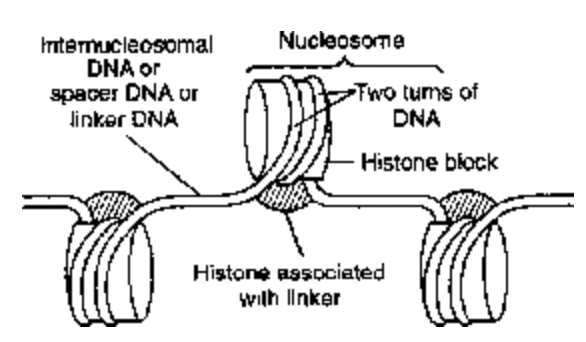
Fig. Structure of nucleosomes
Each nucleosome contains a histone octamer consisting of two of each of the four histones H2A, H2B, H3, and H4 with about 200 pairs of DNA coiled on the outside of the nucleosome. The whole structure has
the shape of a flattened disc with the DNA entering and leaving from the same side, and histone HI sealing off the two turns of DNA.
Besides the dispersed chromatin, certain segments of these fibres are highly folded/condensed forming solenoid structures. These are thicker and more deeply staining. Dispersed chromatin is called euchromatin and folded chromatin is called heterochromatin. Small scattered areas of heterochromatin are called chromocentres. During cell division all chromatin fibres condense mainly by coiling andbsupercoiling into visible thick threads chromosomes.
