

12 Cranial Nerves Made Easy: Brainstem Origins, Functions & Clinical Cases
Cranial nerves, essential for body functions, originate from the forebrain and Brainstem. Here simplifies their names, key functions, and associated nuclei, crucial for understanding conditions like Wallenberg Syndrome. It helps in mastering complex neuroanatomy.
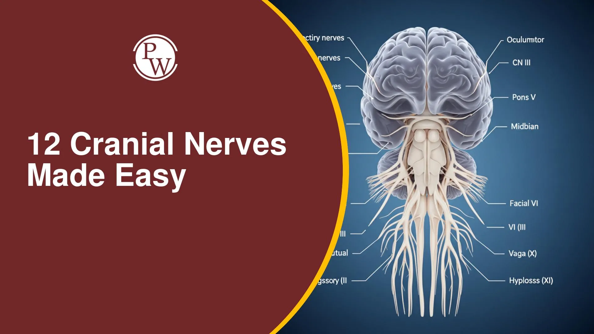

12 Cranial Nerves Made Easy explains how cranial nerves arise and function in a simplified way. Out of 12 cranial nerves, two originate from the forebrain, while the rest emerge from the brainstem (midbrain, pons, and medulla). Understanding their pathways is key to mastering neuroanatomy for exams.
Here highlights key functions, nuclei, and clinical relevance like Wallenberg Syndrome. Important nerves such as the trigeminal and vagus nerve control sensory, motor, and autonomic functions. Learning the front view of the brain stem helps students quickly identify nerve origins and improves retention of complex concepts.
Cranial Nerves
Cranial nerves are 12 pairs of nerves that emerge directly from the brain, not the spinal cord. They control many sensory and motor functions of the head and neck. Two pairs come from the forebrain, while the remaining ten originate from the Brainstem. Understanding these nerves is fundamental for medical and anatomy students.
Cranial Nerve Origins from the Brainstem
The Brainstem is crucial for many vital functions and serves as the origin point for most cranial nerves. It comprises three main parts: the Midbrain, Pons, and Medulla Oblongata.
-
Cranial nerves III and IV (Oculomotor and Trochlear) arise from the Midbrain.
-
Cranial nerve V (Trigeminal) emerges from the Pons.
-
Cranial nerves VI, VII, and VIII (Abducens, Facial, Vestibulocochlear) emerge at the junction of the Pons and Medulla, called the pontomedullary junction.
-
Cranial nerves IX, X, XI, and XII (Glossopharyngeal, Vagus Nerve, Accessory, Hypoglossal) originate from the Medulla Oblongata.
-
Nerves IX, X, XI arise behind the olive, a prominent structure in the medulla.
-
Nerve XII is located in front of the olive, between the pyramid and the olive. This arrangement highlights the precise anatomical pathways of these vital cranial nerves.
Trigeminal Nerves and Their Roles
The Trigeminal Nerves (Cranial Nerve V) are the thickest cranial nerves. They are mixed nerves, meaning they carry both motor and sensory information. They originate primarily from the Pons. The trigeminal nerve has four key nuclei: one motor and three sensory.
-
Motor Nucleus: Located in the pons, it controls the muscles of mastication (chewing). These muscles develop from the first pharyngeal arch.
-
Sensory Nuclei:
-
Main Sensory Nucleus: Also in the pons, it processes touch sensation and vibration from the face.
-
Mesencephalic Sensory Nucleus: Extends into the midbrain and is responsible for proprioception. This includes position sense for the eyeball, tongue, and mandible.
-
Spinal Sensory Nucleus: Extends into the spinal cord, receiving pain and temperature sensations from the face on the same side (ipsilateral).
Masseter Reflex (Jaw Jerk)
The Masseter Reflex (Jaw Jerk) is a monosynaptic reflex involving the Trigeminal Nerves. When the jaw is gently tapped downward, the masseter muscle contracts, causing the mouth to close.
This reflex relies on the mesencephalic sensory nucleus detecting the jaw's position change and signalling the motor nucleus to activate the mastication muscles. It is a quick, involuntary response demonstrating normal nerve function.
Nucleus Tractus Solitarius
The Nucleus Tractus Solitarius (NTS) is a crucial brainstem nucleus for taste sensation. It is located in the lateral medulla. Different cranial nerves carry taste information to the NTS from various parts of the tongue:
-
The Facial Nerve (CN VII) brings taste from the anterior two-thirds of the tongue.
-
The Glossopharyngeal Nerve (CN IX) carries taste from the posterior one-third of the tongue.
-
The Vagus Nerve (CN X) transmits taste sensations from the epiglottis and pharynx. This collective input ensures a comprehensive sense of taste.
Vagus Nerve
The Vagus Nerve (Cranial Nerve X) is the longest cranial nerve, extending from the brainstem down to the abdomen. It plays a wide role in autonomic functions, impacting the heart rate, digestion, and speech.
-
Supply Areas: It supplies structures in the head, neck, thorax, and abdomen, including the larynx, kidneys, and upper ureters.
-
Functions: It controls muscles of the pharynx, palate, and larynx, essential for speech and swallowing. The vagus nerve also forms a complex with the cranial part of the accessory nerve, impacting these functions.
Clinical Features of Lateral Medullary Ischemia or Wallenberg Syndrome
Clinical features of Lateral Medullary Ischemia or Wallenberg Syndrome arise from damage to the lateral part of the medulla oblongata. This condition affects several critical nuclei, leading to distinct symptoms:
-
Vertigo: Due to injury to the vestibular nucleus.
-
Ipsilateral facial pain and temperature loss: Caused by damage to the spinal nucleus of the Trigeminal Nerves.
-
Loss of taste sensation: Results from injury to the Nucleus Tractus Solitarius.
-
Difficulty swallowing and speaking: Occurs due to damage to the Nucleus Ambiguus, affecting muscles of the palate, pharynx, and larynx.
This syndrome highlights the precise functions of the cranial nerves and their nuclei.
12 Cranial Nerves Made Easy FAQs
Which cranial nerves originate from the forebrain?
Cranial nerves I (Olfactory) and II (Optic) originate from the forebrain
Which cranial nerve is the thickest, and where does it originate?
The Trigeminal Nerves (CN V) are the thickest and originate from the Pons.
What does the mesencephalic sensory nucleus of the trigeminal nerve primarily receive?
It primarily receives proprioception, including position sense from the eyeball, tongue, and mandible.
What is the primary function of the Vagus Nerve?
The Vagus Nerve controls various autonomic functions including heart rate, digestion, and muscles for speech and swallowing.
What causes the difficulty in speech and swallowing seen in Wallenberg Syndrome?
This is due to injury to the Nucleus Ambiguus, which controls muscles of the palate, pharynx, and larynx.

Free Learning Resources
PW Books
Notes (Class 10-12)
PW Study Materials
Notes (Class 6-9)
Ncert Solutions
Govt Exams
Our Other Websites
Class 6th to 12th Online Courses
Govt Job Exams Courses
UPSC Coaching
Defence Exam Coaching
Gate Exam Coaching
Other Exams
Know about Physics Wallah
Physics Wallah is an Indian edtech platform that provides accessible & comprehensive learning experiences to students from Class 6th to postgraduate level. We also provide extensive NCERT solutions, sample paper, NEET, JEE Mains, BITSAT previous year papers & more such resources to students. Physics Wallah also caters to over 3.5 million registered students and over 78 lakh+ Youtube subscribers with 4.8 rating on its app.
We Stand Out because
We provide students with intensive courses with India’s qualified & experienced faculties & mentors. PW strives to make the learning experience comprehensive and accessible for students of all sections of society. We believe in empowering every single student who couldn't dream of a good career in engineering and medical field earlier.
Our Key Focus Areas
Physics Wallah's main focus is to make the learning experience as economical as possible for all students. With our affordable courses like Lakshya, Udaan and Arjuna and many others, we have been able to provide a platform for lakhs of aspirants. From providing Chemistry, Maths, Physics formula to giving e-books of eminent authors like RD Sharma, RS Aggarwal and Lakhmir Singh, PW focuses on every single student's need for preparation.
What Makes Us Different
Physics Wallah strives to develop a comprehensive pedagogical structure for students, where they get a state-of-the-art learning experience with study material and resources. Apart from catering students preparing for JEE Mains and NEET, PW also provides study material for each state board like Uttar Pradesh, Bihar, and others
Copyright © 2026 Physicswallah Limited All rights reserved.