Abdominal Anatomy: Understanding Hernia Through Layers, Muscles, Rectus Sheath, and Inguinal Canal

Understanding hernia begins with understanding the ll. The abdomen is not just a hollow cavity; it is protected by multiple layers of skin, fascia, muscles, and aponeuroses arranged in a precise structural pattern. These layers provide strength and support, yet certain natural weak points exist where this support is comparatively thinner. It is at these anatomical weak areas that hernias commonly develop.
Also Read - NEET PG 2026
By studying how the oblique muscles, rectus abdominis, and their aponeuroses form the rectus sheath and by identifying landmarks such as the arcuate line, Spigelian fascia, and the inguinal canal we can clearly understand why some regions are more prone to herniation than others. This approach transforms hernia from a mere surgical condition into a practical application of abdominal wall anatomy.
Also Read - NEET PG 2026 Preparation
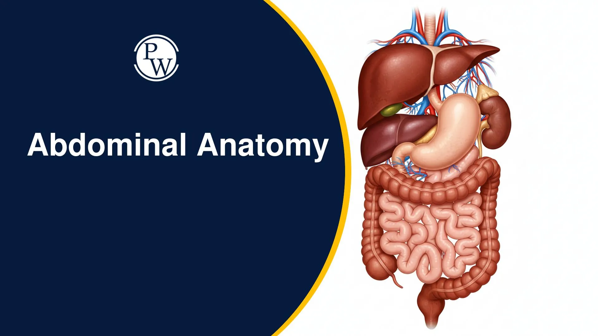
Abdominal Anatomy for Hernia
A hernia fundamentally involves the protrusion of abdominal viscera through an anatomical weakness in the abdominal wall. While hernias can manifest in various body regions, this discussion specifically focuses on those occurring in the abdomen. A thorough understanding of abdominal wall anatomy is paramount to comprehending the formation and types of hernias.
Abdominal Wall Anatomy
To grasp the mechanics of hernia formation, a detailed knowledge of the abdominal wall's structure is essential. We will explore its layers, muscles, aponeuroses, and critical anatomical landmarks.
Layers of the Abdominal Wall (Outside to Inside)
The abdominal wall comprises several distinct layers:
-
Skin
-
Subcutaneous Fat:
-
Camper's Layer: Outer layer.
-
Scarpa's Layer: Inner, fibrous sheet.
-
Abdominal Wall Muscles
-
Aponeuroses
-
Fascia Transversalis
-
Preperitoneal Pad of Fat
-
Peritoneum
-
Intra-abdominal Cavity
Abdominal Wall Muscles
The abdominal muscles are categorized by their location:
-
Lateral Abdominal Wall Muscles:
-
External Oblique Muscle: Fibers run downwards and medially (like hands in pockets).
-
Internal Oblique Muscle: Fibers run upwards and medially (opposite to external oblique).
-
Transverse Abdominis Muscle: Fibers run horizontally.
-
Central Abdominal Wall Muscles:
-
Rectus Abdominis Muscle: Located vertically in the center.
Aponeuroses and Rectus Sheath Formation
Where muscles transition to fibrous tissue, they form aponeuroses:
-
External Oblique Aponeurosis
-
Internal Oblique Aponeurosis: Divides into an anterior leaflet and a posterior leaflet.
-
Transverse Abdominis Aponeurosis
These aponeuroses contribute to the rectus sheath, which encloses the rectus abdominis muscle:
-
Anterior Rectus Sheath: Formed by the external oblique aponeurosis and the anterior leaflet of the internal oblique aponeurosis.
-
Posterior Rectus Sheath: ormed by the transverse abdominis aponeurosis and the posterior leaflet of the internal oblique aponeurosis.
Pathway to the Abdomen
Understanding the layered entry points into the abdomen is crucial:
|
Pathway to the Abdomen |
|
|---|---|
|
Pathway |
Layers (Outside to Inside)
|
|
Lateral Approach |
Skin → Camper's → Scarpa's → External Oblique → Internal Oblique → Transverse Abdominis → Fascia Transversalis → Preperitoneal Pad of Fat → Peritoneum → Intra-abdominal Cavity |
|
Midline Approach |
Skin → Camper's → Scarpa's → Anterior Rectus Sheath → Rectus Abdominis → Posterior Rectus Sheath → Fascia Transversalis → Preperitoneal Pad of Fat → Peritoneum → Intra-abdominal Cavity |
Abdominal Wall Landmarks and Lines
Key landmarks and fibrous lines define abdominal regions:
-
Xiphisternum: Cartilaginous process at the inferior end of the sternum.
-
Umbilicus: Navel.
-
Pubic Symphysis: Cartilaginous joint connecting the left and right pubic bones.
-
Linea Alba: A central fibrous band formed by the fusion of rectus sheaths from xiphisternum to pubic symphysis.
-
Linea Semilunaris: A curved line marking the lateral border of the rectus abdominis muscle, where it meets the lateral abdominal wall muscles.
Posterior Rectus Sheath Variation and Weaknesses
The posterior rectus sheath exhibits a critical variation:
It begins at the xiphisternum but does not cover the entire rectus abdominis muscle. Approximately 2 cm below the level of the umbilicus, the posterior rectus sheath terminates by piercing the rectus abdominis muscle and merging with the anterior rectus sheath. This point is known as the arcuate line.
-
Arcuate Line: The point, typically 2 cm below the level of the umbilicus, where the posterior rectus sheath ends, leaving the lower rectus abdominis muscle uncovered posteriorly. (Memory Tip: Remember 'Arcuate' sounds like 'arch,' indicating a curve or end point for the sheath). This line marks a significant area of posterior abdominal wall weakness.
Another key area of weakness is the Spigelian Fascia:
-
Spigelian Fascia: An area of anatomical weakness where the rectus abdominis muscle and the lateral abdominal wall muscles meet. This small junction is covered only by a thin fascia, leaving it vulnerable to hernia formation. A hernia through this area is termed a Spigelian hernia, commonly occurring at or below the arcuate line. (Memory Tip: Arcuate Line is not the same as Spigelian Fascia).
Sites of Hernia Development
Hernias can emerge at various locations across the abdominal wall:
-
Groin Region:
-
Inguinal Hernia: Above the pubic symphysis or pubic tubercle.
-
Femoral Hernia: Just below the inguinal hernia in the groin.
-
Obturator Hernia: On the medial aspect of the upper thigh.
-
Lateral Abdominal Wall:
-
Lumbar Hernia
-
Spigelian Hernia: Along the linea semilunaris, typically below the umbilicus.
-
Midline (Ventral Hernias): Occur along the linea alba from xiphisternum to pubic symphysis.
-
Epigastric Hernia: Above the umbilicus.
-
Umbilical Hernia: At the umbilicus (including what was previously called paraumbilical hernia).
-
Hypogastric Hernia: Below the umbilicus.
-
Other:
-
Incisional Hernia: Occurs at the site of a prior surgical incision.
Most Common Hernias
Understanding the prevalence of different hernia types is crucial:
-
Overall Most Common Hernia: Inguinal hernia, with the indirect inguinal hernia being more frequent than the direct variety.
-
Most Common Hernia in Females: Still inguinal hernia, predominantly the indirect inguinal hernia.
-
Femoral Hernia: More common in females when compared to males, though less common than inguinal hernias overall.
Inguinal Canal Anatomy
The inguinal canal is a critical passage in the groin region, central to understanding inguinal hernias.
Inguinal Ligament and Superficial Inguinal Ring
The external oblique aponeurosis plays a key role here:
-
Inguinal Ligament: Formed as the inferior border of the external oblique aponeurosis condenses, attaching from the anterior superior iliac spine to the pubic tubercle. (Memory Tip: The inguinal ligament is formed by the external oblique aponeurosis turning back on itself, making it a thick, strong band).
-
Superficial Inguinal Ring: An opening created as the external oblique aponeurosis arches backward near the pubic tubercle. It represents a defect within this aponeurosis.
-
Lacunar Ligament: A fan-shaped extension of the inguinal ligament from the pubic tubercle.
-
Cooper's Ligament: An extension of the lacunar ligament.
-
Femoral Ring: A small opening located near the superficial inguinal ring.
Structure and Definition of the Inguinal Canal & Associated Rings
The fascia transversalis is the innermost continuous fascial layer of the abdominal wall, anteriorly reinforced by the external oblique aponeurosis. The inguinal canal is defined by two key rings:
-
Superficial Inguinal Ring: A defect within the external oblique aponeurosis, formed by its backward turning near the pubic tubercle.
-
Deep Inguinal Ring: A defect found within the fascia transversalis.
Journey of the Vas Deferens
The vas deferens follows a complex path:
-
Ascends from the testes and enters the superficial inguinal ring.
-
Traverses the inguinal canal.
-
Exits from the deep inguinal ring.
-
Immediately makes an abrupt U-turn.
-
Runs between the peritoneum and the fascia transversalis.
-
Passes behind the bladder.
-
Combines with the seminal vesicle (via the seminal duct).
-
Forms the ejaculatory duct.
-
Passes through the prostate gland.
-
Opens into the prostatic urethra at the verumontanum.
Read More: Types of Shock
Anatomical Orientation and Boundaries of the Inguinal Canal
Key landmarks that define the region include the anterior superior iliac spine, pubic tubercle, pubic symphysis, and pubic bone.
The inguinal canal's main layers, from innermost to outermost, are the peritoneum, fascia transversalis (containing the deep inguinal ring as an inverted U-shaped defect), and external oblique aponeurosis (forming the superficial inguinal ring).
The boundaries of the inguinal canal are:
-
Anterior Border: Formed by the external oblique aponeurosis.
-
Inferior Border: Formed by the external oblique aponeurosis.
-
Posterior Border: Formed by the fascia transversalis.
(Memory Tip: The Superficial Inguinal Ring is a "U-turn" or "flip" of the external oblique aponeurosis, while the Deep Inguinal Ring is a "defect" in the fascia transversalis).
Contents of the Inguinal Canal
The inguinal canal houses different structures depending on biological sex:
-
In Males: Vas deferens
-
In Females: Round ligament
In Both Sexes: Ilioinguinal nerve
Abdominal Anatomy for Hernia FAQs
What is the basic definition of a hernia?
What is the significance of the arcuate line in abdominal anatomy?
Which type of hernia is most common overall, and in females specifically?
What is the exam pattern for INI CET 2026?
What structures form the superficial and deep inguinal rings?


Get Free Counselling Today
and Clear up all your Doubts