BDS 1st Year Anatomy Muscles of Mastication - Origin, Insertion, Nerve Supply & Actions

The muscles of mastication are an important topic in BDS 1st Year Anatomy because they play a major role in chewing, speaking, and jaw movements. These muscles work together to control movements of the mandible, such as elevation, depression, protrusion, retraction, and side-to-side motion.
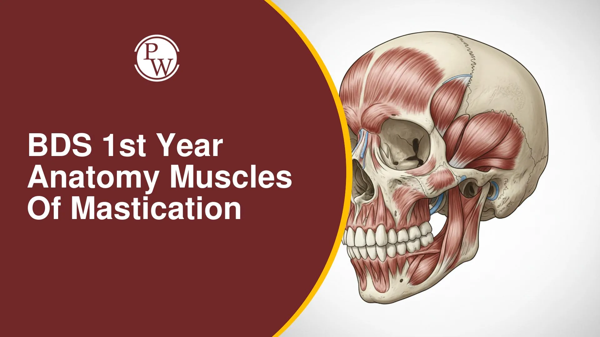
The four primary muscles of mastication are Masseter, Temporalis, Medial Pterygoid, and Lateral Pterygoid. Understanding their origin, insertion, nerve supply, and actions is essential not only for university examinations but also for clinical dental practice and the study of temporomandibular joint (TMJ) movements.
Mandibular Movement During Mastication
During mastication, the mandible undergoes rotatory movement, incorporating side shifts and movements across multiple planes. Observe yourself eating in slow motion: the mandible moves rotarily. Consider how a cow chews; its jaw movements are very evident and rotary. Our movements are similar, though less marked. The effective movement of the lower jaw (mandible) is essential for both eating and speaking.
Embryological Origin and Nerve Supply
The Muscles of Mastication develop from the mesoderm of the first branchial arch. This embryological origin is increasingly relevant in examinations. Most of these muscles are supplied by the mandibular nerve, which innervates them upon exiting the foramen ovale.
Overview of Muscles of Mastication and their Fiber Orientation
Understanding the fiber orientation provides insight into the actions of the muscles of mastication relative to the mandible:
-
Masseter: Located on the outer aspect of the mandible, originating from the zygomatic arch. Its fibers run downward, pulling the mandible upward and slightly forward. The Masseter's fibers are on the outer side of the mandible, visualize pulling the mandible upwards with a rope.
-
Temporalis: A fan-shaped muscle that attaches to the mandible's coronoid process. Its fibers are oriented to pull the mandible upward and backward. Imagine a hand fan fitting into the temporal fossa.
-
Lateral Pterygoid: Distinguished by its horizontal orientation of fibers, which primarily leads to protrusion rather than elevation. It is closely related to the capsule of the Temporomandibular Joint (TMJ). The Lateral Pterygoid is unique because its fiber orientation is distinct from other masticatory muscles.
-
Medial Pterygoid: Located on the inner side of the mandible, opposite to the Masseter. It has a similar fiber orientation to the Masseter (vertical) and pulls the mandible upward and slightly forward.
The orientation of the muscle fibers directly indicates their action. By observing fiber direction, one can infer whether a muscle elevates, retracts, or protrudes the mandible.
Detailed Muscle Analysis
The muscles of mastication are responsible for various movements of the mandible required for chewing, speaking, and swallowing. Understanding the origin, insertion, nerve supply, and actions of these muscles is important for BDS anatomy exams and clinical applications.
1. Masseter Muscle
-
Origin: Composed of three layers:
-
Superficial Head: From the anterior two-thirds of the lower border of the zygomatic arch and adjoining zygomatic process. Fibers run downward and backward at a 45-degree angle.
-
Middle Layer: From the lower border of the posterior one-third of the zygomatic arch.
-
Deep Layer: From the deep surface of the zygomatic arch.
-
Insertion: All fibers insert onto the outer aspect of the ramus of the mandible, with the deep layer attaching to the rest of the ramus.
-
Nerve Supply: Masseteric nerve (branch of the mandibular nerve).
-
Action:
-
Elevation of the mandible (primary action).
-
Slight protrusion.
2. Temporalis Muscle
-
Characteristics: A fan-shaped muscle filling the temporal fossa.
-
Origin: Arises from the temporal fossa (excluding the zygomatic bone) and the temporal fascia.
-
Insertion: Runs vertically downwards and inserts into the coronoid process (anterior border and deep surface) and the anterior border of the ramus of the mandible. The attachment is like a fan holding the mandible.
-
Nerve Supply: Temporal branches of the mandibular nerve.
-
Action:
-
Elevation of the mandible.
-
Retraction of the mandible.
-
Contributes to side-to-side movements.
3. Medial Pterygoid Muscle
It is crucial to note that the Medial Pterygoid muscle has no direct origin from the Medial Pterygoid plate. Both the Medial and Lateral Pterygoid muscles are related to the Lateral Pterygoid plate. The Medial surface of the Lateral Pterygoid plate is the origin for the Medial Pterygoid muscle, while the Lateral surface of the Lateral Pterygoid plate is for the Lateral Pterygoid muscle.
-
Origin:
-
Superficial Head: Arises from the maxillary tuberosity.
-
Deep Head: Arises from the medial surface of the lateral pterygoid plate.
-
Insertion: Fibers run downward and backward, inserting into the medial surface of the angle and adjoining ramus of the mandible, specifically below and behind the mandibular foramen. The Medial Pterygoid is like the Masseter but on the inner aspect of the mandible.
-
Nerve Supply: Nerve to Medial Pterygoid (branch from the main trunk of the mandibular nerve).
-
Action:
-
Elevation of the mandible.
-
Slight protrusion of the mandible.
-
Assists in side-to-side movements.
4. Lateral Pterygoid Muscle
The Lateral Pterygoid muscle is a frequently tested concept due to its unique action. Unlike other muscles of mastication, its fiber orientation is horizontal, allowing it to perform distinct actions.
-
Origin:
-
Upper Head: Arises from the infratemporal fossa.
-
Lower Head: Arises from the lateral surface of the lateral pterygoid plate.
-
Insertion: Fibers run backward and laterally, inserting into the pterygoid fovea (a depression on the condyle), the anterior margin of the articular disc, and the capsule of the TMJ.
-
Nerve Supply: Branches from the anterior division of the mandibular nerve.
-
Action:
-
The Lateral Pterygoid is the only muscle that depresses the mandible (during mouth opening).
-
Protrusion of the mandible.
-
Assists in side-to-side movements.
Clinical Relevance of Muscles of Mastication
Abnormalities related to these muscles can lead to various clinical issues, including TMJ issues, muscle spasm, reduced mouth opening, and conditions like Tetanus and Lockjaw. These muscles are crucial for balanced mastication. Understanding them is essential in dentistry, as prolonged appointments can sometimes lead to mandibular dislocation.
Origin of Pterygoid Muscles
This is a very frequently asked question in exams, and it is crucial not to get confused. Both the Medial Pterygoid Muscle and the Lateral Pterygoid Muscle originate from the Lateral Pterygoid Plate. Specifically, the Medial Pterygoid Muscle originates from the medial surface of the Lateral Pterygoid Plate, and the Lateral Pterygoid Muscle originates from the lateral surface of the Lateral Pterygoid Plate. Do not write Medial Pterygoid Plate as the origin for either muscle.
Importance and Specific Focus Areas
Understanding the muscles of mastication is a very high-yield topic for university and MBBS exams, and is considered the "bread and butter" of dental practice. To understand muscle movements, simply visualize the direction of the muscle fibers; their contraction will dictate the movement, thus removing the need for rote memorization. It is highly recommended to focus more on the Lateral Pterygoid, as many questions, including those in viva and prosthetics viva, have been asked about it in recent years due to its unique actions.
Mandibular Deviation and Hemimandibulectomy
Mandibular deviation refers to lateral deviation, not elevation or protrusion. The Lateral Pterygoid is the muscle primarily responsible for lateral deviation (turning the chin to the side). In cases of Hemimandibulectomy (resection of half the mandible, often due to cancer), the unbalanced muscle pull from the intact side, particularly the Lateral Pterygoid, leads to a noticeable deviation of the mandible towards the operated side.
BDS 1st Year Anatomy Muscles of Mastication FAQs
What are the primary muscles of mastication?
Which nerve supplies the muscles of mastication?
Which muscle is solely responsible for mandibular depression (mouth opening)?
From which embryological structure do the muscles of mastication develop?


Get Free Counselling Today
and Clear up all your Doubts