Traumatic Brain Injury: Types and Difference

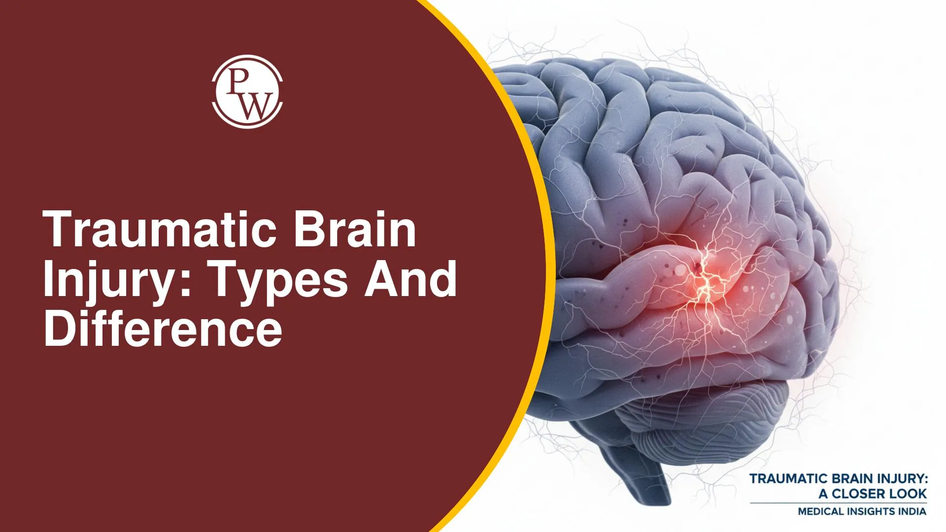
A traumatic brain injury (TBI) signifies a deep impact affecting the brain itself, distinguishing it from minor head injuries like lacerations or skull fractures. While not all head injuries result in brain injury, a TBI presents a more complex medical challenge due to its direct effect on brain function and integrity.
Primary Brain Injury vs. Secondary Brain Injury
Brain injuries are broadly categorized based on their mechanism and timing relative to the initial trauma. This distinction is clear, simple, and crisp.
|
Primary Brain Injury vs. Secondary Brain Injury |
||
|---|---|---|
|
Feature |
Primary Brain Injury |
Secondary Brain Injury
|
|
Definition |
Direct injury to the brain due to impact from trauma |
Injury to the brain due to increased intracranial pressure as a result of primary brain injury |
|
Mechanism |
Direct impact |
Trauma causes injury → inflammation → edema → increased intracranial pressure → further worsening of injury |
|
Nature |
Direct |
Not direct; a response to primary brain injury |
Types of Primary Brain Injury
Primary brain injuries manifest in several forms, each with distinct characteristics and clinical implications. The three predominant types are Concussion Injury, Diffuse Axonal Injury (DAI), and Intracranial Hemorrhage.
Concussion Injury
A concussion is a transient state of confusion following head trauma. It is usually a reversible injury with no structural damage. Diagnosis relies on normal NCCT (Non-Contrast Computed Tomography) findings and the transient confusion itself. Management involves only best supportive care. (Memory Tip: NICE guidelines suggest admitting the patient and observing for 24 hours.)
Diffuse Axonal Injury (DAI)
Diffuse axonal injury (DAI) involves the complete disruption of axons primarily between the central white matter and the peripheral gray matter. It occurs due to sudden decelerating injury, such as in road traffic accidents. The mechanism involves the relatively fixed white matter and mobile gray matter experiencing differential movement, causing axons connecting them to break away at their junction (a "whiplash" effect).
Patients typically present in a coma (non-responsive, GCS 3/5) post-trauma. NCCT findings are usually normal, which is a very early point for understanding DAI. MRI is more diagnostic, showing punctate hemorrhage between the white and gray matter. The prognosis for DAI is very poor, with most patients entering a vegetative state. In advanced CT, specific findings suggesting DAI can include hemorrhagic foci in the corpus callosum and the dorsolateral rostral of the brainstem.
Intracranial Hemorrhage (ICH)
Intracranial hemorrhage (ICH) refers to bleeding within or around the brain parenchyma. Understanding the brain's anatomy is crucial for localizing these bleeds.
Anatomy & Meningeal Layers:
-
Innermost layer: Pia mater (adherent to brain).
-
Surrounding Pia: Arachnoid membrane.
-
Surrounding Arachnoid: Dura mater (adherent to skull).
-
Outermost: Skull bone.
-
CSF Location: In the subarachnoid space (between pia mater and arachnoid membrane).
Locations of Bleeds:
-
Intraparenchymal Hemorrhage: Inside the brain parenchyma.
-
Subarachnoid Hemorrhage (SAH): Between the arachnoid membrane and the pia mater (in the CSF space).
-
Subdural Hemorrhage (SDH): Between the dura mater and the arachnoid membrane.
-
Extradural Hemorrhage (EDH) / Epidural Hemorrhage: Between the dura mater and the skull bone.
The most common traumatic hemorrhage overall is Intraparenchymal Hemorrhage. In the case of extradural hemorrhage, the dura mater's adherence to the skull means any bleed expands inwards, compressing the brain. This compressive force can lead to brain herniation.
Extradural Hemorrhage (EDH) vs. Subdural Hemorrhage (SDH)
These two types of intracranial hemorrhage differ significantly in their etiology, characteristics, and clinical presentation.
|
Extradural Hemorrhage (EDH) vs. Subdural Hemorrhage (SDH) |
||
|
Feature |
Extradural Hemorrhage (EDH) |
Subdural Hemorrhage (SDH)
|
|
Definition |
Collection of blood between the dura and the skull bone |
Collection of blood between the dura and the arachnoid membrane |
|
Mode of Injury |
Usually road traffic accident (high velocity injury); often secondary to temporal fracture (at pterion). |
Usually seen in elderly patients with significant fall or head impact. |
|
Most Common Source of Bleeding |
Middle meningeal artery (arterial bleed) |
Cortical bridging veins (venous bleed) |
|
Hematoma Characteristics |
- Confined to an area (due to dura-skull adhesion). - Expands towards the brain. - Severe downward pressure. - Surgical emergency due to rapid, localized pressure. |
- Occupies a wider surface area (spreads more easily). - Spreads across the brain surface. - Lesser impact on brain parenchyma (less severe pressure symptoms) for the same volume compared to EDH. |
|
Clinical Features (Raised ICP) |
All lesions cause signs/symptoms of raised intracranial pressure (space-occupying effect). |
All lesions cause signs/symptoms of raised intracranial pressure. |
|
Onset of Symptoms |
Sudden, severe onset of symptoms |
Gradual and slow progressive symptoms |
|
Specific Symptoms |
- Severe headache - Projectile vomiting - Blurring of vision - Post-traumatic seizures - Ipsilateral dilated pupil - Contralateral hemiparesis |
- Severe headache - Vomiting - Blurring of vision - (Post-traumatic seizures usually not present) - Ipsilateral dilated pupil - Contralateral weakness |
|
Lucid Interval |
Present |
Absent |
|
Memory Tip |
A lucid interval is eriod of consciousness between two unconscious episodes. It indicates a high-pressure arterial bleed. This occurs because initial bleeding causes unconsciousness, then the hematoma temporarily compresses the bleeding vessel, leading to temporary consciousness. Movement can dislodge the clot, causing re-bleeding and a second episode of unconsciousness. |
Not typically associated with a lucid interval. |
|
NCCT Head Findings |
Biconvex opacity / Lenticular opacity |
Concavoconvex opacity / Crescent opacity |
|
Management |
Emergency Bur-hole surgery (evacuate hematoma, side of dilated pupil). Definitive: Craniotomy to ligate bleeding vessel. |
- Small SDH: Conservative (if asymptomatic). - Large/symptomatic SDH: Craniotomy. |
Secondary Brain Injury
Secondary brain injury is damage to the brain resulting from increased intracranial pressure following a primary brain injury. Primary trauma can lead to cerebral edema, which further elevates intracranial pressure.
Cerebral Perfusion Pressure (CPP)
Cerebral Perfusion Pressure (CPP) is the pressure required to perfuse the brain. It is calculated as:
CPP = Mean Arterial Pressure (MAP) - Intracranial Pressure (ICP)
Normal MAP ranges from 90-110 mmHg, and normal ICP is 5-15 mmHg, yielding a normal CPP of 75-105 mmHg. The minimum CPP required to perfuse the brain is ≥ 65 mmHg (or 60-65 mmHg). If CPP falls below this threshold, the brain experiences hypoxia. Brain neurons are the most sensitive tissue to hypoxia, with irreversible damage occurring within 2-4 minutes of severe oxygen deprivation.
Raised Intracranial Pressure (ICP) and its Impact on CPP
An increase in Intracranial Pressure (ICP) directly leads to a decrease in Cerebral Perfusion Pressure (CPP), as shown by the formula.
This drop in CPP results in inadequate blood supply and hypoxia. To counteract this, the body employs a compensatory mechanism: it increases the Mean Arterial Pressure (MAP) to maintain a stable CPP despite elevated ICP.
Mechanism of MAP Increase for ICP Compensation
Mean Arterial Pressure (MAP) is determined by the formula: MAP = (1/3 * Systolic Blood Pressure) + (2/3 * Diastolic Blood Pressure). To increase MAP, the body primarily elevates Systolic Blood Pressure (SBP).
The body increases pressure within vessels by increasing the volume of blood. Vasoconstriction, while increasing pressure, reduces cerebral blood flow, worsening hypoxia. Therefore, increasing blood volume is more beneficial for cerebral oxygenation.
How Blood Volume is Increased
To increase blood volume and subsequently blood pressure, the body increases Cardiac Output. The speaker's simplified calculation for Cardiac Output is: Cardiac Output = Preload - Afterload. To boost cardiac output, the body increases Preload (the blood volume in the left ventricle before contraction).
Increasing preload requires an increased End Diastolic Filling Time (EDFT) for the left ventricle to fill. This results in a decreased heart rate and an increased systolic blood pressure.
Cushing's Triad
Raised ICP can also exert pressure on the cerebral pontine angle, affecting the respiratory center and causing an irregular respiratory rate. These physiological responses combine to form Cushing's Triad, a critical indicator of raised ICP:
-
Bradycardia (decreased heart rate)
-
Hypertension (increased systolic blood pressure)
-
Irregular Respiratory Rate
(Memory Tip: Understanding the body's compensatory mechanisms against raised ICP helps to comprehend how Cushing's Triad develops, rather than simply memorizing its components.) The presence of Cushing's Triad is a crucial sign of raised Intracranial Pressure, regardless of the underlying cause.
Management of Raised Intracranial Pressure (ICP)
For severe increases in ICP, immediate management strategies include:
-
Positioning: Keep the patient in a propped-up position.
-
Oxygenation: Ensure adequate oxygen supply.
-
Intubation: May be necessary if the patient's condition deteriorates.
-
Hyperventilation: This causes carbon dioxide washout, leading to respiratory alkalosis, which can depress the choroid plexus and reduce CSF production, thereby decreasing ICP. However, hyperventilation is now considered questionable and is generally not recommended as a routine measure.
-
Pharmacological Intervention: Administer IV Mannitol to decrease cerebral edema.
Monitoring Targets for ICP Management
Effective management and a positive patient prognosis in the ICU rely on maintaining specific parameters:
-
Mean Arterial Pressure (MAP): 80 to 90 mmHg.
-
Cerebral Perfusion Pressure (CPP): More than 60 mmHg (below this threshold indicates severe brain hypoxia).
-
Intracranial Pressure (ICP): Less than 20 mmHg (normal is 5-15 mmHg).
-
Sodium (Na+): More than 140 mEq/L.
-
Potassium (K+): More than 4 mEq/L.
-
Partial Pressure of Oxygen (Pa): More than 11 kilopascals.
Achieving these target components is crucial for improving outcomes in traumatic brain injuries.
| NEET PG Surgery Video-Based Questions | Video-Based Questions on Larynx for NEET PG |
| Medicine Video-Based Questions to Crack NEET PG | NEET PG 2026 Pediatrics Video-Based Questions |
Traumatic Brain Injury FAQs
What is the primary difference between primary and secondary brain injury?
What are the key characteristics that differentiate Extradural Hemorrhage (EDH) from Subdural Hemorrhage (SDH)?
Why is Diffuse Axonal Injury (DAI) considered to have a very poor prognosis, despite often showing normal findings on NCCT?
How does the body compensate for increased Intracranial Pressure (ICP) to maintain Cerebral Perfusion Pressure (CPP)?
What is Cushing's Triad, and what is its clinical significance?


Get Free Counselling Today
and Clear up all your Doubts