







NEET Human Reproduction and Reproductive Health Notes by PW
 Mehjabeen Hussain14 Oct, 2026
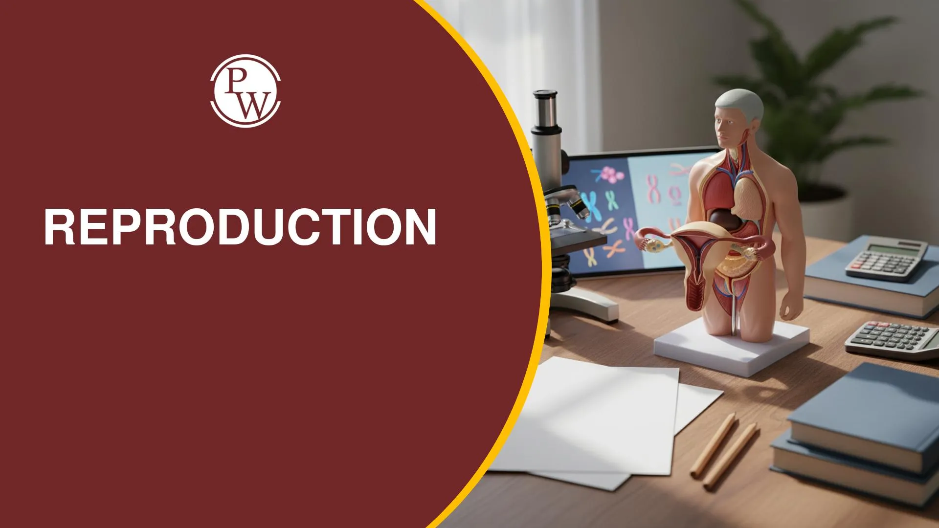
Human reproduction questions often test your ability to connect reproductive structures with their functions and distinguish between processes such as spermiogenesis, spermiation, fertilisation, and implantation. You also need to understand hormonal changes during the menstrual cycle, early embryonic development, contraceptive mechanisms, and the applications of assisted reproductive techniques.
For focused revision, Physics Wallah’s Human Reproduction and Reproductive Health notes help you revisit the chapter’s key concepts, reproductive pathways, hormone functions, and important biological differences. Use these concepts to strengthen your recall and practise applying them to NEET Biology questions.
Major Events of Human Reproduction
Humans are viviparous animals that show internal fertilisation. The reproductive process involves gamete formation, sperm transfer, fertilisation, implantation, pregnancy, and childbirth.
The main sequence of human reproduction is:
Gametogenesis → Insemination → Fertilisation → Implantation → Gestation → Parturition
-
Gametogenesis: Formation of male and female gametes. It includes spermatogenesis and oogenesis.
-
Insemination: Transfer of sperm into the female reproductive tract.
-
Fertilisation: Fusion of the sperm and ovum to form a zygote.
-
Implantation: Attachment of the blastocyst to the uterine endometrium.
-
Gestation: The period of development during pregnancy, lasting approximately nine months.
-
Parturition: The process of childbirth.
Memory tip: Remember the sequence from gamete formation to childbirth: gametogenesis, insemination, fertilisation, implantation, gestation, and parturition.
Male Reproductive System
The male reproductive system consists of the testes, accessory ducts, accessory glands, penis, and scrotum. The testes produce sperm and secrete testosterone, while the ducts transport sperm through the reproductive tract.
Testes and Scrotum
The testes are the primary male reproductive organs. They are located in the scrotum, outside the abdominal cavity, because sperm formation requires a temperature approximately 2–2.5°C below body temperature.
Each testis is approximately 4–5 cm long and 2–3 cm wide. The tunica albuginea divides each testis into about 250 testicular lobules. Each lobule contains one to three seminiferous tubules, where spermatogenesis occurs.
Sperm Transport Pathway
Sperm travel through the following structures:
Seminiferous tubules → Rete testis → Vasa efferentia → Epididymis → Vas deferens → Ejaculatory duct → Urethra
-
Seminiferous tubules: Sites of sperm formation.
-
Rete testis: Collects sperm from the seminiferous tubules.
-
Vasa efferentia: Carry sperm towards the epididymis.
-
Epididymis: Located along the posterior surface of each testis; sperm mature and are stored here.
-
Vas deferens: Transports sperm towards the ejaculatory duct and loops over the urinary bladder.
-
Ejaculatory duct: Formed by the union of the vas deferens and the duct of the seminal vesicle.
-
Urethra: A common urinogenital duct that carries urine or semen out of the body at different times.
Cells of the Testis
Three important cell types are associated with the testes.
-
Spermatogonia: Diploid male germ cells that divide and give rise to cells that enter spermatogenesis.
-
Sertoli cells: Also called nurse or sustentacular cells, they nourish developing sperm, support their development, and remove residual cytoplasm.
-
Leydig cells: Also called interstitial cells, they lie outside the seminiferous tubules and produce testosterone under the influence of luteinising hormone (LH).
Male Accessory Glands
The male reproductive system contains three types of accessory glands:
-
Seminal vesicles: Paired glands located behind the urinary bladder.
-
Prostate gland: A single gland located below the urinary bladder.
-
Bulbourethral glands: Paired glands located beside the urethra.
Their secretions contribute to seminal plasma, which contains substances such as fructose, enzymes, prostaglandins, calcium, and mucus. These secretions support sperm movement and function.
Semen = Sperm + Seminal plasma
Spermatogenesis
Spermatogenesis is the process through which diploid spermatogonia develop into haploid spermatozoa. It begins at puberty and may continue into older age.
Stages of Spermatogenesis
The process involves mitosis, meiosis, and the transformation of spermatids into sperm.
-
Spermatogonia: Diploid cells that divide by mitosis.
-
Primary spermatocyte: Develops from a spermatogonium and undergoes meiosis I.
-
Secondary spermatocytes: Two haploid cells formed after meiosis I.
-
Spermatids: Four haploid cells formed after the two secondary spermatocytes undergo meiosis II.
-
Spermatozoa: Mature sperm formed when spermatids differentiate through spermiogenesis.
Spermiogenesis is the conversion of spermatids into spermatozoa. Spermiation is the release of mature sperm into the lumen of the seminiferous tubules.
One primary spermatocyte produces four haploid spermatids, which develop into four spermatozoa.
Structure of a Sperm
A sperm consists of four main parts:
-
Head: Contains the nucleus and acrosome. The acrosome contains enzymes that help sperm penetrate the oocyte coverings.
-
Neck: Contains centrioles.
-
Middle piece: Contains mitochondria that provide energy for movement.
-
Tail: Helps the sperm move towards the oocyte.
Hormonal Control of Spermatogenesis
Hormonal regulation involves the hypothalamus, anterior pituitary gland, and testes.
-
GnRH: Released by the hypothalamus; stimulates the anterior pituitary.
-
FSH: Acts on Sertoli cells and supports spermatogenesis.
-
LH: Acts on Leydig cells, stimulating testosterone production.
-
Testosterone: Supports sperm production and male reproductive functions.
-
Inhibin: Produced by Sertoli cells and helps regulate FSH secretion.
Memory tip: LH acts on Leydig cells, while FSH acts on Sertoli cells.
Female Reproductive System
The female reproductive system consists of the ovaries, fallopian tubes, uterus, cervix, vagina, external genitalia, and mammary glands. The ovaries produce ova and secrete the hormones oestrogen and progesterone.
Ovary and Fallopian Tube
The ovary is the primary female reproductive organ. Its cortex contains ovarian follicles, while its medulla contains blood vessels and other supporting structures.
The fallopian tube has three main parts:
-
Infundibulum: The funnel-shaped region closest to the ovary. It bears finger-like projections called fimbriae.
-
Ampulla: The wider region of the fallopian tube and the usual site of fertilisation.
-
Isthmus: The narrow region near the uterus.
The pathway is:
Infundibulum → Ampulla → Isthmus → Uterus
Uterus and Its Layers
The uterus is an inverted, pear-shaped organ where implantation and embryonic development occur. Its wall consists of three layers:
-
Perimetrium: The outermost layer.
-
Myometrium: The smooth muscle layer responsible for powerful contractions during labour.
-
Endometrium: The vascular inner lining that undergoes changes during the menstrual cycle and supports implantation.
The cervix is the narrow terminal part of the uterus. The cervical canal opens into the vagina, and the cervix and vagina together form the birth canal.
Mammary Glands
Mammary glands are modified sweat glands. Each gland contains approximately 15–20 lobes, which contain structures involved in milk production and transport.
The pathway of milk is:
Mammary alveoli → Mammary tubules → Mammary ducts → Mammary ampulla → Lactiferous duct → Nipple
Two hormones regulate lactation:
-
Prolactin: Stimulates milk production.
-
Oxytocin: Causes milk ejection by contracting myoepithelial cells.
Colostrum is the first yellowish milk produced after childbirth. It is rich in IgA antibodies, which help provide immune protection to the newborn.
Oogenesis
Oogenesis is the process of female gamete formation. Unlike spermatogenesis, it begins during embryonic development.
Primary oocytes remain arrested in prophase I until puberty. Many ovarian follicles degenerate through a process called follicular atresia. At puberty, approximately 60,000–80,000 primary follicles remain in each ovary.
Stages of Follicular Development
The sequence of follicular development is:
Primary follicle → Secondary follicle → Tertiary follicle → Graafian follicle
During oogenesis, the primary oocyte completes meiosis I to form a secondary oocyte and a polar body. The secondary oocyte begins meiosis II but remains arrested at metaphase II.
Meiosis II is completed only after sperm entry.
Spermatogenesis vs Oogenesis
The two processes differ in their timing and products.
-
Spermatogenesis: One primary spermatocyte produces four haploid spermatids that develop into spermatozoa.
-
Oogenesis: One primary oocyte produces one functional female gamete and polar bodies.
The unequal division of cytoplasm during oogenesis allows the functional female gamete to retain most of the cytoplasm.
Menstrual Cycle
The menstrual cycle involves coordinated changes in the ovaries and uterus. Hormones regulate follicular development, ovulation, and changes in the endometrium.
A typical menstrual cycle lasts about 28 days, although cycle length can vary. In a standard 28-day cycle, ovulation occurs around Day 14.
Phases of the Menstrual Cycle
1. Menstrual phase
If pregnancy does not occur, the corpus luteum degenerates and progesterone levels fall. The uterine lining breaks down and is discharged as menstrual flow.
2. Follicular phase and proliferative phase
FSH promotes the growth and development of ovarian follicles. Developing follicles secrete oestrogen, which supports the repair and growth of the endometrium. This uterine stage is called the proliferative phase.
3. Ovulatory phase
A high oestrogen level triggers a surge in LH. This LH surge causes the Graafian follicle to rupture and release the secondary oocyte. This event is called ovulation.
4. Luteal phase and secretory phase
After ovulation, the ruptured follicle develops into the corpus luteum. It secretes progesterone, which maintains the endometrium in preparation for possible implantation. The corresponding uterine stage is called the secretory phase.
If fertilisation and pregnancy do not occur, the corpus luteum degenerates, progesterone levels decrease, and menstruation begins.
Important hormone relationships:
-
FSH promotes follicular growth.
-
The LH surge triggers ovulation.
-
Oestrogen supports endometrial growth during the proliferative phase.
-
Progesterone maintains the endometrium during the secretory phase.
Fertilisation and Early Embryonic Development
Human fertilisation usually occurs in the ampullary region of the fallopian tube. Before fertilisation, sperm undergo capacitation in the female reproductive tract, preparing them to penetrate the oocyte coverings.
Oocyte Coverings
From the outside towards the oocyte plasma membrane, the coverings and spaces are arranged as follows:
Corona radiata → Zona pellucida → Perivitelline space → Oocyte plasma membrane
Sperm bind to ZP3 receptors on the zona pellucida. The acrosomal reaction releases enzymes, including hyaluronidase and zona lysin, that help the sperm penetrate the oocyte coverings.
Events During Fertilisation
After sperm entry:
-
The secondary oocyte completes meiosis II.
-
The ovum and second polar body are formed.
-
The sperm and ovum nuclei fuse, forming a diploid zygote.
Mechanisms that prevent polyspermy ensure that additional sperm do not enter the oocyte. The cortical reaction modifies the zona pellucida and helps prevent further sperm entry.
Cleavage and Blastocyst Formation
The zygote undergoes successive mitotic divisions called cleavage. The early stages include:
Zygote → 2-cell stage → 4-cell stage → 8-cell stage → 16-cell stage (morula) → Blastocyst
The morula develops into a blastocyst, which has two important components:
-
Trophoblast: The outer cell layer that helps with implantation and contributes to the formation of chorionic villi.
-
Inner cell mass: The group of cells that develops into the embryo.
Implantation occurs when the blastocyst attaches to the uterine endometrium.
Placenta and Its Functions
The placenta develops through interactions between the fetal chorionic villi and the maternal endometrium. It connects the developing fetus with the mother and supports the exchange of nutrients, gases, and waste products.
The placenta also secretes hormones, including:
-
Human chorionic gonadotropin (hCG): Maintains the corpus luteum during pregnancy.
-
Human placental lactogen (hPL): Supports maternal metabolic adaptations during pregnancy.
-
Oestrogen and progesterone: Support pregnancy and changes in reproductive tissues.
-
Relaxin: Helps prepare the maternal reproductive system for childbirth.
Contraceptive Methods
Contraception helps prevent pregnancy. An ideal contraceptive should be effective, reversible, affordable, user-friendly, and associated with minimal side effects.
Natural Methods
Natural methods avoid the use of devices or contraceptive hormones.
-
Periodic abstinence: Avoids intercourse during the fertile period, approximately Days 10–17 of a standard menstrual cycle.
-
Withdrawal or coitus interruptus: Involves withdrawing the penis before ejaculation. It has a relatively high failure rate.
-
Lactational amenorrhoea: May prevent ovulation during the first six months after childbirth when breastfeeding suppresses reproductive hormonal activity. Its effectiveness depends on the conditions being met.
Barrier Methods
Barrier methods prevent sperm from reaching the ovum. Examples include condoms, diaphragms, cervical caps, and vaults.
Condoms also reduce the risk of sexually transmitted infections (STIs), making them important for both pregnancy prevention and sexual health.
Intrauterine Devices (IUDs)
IUDs are placed inside the uterus. They are classified according to their composition and action.
Inert IUDs
Example: Lippes loop.
These devices increase sperm phagocytosis within the reproductive tract.
Copper-releasing IUDs
Examples: CuT, Cu7, and Multiload 375.
Copper ions reduce sperm motility and fertilising capacity.
Hormone-releasing IUDs
Examples: Progestasert and LNG-20.
These devices make the endometrium unsuitable for implantation and thicken cervical mucus, which makes sperm movement more difficult.
Oral Pills, Implants, and Emergency Contraception
Oral contraceptive pills prevent pregnancy by interfering with reproductive processes. Their actions can include suppressing ovulation, thickening cervical mucus, and making the endometrium unsuitable for implantation.
Saheli (Centchroman) is a non-steroidal oral contraceptive taken once a week. It affects normal endometrial development without suppressing ovulation.
Emergency contraceptives can prevent pregnancy after unprotected intercourse and should be used as soon as possible within the recommended time window. The material covered here identifies 72 hours as the relevant window. Emergency contraception is not the same as an abortion pill.
Surgical Contraceptive Methods
Permanent methods prevent gametes from meeting by blocking their transport.
-
Vasectomy: A male sterilisation procedure in which the vas deferens is cut or blocked.
-
Tubectomy: A female sterilisation procedure in which the fallopian tubes are cut or blocked.
These procedures prevent the transport of gametes while generally preserving the production of reproductive hormones.
Sexually Transmitted Infections (STIs)
Sexually transmitted infections are infections that can spread through sexual contact. Important examples include:
-
Chlamydiasis
-
Gonorrhoea
-
Syphilis
-
Genital herpes
-
Genital warts
-
AIDS caused by HIV
-
Hepatitis B
-
Trichomoniasis
Many bacterial STIs can be cured with appropriate treatment, particularly when detected early. HIV infection, genital herpes, and hepatitis B are not completely curable, although medical treatment can help manage these infections.
Condom use, safer sexual practices, early diagnosis, and timely medical consultation help reduce the risk of STIs and their complications.
Human Reproduction and Reproductive Health requires a clear understanding of reproductive anatomy, gametogenesis, hormonal regulation, the menstrual cycle, fertilisation, embryonic development, contraception, STIs, and assisted reproductive techniques. Focus on the correct sequences, hormone functions, reproductive pathways, and differences between similar processes. Physics Wallah’s Human Reproduction and Reproductive Health notes can support your revision and help you strengthen your understanding while practising NEET Biology questions.
NEET Resources You Might Like:
|
Resource |
Link |
|
NEET Syllabus |
|
|
NEET PYQs |
|
|
NEET Mind Maps |
|
|
NEET Sample Papers |
|
|
NEET Formula |
|
|
NEET MCQs |
|
|
NEET Diagrams |
Elevate your NEET preparation with Physics Wallah’s NEET Online Courses. Get access to comprehensive live classes, expert guidance, quality study materials, practice tests, and more—all designed to help you prepare effectively for the NEET exam. Explore Now!
FAQs
What Is the Difference Between Spermiogenesis and Spermiation?
Where Does Fertilisation Occur in Humans?
What Resources Does PW Provide for NEET Preparation?


Have questions about your prep?
Our experts can answer all your questions






 Live & recorded classes available at easeDashboard for progress trackingLakhs of practice questions