








NEET Structural Organisation in Animals: Animal Tissues, Frog and Cockroach Notes by PW
 Mehjabeen Hussain12 Oct, 2026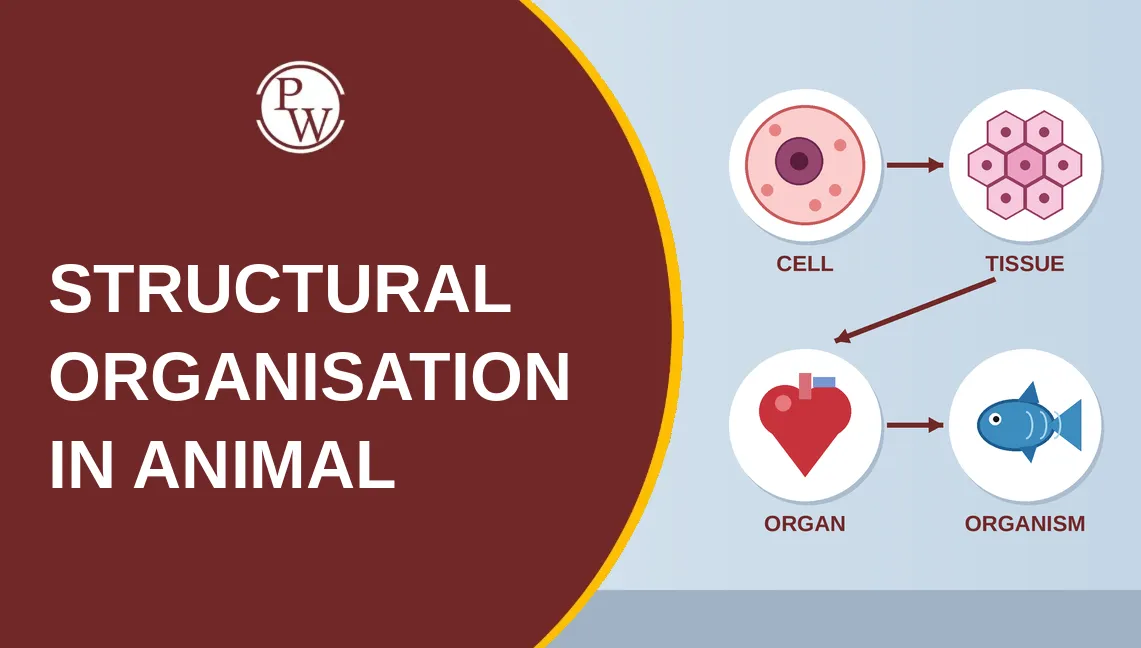
Animal tissue questions often test how well you can distinguish between similar structures, identify their locations, and connect each tissue with its function. Frog and cockroach questions also require familiarity with anatomical features and processes, including circulation, respiration, excretion, and reproduction.
For targeted revision, Physics Wallah’s Structural Organisation in Animals notes bring together the chapter’s key tissue classifications and animal anatomy concepts. Use them to revisit important characteristics, compare similar structures, and recall the biological details needed to answer NEET questions accurately.
Animal Tissues and Levels of Organisation
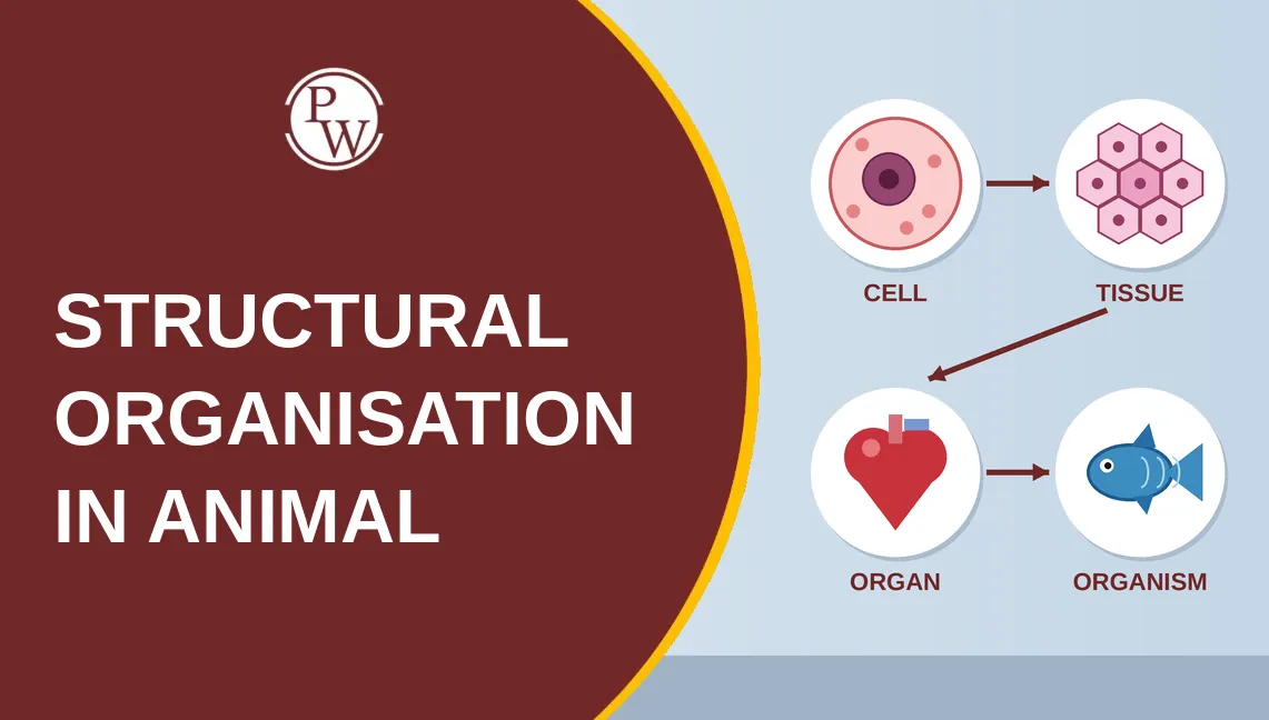
Animals are multicellular, heterotrophic organisms that generally obtain food through holozoic nutrition. Their bodies show different levels of organisation, progressing from cells to tissues, organs, and organ systems.
Cells → Tissues → Organs → Organ Systems
For example, the digestive system contains organs such as the oesophagus, stomach, and intestines. Each organ contains different tissues that work together to perform specific functions.
A tissue is a group of similar cells, along with their intercellular material, that perform a particular function. The extracellular matrix may contain substances such as glycoproteins and mucopolysaccharides.
The four basic types of animal tissues are:
-
Epithelial tissue: Covers body surfaces and lines cavities; it also performs protection, secretion, and absorption.
-
Connective tissue: Supports, binds, and connects different body structures.
-
Muscular tissue: Produces movement through contraction.
-
Neural tissue: Receives stimuli and coordinates responses.
Epithelial Tissue
Epithelial tissue, or epithelium, covers body surfaces and lines internal cavities and organs. Its cells are closely packed, leaving very little intercellular space. The epithelium rests on a non-cellular basement membrane that separates it from the underlying connective tissue.
The basement membrane contains glycoproteins and other extracellular components. It is associated with the epithelium and the connective tissue beneath it.
Epithelium is avascular, meaning it does not contain blood vessels. Oxygen and nutrients reach its cells by diffusion from the underlying vascular connective tissue. Epithelial tissue also has a high capacity for regeneration and performs protective, absorptive, and secretory functions.
The basal surface faces the basement membrane, while the apical or free surface faces the external environment, a body cavity, or a fluid-filled space.
Epithelial Tissue: Important Features
-
Compact arrangement: Epithelial cells are closely packed together.
-
Basement membrane: Supports the epithelium and separates it from underlying connective tissue.
-
Avascular nature: Nutrients and oxygen reach the cells through diffusion.
-
Regeneration: Many epithelial tissues can replace damaged or lost cells.
Free-Surface Modifications
Certain epithelial cells have specialised surface structures that support their functions.
Microvilli: These are small, finger-like projections that increase the surface area available for absorption or reabsorption. They are prominent in the small intestine and proximal convoluted tubules of the kidneys.
Cilia: These are hair-like projections that move substances along the epithelial surface. For example, cilia help move mucus in parts of the respiratory tract and assist in the movement of the ovum through the fallopian tube.
Classification of Epithelium
Epithelial tissue is broadly classified into simple and compound epithelium based on the number of cell layers.
Simple Epithelium
Simple epithelium consists of a single layer of cells. It commonly performs functions such as diffusion, filtration, absorption, and secretion.
Based on cell shape, simple epithelium is classified into:
-
Simple squamous epithelium
-
Simple cuboidal epithelium
-
Simple columnar epithelium
Simple Squamous Epithelium
Simple squamous epithelium consists of thin, flat cells with centrally placed flattened nuclei. It is also called pavement epithelium because the cells resemble tiles arranged on a floor.
Its thin structure makes it suitable for diffusion and filtration.
Important locations include:
-
Alveoli of the lungs
-
Blood capillaries
-
Glomerulus
-
Bowman’s capsule
Simple Cuboidal Epithelium
Simple cuboidal epithelium consists of cube-shaped cells with generally round, centrally located nuclei. It performs absorption and secretion.
Important locations include:
-
Thyroid follicles
-
Small ducts of glands
-
Distal convoluted tubules of the kidneys
-
Germinal epithelium of the ovaries and testes
Brush-bordered cuboidal epithelium occurs in the proximal convoluted tubules of the kidneys. Ciliated cuboidal epithelium is found in smaller bronchioles.
Simple Columnar Epithelium
Simple columnar epithelium consists of tall, column-shaped cells. Their nuclei are generally oval and located towards the basal region.
This tissue performs absorption and secretion and occurs in parts of the gastrointestinal tract, including the stomach and small intestine.
Important features include:
-
Brush border: Increases surface area for absorption, particularly in the small intestine.
-
Goblet cells: Secrete mucus that lubricates and protects epithelial surfaces.
-
Ciliated columnar epithelium: Helps move substances along the surface and occurs in locations such as the fallopian tube and parts of the respiratory tract.
Compound Epithelium
Compound epithelium consists of multiple layers of cells and mainly provides protection against mechanical stress, abrasion, and other forms of damage. Compared with simple epithelium, it is generally less specialised for absorption and secretion.
Based on its characteristics and location, compound epithelium may be keratinised, non-keratinised, or transitional.
Keratinised epithelium: Occurs on dry surfaces such as the outer layer of the skin. Keratin helps protect the surface and reduces water loss.
Non-keratinised epithelium: Occurs on moist surfaces exposed to friction, including the buccal cavity, pharynx, oesophagus, and vagina.
Transitional epithelium: Can stretch and is found in parts of the urinary tract, including the urinary bladder.
Muscular Tissue
Muscular tissue consists of specialised cells capable of contraction. It enables body movement, movement of substances through internal organs, and pumping of blood.
Muscle fibres contain myofibrils, which are made up of contractile proteins, mainly actin and myosin.
The general structural organisation is:
Muscle → Fascicle → Muscle fibre → Myofibril → Actin and myosin
Muscular tissue exhibits important properties such as contractility, excitability, extensibility, and elasticity.
Skeletal Muscle
Skeletal muscle is generally attached to bones and helps produce voluntary body movements.
Its key characteristics are:
-
Striated appearance
-
Long, cylindrical fibres
-
Multiple nuclei located towards the periphery
-
Voluntary control
-
Relatively rapid contraction and fatigue under sustained activity
Smooth Muscle
Smooth muscle is found in the walls of internal organs, including the digestive tract and blood vessels.
Its key characteristics are:
-
Non-striated appearance
-
Spindle-shaped cells
-
A single, centrally located nucleus
-
Involuntary control
Smooth muscle helps move substances through organs and regulate the diameter of structures such as blood vessels.
Cardiac Muscle
Cardiac muscle forms the muscular wall of the heart.
Its key characteristics are:
-
Striated appearance
-
Branched muscle fibres
-
Usually one centrally located nucleus per cell
-
Involuntary, rhythmic contractions
-
Intercalated discs that connect adjacent cells and support coordinated contraction
Neural Tissue
Neural tissue receives stimuli, processes information, and coordinates responses. It consists mainly of neurons and neuroglial cells.
Neurons
A neuron is the structural and functional unit of the nervous system. It receives and transmits nerve impulses.
The main parts of a typical neuron are:
-
Cell body: Contains the nucleus and most of the cell organelles.
-
Dendrites: Receive signals from other cells.
-
Axon: Conducts nerve impulses away from the cell body.
Neuroglial Cells
Neuroglial cells support, protect, and maintain neurons. They contribute to the proper functioning of neural tissue.
Important myelin-forming cells include:
-
Schwann cells: Form myelin around axons in the peripheral nervous system.
-
Oligodendrocytes: Form myelin around axons in the central nervous system.
Myelin helps increase the speed of nerve impulse conduction along myelinated axons.
Frog: Important Anatomical Facts
The frog discussed in standard school-level zoology is commonly identified as Rana tigrina, now generally classified as Hoplobatrachus tigerinus. It belongs to phylum Chordata and class Amphibia.
Frogs are ectothermic animals, meaning their body temperature depends largely on environmental conditions. They may undergo aestivation during hot, dry periods and hibernation during cold conditions.
External Features of a Frog
The frog has a head and trunk, with no distinct neck. Its skin is smooth, moist, and covered with mucus. The skin also plays an important role in respiration and water absorption.
Important external features include:
-
Tympanum: Receives sound vibrations.
-
Nictitating membrane: Protects the eye, particularly in water.
-
Forelimbs: Have four digits.
-
Hindlimbs: Have five webbed digits and are adapted for jumping and swimming.
Male frogs have vocal sacs in many species, which help amplify mating calls. During the breeding season, males may also develop nuptial pads on the first digit of the forelimbs.
Frogs generally show external fertilisation, with eggs and sperm released into water.
Circulatory System of a Frog
The frog has a three-chambered heart consisting of two atria and one ventricle. It shows incomplete double circulation because oxygenated and deoxygenated blood may mix in the single ventricle.
Important structures include:
-
Sinus venosus: Receives deoxygenated blood before it enters the right atrium.
-
Right atrium: Receives deoxygenated blood.
-
Left atrium: Receives oxygenated blood.
-
Ventricle: Pumps blood into the arterial system.
-
Conus arteriosus: Carries blood away from the ventricle.
Frog red blood cells are nucleated, unlike mature human red blood cells.
The frog also has hepatic and renal portal systems, which direct blood through the liver and kidneys, respectively, before it returns to the general circulation.
Excretory and Reproductive Systems of a Frog
The kidneys remove nitrogenous waste from the blood. Adult frogs are generally ureotelic, meaning they excrete nitrogenous waste mainly in the form of urea.
The urinary and reproductive systems have anatomical connections that differ between males and females.
In male frogs, the ureters also function as urinogenital ducts, carrying urine and sperm. In females, the oviducts carry eggs, while the ureters carry urine.
The cloaca is a common chamber into which the digestive, urinary, and reproductive systems open.
Cockroach: Important Anatomical Facts
The American cockroach, Periplaneta americana, belongs to phylum Arthropoda and class Insecta. It is nocturnal, omnivorous, and adapted for rapid movement.
External Features of a Cockroach
The body is divided into three main regions:
-
Head
-
Thorax
-
Abdomen
The body is covered by a chitinous exoskeleton. Each body segment may have hardened plates called sclerites. The dorsal plates are terga, the ventral plates are sterna, and the lateral plates are pleura.
The pronotum is a prominent plate associated with the prothorax and partly covers the head when viewed from above.
Legs and Wings
The cockroach has three pairs of legs attached to the thoracic segments.
It also has two pairs of wings:
-
Forewings: Arise from the mesothorax and are leathery, forming protective tegmina.
-
Hindwings: Arise from the metathorax and are membranous, helping with flight.
The chewing and biting mouthparts are adapted for feeding on a wide range of food materials.
Abdomen and External Appendages
The adult cockroach typically has ten visible abdominal segments, although some structures are modified or partly concealed.
Both sexes possess a pair of anal cerci. Male cockroaches also possess a pair of short anal styles, which are absent in females.
These features help distinguish male and female cockroaches during anatomical examination.
Digestive System of a Cockroach
The digestive tract is divided into three regions:
-
Foregut: Includes the crop, which stores food, and the gizzard, which grinds it.
-
Midgut: The main site of digestion and absorption.
-
Hindgut: Involved in water and ion absorption and the elimination of waste.
Six to eight hepatic caeca occur at the junction of the foregut and midgut. They release digestive secretions that help break down food.
Respiratory and Circulatory Systems
The cockroach has a tracheal respiratory system. Air enters through openings called spiracles and passes through branching tracheae that deliver oxygen directly to body tissues.
There are ten pairs of spiracles: two thoracic pairs and eight abdominal pairs.
The circulatory system is open. The dorsal vessel includes a tubular heart with thirteen chambers in the standard school-level description.
Excretory System
Malpighian tubules are the principal excretory structures of the cockroach. They remove nitrogenous waste from the haemolymph and help maintain water and ion balance.
Cockroaches are uricotelic, meaning they excrete nitrogenous waste mainly as uric acid. This helps conserve water.
Nervous System and Vision
The cockroach has a double, ventral, solid nerve cord with segmental ganglia. Its nervous system includes three thoracic ganglia and six abdominal ganglia in the standard anatomical description.
The compound eyes contain numerous visual units called ommatidia. Each compound eye has approximately 2,000 ommatidia, and the resulting vision is described as mosaic vision.
Reproduction and Development
Cockroaches reproduce sexually and show internal fertilisation. Females produce an egg case called an ootheca, which is formed with the help of secretions from the colleterial glands.
An ootheca typically contains 14–16 eggs arranged in two rows.
Cockroaches show paurometabolous development. The young nymph resembles a smaller, wingless adult and develops into an adult through successive moults. The nymph may undergo around 13 moults before reaching adulthood.
Revising Structural Organisation in Animals requires a clear understanding of tissue types, their locations and functions, and the anatomical features of frogs and cockroaches. Pay particular attention to cell junctions, muscle characteristics, circulation, excretion, reproductive structures, and important numerical facts. Physics Wallah’s Structural Organisation in Animals notes can help you revise these concepts and strengthen your recall while practising Biology questions.
NEET Resources You Might Like:
|
Resource |
Link |
|
NEET Syllabus |
|
|
NEET PYQs |
|
|
NEET Mind Maps |
|
|
NEET Sample Papers |
|
|
NEET Formula |
|
|
NEET MCQs |
|
|
NEET Diagrams |
Elevate your NEET preparation with Physics Wallah’s NEET Online Courses. Get access to comprehensive live classes, expert guidance, quality study materials, practice tests, and more—all designed to help you prepare effectively for the NEET exam. Explore Now!
FAQs
What Are the Four Basic Types of Animal Tissues?
Why Is Epithelial Tissue Avascular?
What Resources Does PW Provide for NEET Preparation?








Have questions about your prep?
Our experts can answer all your questions
 Live & recorded classes available at easeDashboard for progress trackingLakhs of practice questions