





Human Reproduction Class 12 Biology: Complete Notes for CUET 2027
 Mishika Gupta21 Oct, 2026
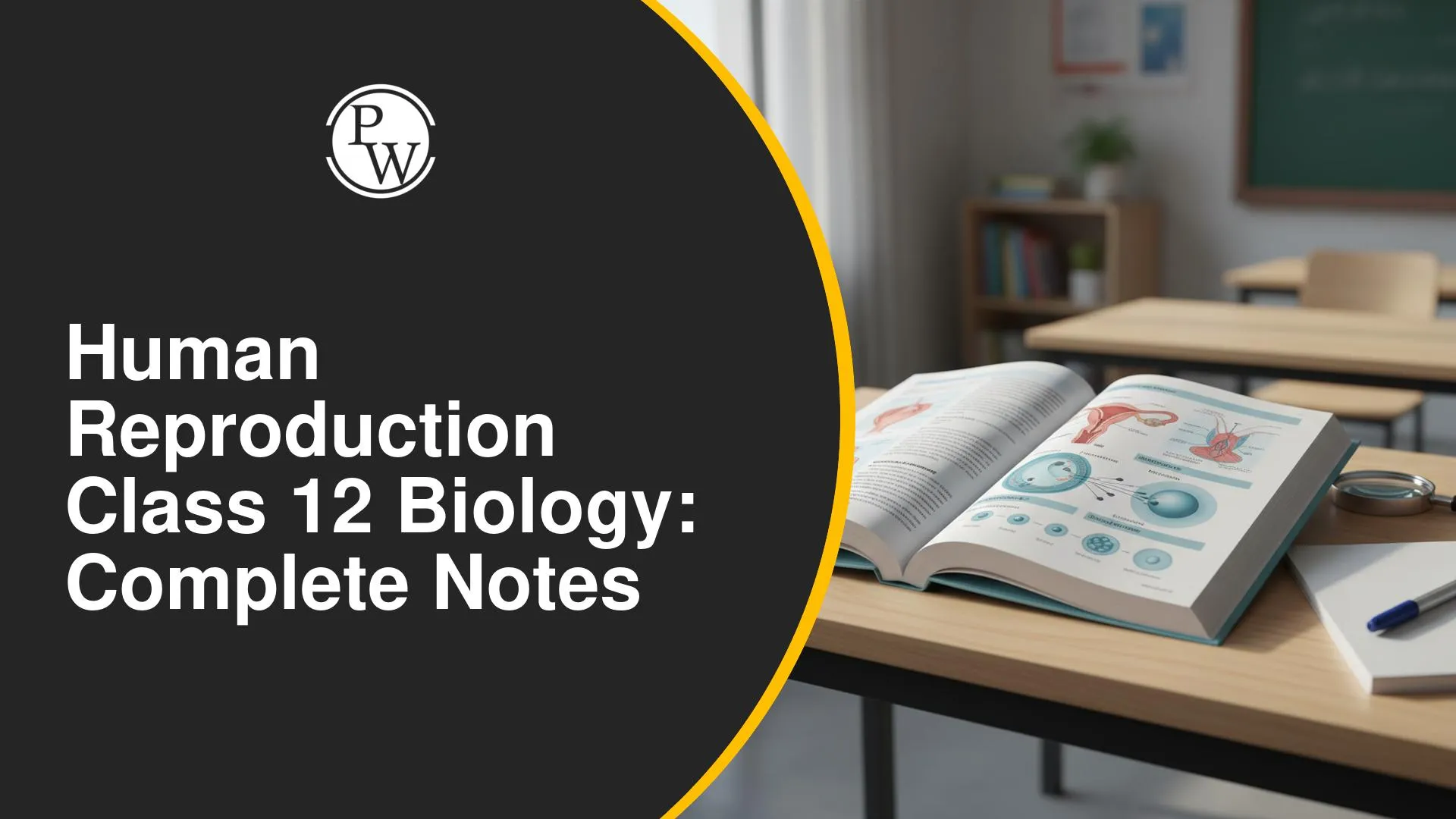
With CUET 2027 preparation, do you find yourself forgetting the stages of human reproduction or mixing up the functions of different reproductive hormones? Revising the Human Reproduction chapter from Class 12 Biology can be difficult when there are so many terms, processes and diagrams to remember.
Physics Wallah provides detailed notes to help you review these concepts and understand the key stages of human reproduction. Revising spermatogenesis, oogenesis, reproductive anatomy, the menstrual cycle, fertilisation, implantation, placental functions, parturition and lactation can help you strengthen your Class 12 Biology concepts and prepare more effectively for CUET 2027.
Definition And Modes Of Reproduction
Reproduction is the biological process through which organisms produce offspring and continue their species across generations.
|
Mode |
Main Feature |
Example |
|
Asexual Reproduction |
Does not require fusion of male and female gametes |
Bacteria and fungi |
|
Sexual Reproduction |
Involves formation and fusion of male and female gametes |
Humans |
Humans show sexual reproduction. The male gamete is the sperm, and the female gamete is the egg or oocyte. The organs that produce gametes are called gonads. The testis is the male gonad, while the ovary is the female gonad.
Major Events Of Human Reproduction
Human reproduction occurs through a sequence of coordinated events:
-
Gametogenesis: Formation of gametes
-
Insemination: Transfer of semen into the female reproductive tract
-
Fertilisation: Fusion of male and female gametes
-
Cleavage: Repeated divisions of the zygote
-
Blastocyst Formation
-
Implantation: Embedding of the blastocyst in the uterine wall
-
Gestation: Development of the embryo and fetus
-
Parturition: Delivery of the fetus
-
Lactation: Milk production after childbirth
Memory Tip: Remember the sequence as Gametes, Insemination, Fertilisation, Cleavage, Blastocyst, Implantation, Gestation, Parturition and Lactation.
Gametogenesis: Spermatogenesis And Oogenesis
Gametogenesis means the formation of gametes. In males, it is called spermatogenesis, while in females it is called oogenesis.
|
Feature |
Spermatogenesis |
Oogenesis |
|
Beginning |
At puberty |
During the embryonic period |
|
Gamete |
Sperm |
Egg or oocyte |
|
Production |
Continues throughout adult life |
Primary oocytes form before birth |
|
Important endpoint |
Continues through life, although it may decline with age |
Menopause marks the end of the reproductive cycle |
Spermatogenesis begins at puberty and occurs in the seminiferous tubules of the testes. Oogenesis begins during embryonic development. Primary oocytes remain arrested until hormonal changes at puberty allow their development to continue. Menarche marks the beginning of menstruation, while menopause marks its permanent cessation.
Male Reproductive System
The testes are the primary male reproductive organs. They produce sperm and male sex hormones, mainly testosterone. During embryonic development, the testes form in the abdominal region and later descend into the scrotum through the inguinal canal. Spermatogenesis requires a temperature approximately 2 to 2.5 degrees Celsius lower than body temperature. If the testes fail to descend, the condition is called cryptorchidism.
Testicular Structure
The testis is divided into approximately 250 testicular lobules. Each lobule contains one to three highly coiled seminiferous tubules.
The seminiferous tubules are the site of sperm production.
-
Spermatogonia are sperm mother cells that give rise to sperm through spermatogenesis.
-
Sertoli cells nourish and support developing sperm. They also remove damaged cells and secrete inhibin.
-
Leydig cells, located in the interstitial space, secrete testosterone.
The sperm transport pathway is:
Seminiferous Tubules → Rete Testis → Vasa Efferentia → Epididymis → Vas Deferens → Ejaculatory Duct → Urethra
The epididymis has three parts: caput, corpus and cauda. It helps in sperm maturation, storage and transport.
Semen And Accessory Glands
Insemination transfers semen, not pure sperm. Semen consists of sperm and seminal plasma.
The accessory glands are:
-
Two seminal vesicles
-
One prostate gland
-
Two bulbourethral glands, also called Cowper’s glands
Their secretions combine with sperm to form semen. Seminal vesicles provide most of the seminal plasma. The prostate contributes fluid that supports sperm activity, while bulbourethral glands provide lubrication.
Penis And Urethra
The penis is an erectile copulatory intromittent organ. It contains erectile tissues, not bone. Erection occurs when these tissues fill with blood and is controlled by the autonomic nervous system.
-
The penis includes the shaft, glans penis, urethra and urethral meatus. Its erectile tissues are the single corpus spongiosum and paired corpora cavernosa.
-
The urethra is a urogenital duct because it carries urine and male reproductive fluid. However, urine and semen are not released simultaneously.
Female Reproductive System
The ovary is the primary female sex organ. It produces eggs and releases estrogen and progesterone. The female reproductive system also includes the fallopian tubes, uterus, cervix, vagina, external genitalia and mammary glands.
Fallopian Tube And Uterus
Each fallopian tube has three parts:
-
Infundibulum: Funnel-shaped end with finger-like fimbriae
-
Ampulla: Wider region where fertilisation occurs
-
Isthmus: Narrow region close to the uterus
The uterus has three layers:
-
Perimetrium: Outer protective layer
-
Myometrium: Smooth muscle layer responsible for uterine contractions
-
Endometrium: Inner glandular layer where implantation occurs
The cervix is the lower narrow part of the uterus. The cervical canal and vagina together form the birth canal.
The external female genitalia are collectively called the vulva. They include the mons pubis, labia majora, labia minora, vestibule, hymen, Bartholin’s glands and clitoris.
The hymen is not a reliable indicator of virginity because its structure varies and it may not rupture during first intercourse.
Menstrual Cycle
The menstrual cycle and its hormonal regulation involve a series of monthly changes in the ovaries and uterus. An ideal cycle lasts 28 days and has four phases.
|
Phase |
Main Events |
|
Menstrual Phase |
Endometrial breakdown and bleeding |
|
Follicular Phase |
Follicular growth and endometrial proliferation |
|
Ovulation |
Release of the secondary oocyte |
|
Luteal Phase |
Corpus luteum formation and progesterone secretion |
The hypothalamus releases GnRH, which stimulates the anterior pituitary to release FSH and LH.
-
FSH promotes follicular growth.
-
Theca interna cells produce androgens, which granulosa cells convert into estrogens.
-
A sustained high estrogen level causes the LH surge.
-
The LH surge causes follicular rupture and ovulation.
-
The corpus luteum produces progesterone.
-
Progesterone maintains and prepares the endometrium for implantation.
Ovulation Day ≈ Total Cycle Length − 14
Therefore, ovulation generally occurs around day 14 in a 28-day cycle and around day 18 in a 32-day cycle, although the exact day can vary.
Fertilisation And Early Development
Fertilisation occurs in the ampullary region of the fallopian tube. The sperm passes through the corona radiata and zona pellucida. Enzymes in the acrosome help the sperm penetrate the surrounding layers.
Fertilisation requires the sperm and oocyte to reach the same place at the same time.
After one sperm enters, changes prevent additional sperm from entering. This prevents polyspermy. The secondary oocyte then completes Meiosis II, and the male and female pronuclei fuse to form a diploid zygote.
The developmental sequence covered in Biology previous year question papers is:
Zygote → Cleavage → Morula → Blastocyst → Implantation → Embryo → Fetus
During cleavage, the zygote forms blastomeres. The 16-cell stage is called the morula. A cavity then forms, producing the blastocyst.
The blastocyst contains:
-
Trophoblast: Contributes to placenta formation
-
Inner cell mass or embryoblast: Forms the embryo
-
Blastocoel: Fluid-filled cavity
The blastocyst attaches to the endometrium during implantation. The trophoblast forms chorionic villi, which interact with the maternal uterine wall to form the placenta.
Placenta, Pregnancy, And Parturition
The placenta connects the mother and developing fetus. It allows the exchange of oxygen, carbon dioxide, nutrients, minerals and wastes. The umbilical cord connects the fetus to the placenta.
The placenta also acts as an endocrine tissue and produces:
-
Human chorionic gonadotropin (hCG)
-
Human placental lactogen (hPL)
-
Estrogen
-
Progesterone
Pregnancy tests detect hCG in urine, while understanding the hormone’s role in pregnancy is an important part of Human Reproduction for CUET Biology.
Parturition is childbirth. Signals from the fetus and placenta stimulate oxytocin release from the posterior pituitary. Oxytocin causes contractions of the myometrium. Stronger contractions cause further oxytocin release, forming a positive feedback mechanism.
Lactation And Colostrum
Lactation involves milk production after childbirth, while colostrum provides essential antibodies that support a newborn’s early immunity.
Lactation is milk production after childbirth.
-
Prolactin stimulates milk production.
-
Oxytocin causes milk ejection.
-
Oxytocin also supports uterine contractions during childbirth.
The first milk produced after childbirth is colostrum. It is yellowish and rich in antibodies, especially immunoglobulin A. Colostrum provides early immunity to the newborn.
A strong grasp of human reproduction requires you to connect the functions of reproductive organs with hormonal changes, fertilisation, embryonic development and childbirth. Revising these concepts alongside important terms and biological processes can help you retain key information for Class 12 Biology and CUET 2027. Physics Wallah CUET Online Coaching helps you revisit these topics, clarify important concepts and strengthen your exam preparation.
Human Reproduction Class 12 Biology FAQs
Where Does Fertilisation Occur In Humans?
What Is The Difference Between Spermatogenesis And Oogenesis?
Which Cells Produce Testosterone?
What Causes Ovulation?
What Is The Role Of The Placenta?


Have questions about your prep?
Our experts can answer all your questions



 Live & recorded classes available at easeDashboard for progress trackingLakhs of practice questions