




NSEJS Botany Cell Cycle and Cell Division: Mitosis, Meiosis, Checkpoints & Cell Cycle
The Cell Cycle and Cell Division are fundamental concepts in biology that explain how cells grow, replicate, and produce new cells. The cell cycle consists of interphase (G₁, S, and G₂ phases) and the M phase, where mitosis or meiosis takes place. Understanding the stages of the cell cycle, checkpoints, mitosis, meiosis, and their significance is essential for scoring well in the NSEJS Botany section.

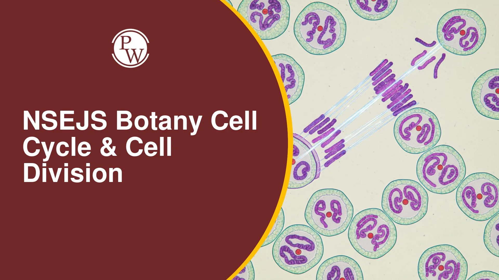
Cell Cycle and Cell Division are among the most important topics in the NSEJS Botany syllabus. Every living organism grows, repairs damaged tissues, and reproduces because its cells divide in a controlled and organised manner. Before a cell divides, it passes through a series of stages known as the cell cycle, during which it grows, replicates its DNA, and prepares for division.
This chapter covers the structure and phases of the cell cycle, cell cycle checkpoints, mitosis, meiosis, chromosome behaviour, and the significance of these processes in living organisms. It also explains how cells regulate division and how errors in this process can lead to diseases such as cancer. A strong understanding of these concepts is essential not only for NSEJS but also for other Olympiads, NEET, and higher-level biology studies.
Also Read:
Understanding Cell Division and the Cell Cycle
Before learning about mitosis and meiosis, it is important to understand what cell division and the cell cycle actually mean. Although these terms are closely related, they describe different biological processes.
What is Cell Division?
Cell division is the process by which a parent cell divides to produce two new daughter cells. As a cell grows, its cytoplasmic and nuclear contents increase, causing the cell to become larger. Once it reaches a stage where it can no longer efficiently carry out its metabolic activities, it divides into new cells. This helps maintain a favourable surface area-to-volume ratio, allowing the cells to function properly.
Cell division is essential for growth, tissue repair, replacement of damaged cells, and reproduction in living organisms.
What is the Cell Cycle?
The cell cycle is the complete sequence of events that a cell undergoes from the time it is formed until it divides into two daughter cells. It includes periods of growth, DNA replication, preparation for division, and the actual process of cell division.
In simple terms, cell division is one stage of the cell cycle, whereas the cell cycle includes every event that prepares a cell for successful division.
Historical Background
Scientists have studied cell division for more than two centuries, leading to several important discoveries.
-
Cell division was first observed by Prevost and Dumas in 1824.
-
In 1950, Alma Howard and Stephen Pelc identified the different phases of the cell cycle and classified them into G₁, S, G₂, and M phases. This classification is still followed in modern biology.
Giant Chromosomes
Giant chromosomes are unusually large chromosomes that are formed due to repeated DNA replication or extensive chromosomal coiling. Their large size makes them easier to study under a microscope and helps scientists understand chromosome structure and gene activity.
The two major types of giant chromosomes are:
-
Polytene chromosomes – Found in the salivary glands of Drosophila (fruit fly). They are formed by repeated DNA replication without cell division and are commonly used in genetic studies.
-
Lampbrush chromosomes – Found in the oocytes (egg cells) of frogs, newts, birds, and reptiles. These chromosomes have loop-like structures that are actively involved in RNA synthesis during egg development.
Regulation of Cell Division: Why and When a Cell Divides
A cell's division is tightly regulated:
-
In Plants: Cell division is initiated by a phytohormone called Cytokinin.
-
In Animals:
-
Factors (Proteins): Ras and Ras factors act as signals to initiate cell division. (Memory Tip: When the term "factor" is used in this context, it generally refers to a protein.)
-
Karyoplasmic Index (KI): KI = Volume of Nucleoplasm / Volume of Cytoplasm. A cell divides if its KI value is equal to or less than one, or more specifically, close to one.
-
Cyclin and CDK (Cyclin-Dependent Kinase enzyme): Cyclin is a protein, and CDK is an enzyme. Their binding, requiring ATP, facilitates the cell's progression from one phase to another within the cell cycle.
Cell Division Timings
Standard doubling times:
-
Yeast: Doubles every 90 minutes.
-
Bacteria: Doubles every 20 minutes.
-
Human Cells: Doubles every 24 hours.
Example Problem: A test tube of bacteria fills completely in one hour. Since bacteria double every 20 minutes, the test tube was half-filled 20 minutes before it was completely full (at the 40-minute mark).
The Cell Cycle Phases
The Cell Cycle is broadly divided into two main phases:
1. Interphase (Preparatory Phase)
This is the longest phase, lasting approximately 23 hours in human cells. It prepares the cell for division and is divided into three sub-phases:
-
G1 Phase (Gap 1 Phase):
-
The longest sub-phase (approx. 11-13 hours), metabolically very active, with rapid cell growth.
-
Chromosomes are unduplicated.
-
Key events: Cell size increases, cell organelles double, Cyclin and CDK levels ensured, ATP formed, proteins and enzymes synthesized. (Memory Tip: Like filling a packet with more items, the cell's volume increases as its components duplicate.)
-
G0 Phase (Quiescent Stage / Resting Phase):
-
Cells temporarily or permanently exit the cell cycle from G1 phase and stop dividing. They are metabolically active but not proliferating.
-
Examples of cells in G0: Blood cells, Muscle cells, Nerve cells.
-
S Phase (Synthetic Phase / Replicative Phase):
-
Lasts approx. 6-7 hours.
-
Key events: DNA replication/synthesis, histone proteins double, centrioles double.
-
The DNA content doubles (e.g., from 2C to 4C), while the chromosome number remains the same (e.g., 46 chromosomes, each now with two sister chromatids).
-
G2 Phase (Gap 2 Phase):
-
The shortest sub-phase (approx. 3-4 hours), metabolically active, with continued growth.
-
Key events: Remaining cell organelles duplicate, further protein synthesis and preparation for M-phase occur.
2. M-phase (Mitotic Phase)
This is the shortest phase (approx. 1 hour), where M stands for Mitosis or Meiosis.
Cell Cycle Checkpoints
Checkpoints ensure the cell cycle progresses correctly:
-
G1 Checkpoint (Restriction Checkpoint):
-
Checks for DNA damage, proper Cyclin-CDK synthesis, and availability of nutrients and growth factors. (Memory Tip: Like a parent checking homework before allowing play, the G1 checkpoint ensures all preparations are complete before entering S phase.)
-
G2 Checkpoint:
-
Checks for complete and accurate DNA replication, adequate cell size, and proper synthesis of necessary proteins (e.g., tubulin protein). Errors can lead to programmed cell death.
-
M Checkpoint (Metaphase Checkpoint):
-
Checks for proper attachment of spindle fibers to the kinetochores of chromosomes. Spindle fibers attach to kinetochores (protein structures on the centromere) to ensure proper chromosome separation.
Types of Cell Divisions
There are different modes of cell division:
1. Amitosis (Direct Division)
-
Characteristics: Karyokinesis and Cytokinesis occur simultaneously without distinct phases, often resulting in unequal distribution of genetic material.
-
Examples: Bacterial cells, Amoeba, Mitochondria, and Chloroplasts.
2. Mitosis (Indirect / Equational Division)
-
Characteristics: Ensures identical and equal sets of chromosomes in daughter cells. Occurs in somatic cells. A diploid mother cell (2n, 2C) undergoes division, resulting in two daughter cells (2n, 2C).
3. Meiosis (Reductional Division)
-
Characteristics: Reduces chromosome number by half. Occurs in germ cells to produce haploid gametes. A diploid germ cell (2n, 2C) produces four haploid cells (n, 1C) after two rounds of division.
Mitosis vs. Meiosis: Key Differences
Mitosis and meiosis are the two main types of cell division, but they serve different purposes. Mitosis helps an organism grow, repair damaged tissues, and replace worn-out cells, whereas meiosis is responsible for producing reproductive cells (gametes) and creating genetic variation.
In mitosis, a diploid parent cell (2n) divides once to produce two genetically identical diploid daughter cells. Since the chromosome number remains unchanged, mitosis is also known as equational division.
In meiosis, a diploid parent cell undergoes two successive divisions. During Meiosis I, the chromosome number is reduced by half, and Meiosis II separates the sister chromatids. As a result, one diploid cell produces four genetically different haploid cells (n). Because the chromosome number is reduced, meiosis is called a reductional division.
Mitosis Overview
Mitosis is the process of cell division that produces two identical daughter cells. It plays an important role in growth, tissue repair, and replacing damaged or old cells.
The process of mitosis was first described by Eduard Strasburger in plants and Walther Flemming in animals, who also introduced the term "mitosis." It mainly occurs in somatic (body) cells.
Mitosis consists of two major steps:
-
Karyokinesis – Division of the nucleus.
-
Cytokinesis – Division of the cytoplasm, resulting in two separate daughter cells.
Karyokinesis takes place in four successive stages:
-
Prophase
-
Metaphase
-
Anaphase
-
Telophase
Chromosome Duplication During Mitosis
Before mitosis begins, DNA is replicated during the S phase of interphase. Each chromosome forms two identical sister chromatids joined together at the centromere.
During mitosis, these sister chromatids separate and move to opposite poles of the cell. This ensures that each daughter cell receives an identical set of chromosomes, maintaining the same chromosome number as the parent cell.
Stages of Mitosis
Mitosis is a continuous process of nuclear division that ensures each daughter cell receives an identical set of chromosomes. It is divided into four main stages: Prophase, Metaphase, Anaphase, and Telophase. These stages are followed by cytokinesis, which divides the cytoplasm and completes cell division.
1. Prophase
Prophase is the first stage of mitosis, during which the cell prepares for chromosome separation.
Key events include:
-
Chromatin fibres condense into clearly visible chromosomes.
-
The nuclear membrane and nucleolus gradually disappear.
-
Centrioles move towards opposite poles in animal cells.
-
Spindle fibres begin to form and prepare for chromosome movement.
2. Metaphase
Metaphase is the stage where chromosomes are most condensed and easiest to observe under a microscope.
During this phase:
-
Chromosomes align along the equatorial plane, forming the metaphase plate.
-
Spindle fibres attach to the centromeres through specialised structures called kinetochores.
-
This stage is commonly used to study chromosome number and structure.
3. Anaphase
Anaphase begins when the centromeres split.
The main events are:
-
Sister chromatids separate from each other.
-
Each chromatid is pulled towards opposite poles by spindle fibres.
-
In animal cells, cytokinesis begins towards the end of this stage.
-
Chromosomes appear in different shapes, such as V-, L-, J-, or I-shaped, depending on the position of the centromere.
4. Telophase
Telophase is the final stage of mitosis and is often considered the reverse of prophase.
During telophase:
-
Chromosomes uncoil back into chromatin.
-
The nuclear membrane and nucleolus reappear.
-
Spindle fibres disappear.
-
Cytokinesis is completed, resulting in the formation of two genetically identical daughter cells.
PW provides Olympiad exam content, including Olympiad Exams Updates, sample papers, mock tests, guidance sessions, and more. Also, enroll today in the Olympiad Online Batches for preparation.
NSEJS Botany Cell Cycle & Cell Division FAQs
What is the primary difference between Cell Division and Cell Cycle?
Which phase of the cell cycle involves DNA replication, and what is its significance?
What are cell cycle checkpoints, and why are they important?
How does crossing over contribute to genetic variation, and during which stage does it occur?