
Complex Permanent Tissue
Anatomy Of Flowering Plants of Class 11
About Permanent Tissues type of Permanent Tissues
The tissue whose cells have lost their divisional capacity and have assumed a definite shape and size in response to varied mechanical and physiological functions, is called permanent tissue. .
On the basis of constituent cells, permanent tissues are classified into three groups: Simple tissues, Complex tissues and Special tissues or secretary tissues.
SIMPLE PERMANENT TISSUES
The permanent tissue, whose cells are homogeneous i.e., having similar origin, structure and function, is called simple tissue. On the basis of the structure of constituent cells, simple tissues are of three types, parenchyma, collenchyma and sclerenchyma.
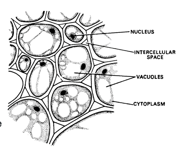
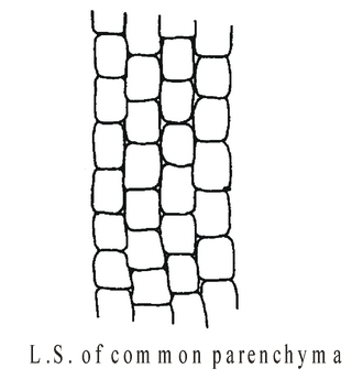
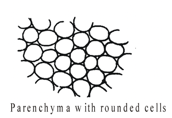
PARENCHYMA
Parenchyma (Gk. para = beside; en-chein = to pour; i.e., some semi liquid substance poured beside other solid tissues; Grew 1682) is the most basic type of differentiated tissue from which other types have evolved.
Characteristics
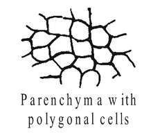
- The cells are living and may be rounded, oval, rectangular, star shaped but usually polygonal. Number of sides in a polygonal cell is usually 14 but in Elodea, it may be 17.
- Cells are loosely arranged with many intercellular spaces, either schizogenous or lysigenous (exception epidermis/epiblema, endodermis, pericycle and pith rays, where cells are compactly arranged).
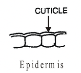
- The cell wall is thin and is made up of cellulose, hemicellulose and pectin. Though the cell wall of xylem parenchyma is thick, that of epidermal cells is cutinised and that of endodermal cells are suberised.
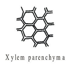
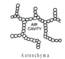
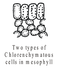
MODIFICATIONS OF PARENCHYMATo perform functions other than normal (storage of food), parenchyma gets modified into following types: Chlorenchyma: Parenchyma cells having chloroplast; found in mesophyll of leaves; meant for photosynthesis. |
Fig. Parenchyma |
Aerenchyma: Parenchyma with air filled spaces; found in hydrophytes, meant for buoyancy.
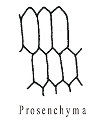
Prosenchyma: Parenchyma cells are much longer than their breadth and slightly thick walled to provide mechanical support. It can occur anywhere in the plant, more common in pericycle.
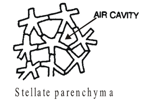
Stellate parenchyma: Parenchyma with star shaped cells; found in the leaf base of banana and Canna, meant for expansion of leaf base. Provides lightness as well as strength.
Idioblast: Specialized parenchyma whose cells secrete some secretions like mucilage, gums, resins, alkaloids, terpenes, etc. e.g., Citrus, Eucalyptus, etc.
|
|
|
|
|||
|
|
|
|
|
||
|
|
|
|
ORIGIN
Parenchyma originates from the cells of meristem by loosening their divisional capacity.
FUNCTIONS
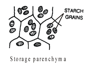
- Storage of food is the main function.
- Storage of water especially in xerophytes.
- Formation of interfascicular cambium and cork cambium in dicot stems.
- Formation of cambium and cork cambium in dicot roots.
- Formation of accessory cambium in monocot stems which exhibit secondary growth.
- Formation of wound cambium to heal up wounds.
- Vegetative reproduction.
- Turgid parenchyma cells provide some sort of mechanical support and maintain shape of the plant body.
- Parenchyma associated with the vascular tissues (xylem and phloem) plays an important role in the conduction of sap and transportation of food.
COLLENCHYMA
Collenchyma (Gk. Colla: glue, referring to its characteristic shining wall; Schleiden, (1839) is a specialized supporting tissue which has living cells and possesses considerable tensile strength. It is usually found in stems beneath the epidermis as a complete cylinder or in the form of longitudinal strips.
Structure
- Cells of collenchyma are elongated, tubular and are arranged along the long axis of the stem but in T.S. they appear as circular, oval or polyhedral.
- The peculiarity of collenchyma cells is the unevenly thickened cell wall i.e., thickenings are restricted to the corners of the cells.
- The thickening materials are mostly pectin in addition to cellulose and hemicellulose.
- The cells are living and the protoplast is highly vacuolated. Therefore, cytoplasm and nucleus become peripheral.
- Cytoplasm has all the cell organelles including chloroplasts.
Different Types of Collenchyma
On the basis of mode of wall thickening, collenchyma may be classified into the following three types:
- Lamellar: In this type, the thickenings are more heavily deposited on the tangential than on the radial cell walls. This type of collenchyma occurs in the stem of Raphanus, Helianthus, Rheum, etc.
- Angular: In this type, the thickenings are primarily deposited at the corners or angles of the cells. Angular collenchyma, the most common type of collenchyma is found in the stems of Datura, Lycopersicum, Cucurbita, Solanum, Ficus, Vitis, Morus, Polygonum, etc.
- Lacunar: In this type, the thickenings are primarily deposited around the intercellular spaces, e.g., in aerial roots of Monstera and petioles of Malva, Asclepias, etc.
About Complex Permanent Tissue type of Complex Permanent Tissue
The permanent tissues whose cells are heterogeneous i.e., cells having dissimilar origin and structure but performing common functions, is called complex tissue.
A complex tissue is a collection of different types of cells which forms a structural unit and perform a specific function. Xylem and phloem, present in all vascular plants, are complex tissues.
Xylem
- Xylem (Gk. Xylos: wood; Nageli, 1858; term hadrom for xylem by Haberlandt, 1914) is a complex tissue, composed of several types of living and dead cells. It is primarily concerned with the conduction of water and minerals and also provides mechanical support to the plant. As a conducting strand, xylem forms a continuous channel through roots, stems, leaves, flowers and fruits.
- The three main components of xylem are: Tracheary elements, xylem parenchyma and xylem fibres.
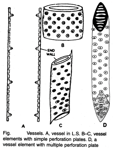
Tracheary Elements
- Tracheary elements are highly specialized cells, principally concerned with the conduction of water.
- Two kinds of tracheary elements occuring in xylem are tracheids and vessels. Both are dead cells and are devoid of living protoplasts at maturity. They are more or less elongated cells with lignified secondary walls.
-
Tracheids are present in all vascular plants, whereas vessels are confined to angiosperms. However, there are some primitive angiosperms where vessels are absent. Such vesselless angiosperms belongs to families : (i) Winteraceae (e.g., Wintera), (ii) Tetracentraceae (e.g., Tetracentron),
(iii) Trochodendraceae (e.g., Trochodendron) whereas some advanced gymnosperms (e.g., Gnetum, Ephedra, Welwitschia) exhibit the occurrence of vessels. Besides, vessels are absent in stem and leaves of Yucca and Dracaena. - Tracheids originate from single cells and are imperforate cells with pit pairs on their common walls. On the other hand, vessel members, which are joined into long continuous tubes of varying lengths, originate from longitudinal files of cells. They have dissolved transverse walls and the sap moves from one vessel member to another through these walls.
- The secondary walls of tracheary elements have a wide variety of thickening patterns. The tracheary elements of the first formed xylem (protoxylem) have secondary wall thickenings in the form of rings (annular) or helices (spiral). In the later formed metaxylem and secondary xylem, secondary walls usually show scalariform or reticulate thickenings.
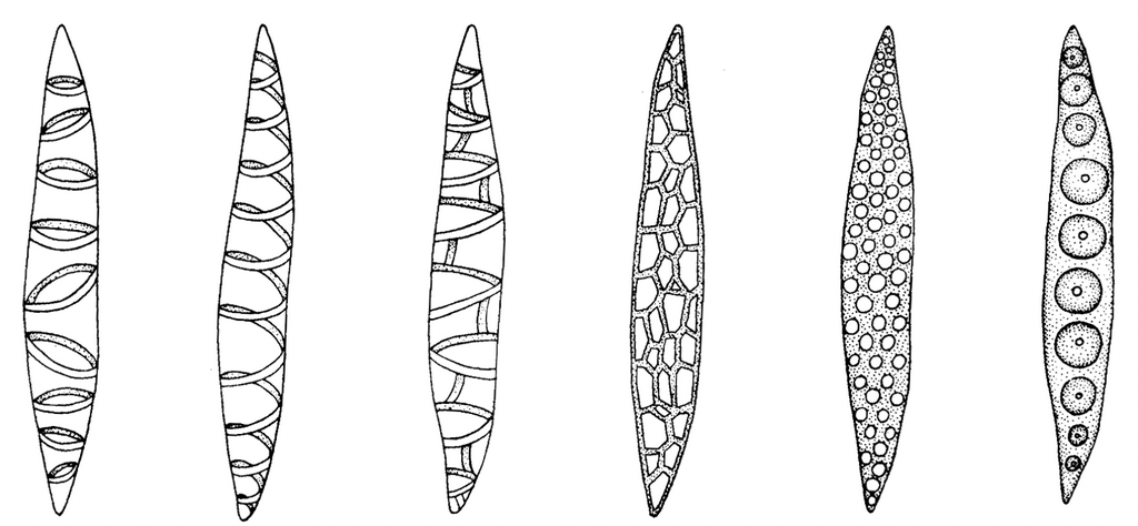
Annular Spiral Scalariform Reticulate Pitted
Fig. L.S. of a tracheid showing simple pits
Xylem Parenchyma
- Parenchymatous cells associated with xylem are called xylem parenchyma.
- These are present in the primary and secondary xylem. In the primary xylem, the walls of the parenchyma cells are thin and made up of cellulose, whereas in the secondary xylem, the walls are often thickened.
- The main function of xylem parenchyma is storage of food materials. It also helps in the lateral conduction of water and minerals.
Xylem Fibres
- Xylem fibres are supporting sclerenchymatous cells and provide mechanical strength to the plant body.
- They have extremely thick lignified walls and narrow lumen.
Transfer Cells
- Besides the above mentioned cell types, yet another type of cells are associated with xylem which are called transfer cells.
- These cells have dense cytoplasmic contents, large nuclei, numerous mitochondria and endoplasmic reticulum. They are characterized by wall ingrowths of non lignified secondary wall. Plasmodesmata are also present on the walls of the two adjacent transfer cells. Transfer cells are associated with the internal transfer of solutes.
Phloem
Like xylem, phloem (Nageli, 1858 term leptom for phloem by Haberlandt, 1914) is also a specialized complex tissue mainly responsible for the translocation of food materials. The basic components of phloem are:
Sieve Elements
- The translocating elements of phloem are collectively known as sieve elements.
- There are two kinds of sieve elements: (i) sieve cells and (ii) sieve tube elements or sieve tubes.
- Sieve cells, which occur in pteridophytes and gymnosperms, are single cells with specialized sieve areas usually in their lateral walls.
- Sieve tubes, which occur in angiosperms, are longitudinal file of cells (sieve tube components) with more specialized sieve area (sieve plate) usually in or near end wall.
- The most characteristic feature of sieve tubes is the presence of thickened cross walls between two adjacent sieve tube members, which are known as sieve plates. A sieve plate has a large number of perforations.
- There are two types of sieve plates, simple and compound. Simple sieve plates are transversely placed and consists of sieve area while the compound sieve plates are somewhat obliquely inclined with several sieve areas. Long sieve tube elements usually have a compound sieve plate, whereas the short elements have simple sieve plates.
- Degeneration of nucleus starts during differentiation of sieve elements. In mature sieve elements, there is a thin layer of parietal cytoplasm and a large central vacuole. A slimy substance of proteinaceous nature is also present. The slime originates in the cytoplasm in the form of discrete bodies, known as slime bodies, that occur either singly or in multiples and are variously shaped. In young sieve elements, these bodies consists of aggregates of tubules, designated as P-proteins.
- During the maturation of sieve elements, these tubules are dispersed in the cytoplasm to form groups of striated fibrils, designated as P2 proteins. The walls of sieve elements vary in thickness but they are usually thicker than those of the neighbouring parenchyma cells. Sieve elements usually have primary walls which consist mainly of cellulose.
- However, sometimes special thickenings, known as nacreous thickenings also develop. These thickenings have glistening properties and sometimes they almost occlude the cell lumen. Non functional sieve tubes may become filled with tylosoids (proliferations from contiguous parenchyma cells) in some plants.
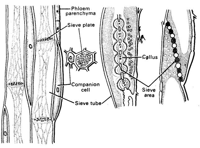
Fig. Parts of phloem
Companion Cells
- With the sieve tube elements of angiosperms, highly specialized parenchyma cells are associated which are called companion cells.
- Normally, only one companion cell is associated with each sieve tube cell in the primary phloem of herbaceous plants. But in the secondary phloem of woody plants many short companion cells are associated with each sieve tube member.
- They are living cells having dense granular cytoplasm, a prominent elongated nucleus and numerous small vacuoles.
- Companion cells, along with phloem parenchyma, play an important role in the maintenance of a pressure gradient in the sieve tube. They form a link between sieve tube members and other cells and control the passage of materials.
Phloem Parenchyma
- They are typical parenchyma cells found associated with sieve tube cells.
- The parenchyma cells of the primary phloem are longitudinally oriented like sieve elements in the vascular tissue.
- In the secondary phloem, they occur as two systems, the vertical and horizontal systems. The vertical system runs longitudinally along with the sieve elements and the horizontal system forms phloem rays that run horizontally across the organ.
- The phloem parenchyma cells are mainly concerned with storage of starch, fat and other organic food materials, accumulation of tannins, resins, crystals, mucilage, etc. and lateral transport of soluble food materials and water. Phloem parenchyma is absent in monocots.
Phloem Fibres or Bast Fibres
- They are typically elongated cells with interlocked ends and lignified walls having simple pits.
- They differ from xylem fibres in having small simple pits.
- Besides providing support, phloem fibres also play a role in the transport of food materials.
- Phloem fibres of plants like jute, flax and hemp are retted in water and extracted for making ropes and coarse textiles.
Phloem Transfer Cells
The phloem cells which are responsible for short distance transport of metabolites are called phloem transfer cells. These cells are characterized by wall ingrowths and a cytoplasm rich in organelles. They have a specialized type of non lignified secondary cell wall deposited on the inner side of the primary wall. Plasmodesmata usually occur between the two adjacent transfer cells.
Differences between Xylem and Phloem
|
Xylem |
Phloem |
|
|
|
|
|
|
|
|
|
|
|
|
|
|
|
|
SPECIAL TISSUES OR SECRETORY TISSUES
The tissue that is concerned with the secretion of gums, resins, volatile oils, nectar, latex and other such substances is called secretory tissue. Secretory tissue may be external or internal in origin.
external secretory tissue
The external secretory tissues are epidermal in origin or derived from the sub epidermal cells. Some common external secretory tissues are as follows:
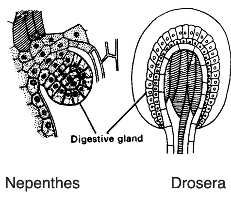
Glandular Trichomes
They are the simplest type of secretory structure. A glandular trichome has a unicellular or multicellular stalk and a head composed of one or more cells which produce secretions.
|
Nepenthes Drosera Fig. Digestive glands |
Hydathodes
- They are the water exuding structures of plants inhabiting the humid tropics.
- These hydathodes are found in the leaves of many angiosperms and are located on leaf margins (e.g., Tropaeolum) or at the tip (e.g., Colocasia).
- The cells of the mesophyll adjacent to the vascular bundle proliferate to give rise to epithem. The cells of the epithem are thin walled, elongated with dense cytoplasm and deficient in chloroplasts. They have a well developed system of intercellular spaces and are in close contact with terminal tracheary elements.
- Overlying the epithem are present two guard cells. The water that moves out of the tracheids under conditions of high root pressure and humidity is ultimately discharged through the terminal pore of the hydathode.

Fig. Hydathode
Nectaries
- They are commonly associated with flower but they also occur on vegetative parts.
- The cells of nectaries have dense cytoplasm and contain numerous mitochondria and abundant endoplasmic reticulum. They secrete nectar, e.g. Euphorbia pulcherrima.

Fig. Nectaries
INTERNAL SECRETORY TISSUE
There is a wide variety in internal secretory structures. They may consists of a single cell, a small group of cells or a whole tissue. Some common internal secretory structures are:
secretory cells
- They are specialized cells dispersed in the ground parenchyma. These cells contain a variety of contents, such as oils, resins, tannins, gums, mucilages and crystals of various types.
- The secretory cells which are distinct from neighbouring cells are called idioblasts.
Glands
- Several plants like Citrus have internal glands. These glands originate either schizogenously by breakdown of middle lamella or lysogenously by lysis of some cells. Schizogenous cavities are usually lined by intact cells, whereas lysogenous cavities have partly disintegrated cells along the periphery.
Resin Ducts
They are long intercellular spaces lined with epithelial cells which produce resins. They form complex vertical and horizontal systems within the plant body, e.g. Pinus.
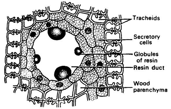
Fig. Resin secreting glands
Laticifers
They are specialized parenchyma cells which secrete a viscous fluid, known as latex. These tissues are present throughout the ground tissue of plant and contain organic substances of much importance, e.g., papain enzyme (in papaya), chikle (chewing gum) rubber, etc. On the basis of their structure and development, the following types of laticifers are recognized:
- Non articulated laticifers: They are multinucleate cells of immeasurable length. They develop from single cells which are capable of growing independently. Such cells grow more rapidly than the neighbouring cells, e.g., Calotropis, Nerium, Euphorbia, Ficus, Catharanthus, etc.
- Articulated laticifers: They are made up of a number of cells which are joined end to end to form longitudinal files of cells. There is complete or partial dissolution of separating end walls of continuous cells early in the ontogeny. These laticifers may be unbranched or branched, e.g., Papaver, Argemone, Sonchus, Carica, etc.
- In Hevea (rubber), these laticifers occur in secondary phloem and in papaya, both in xylem and phloem.
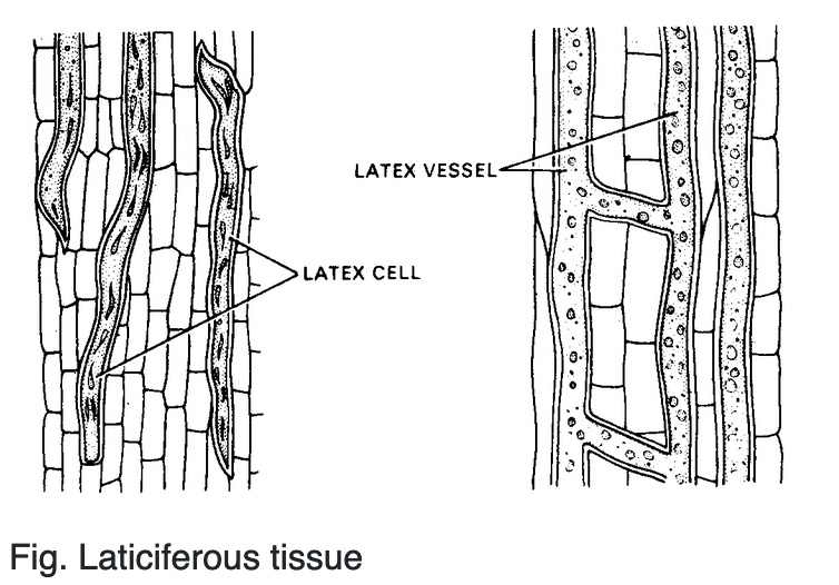
Fig. Laticiferous tissue