

NCERT Solutions for Class 11 Biology Chapter 18 Question Answer PDF
NCERT Solutions for Class 11 Biology Chapter 18: Chapter 18 of Class 11 Biology, "Neural Control and Coordination," focuses on how the nervous system and endocrine system coordinate various physiological processes in the human body. It covers the structure and functioning of neurons, nerve impulses, and synaptic transmission.
The chapter delves into the central and peripheral nervous systems, reflex actions, and sensory organs. Special emphasis is given to the brain and spinal cord, highlighting their role in maintaining coordination. Topics like chemical coordination through hormones and disorders of the nervous system are also discussed. The chapter aims to build a foundation for understanding human physiology and control mechanisms.NCERT Solutions for Class 11 Biology Chapter 18 Overview
Chapter 18 of Class 11 Biology, "Neural Control and Coordination," highlights the importance of the nervous and endocrine systems in regulating body functions. It explains how neurons transmit signals, enabling quick responses to stimuli and maintaining homeostasis. The chapter explores the structure and functions of the brain, spinal cord, and sensory organs, demonstrating their critical role in coordination and reflex actions. By understanding neural mechanisms and hormonal regulation, students grasp the intricate balance required for body functions. This knowledge is vital for comprehending human physiology, diagnosing disorders, and appreciating the role of coordination in survival and adaptation.NCERT Solutions for Class 11 Biology Chapter 18 PDF
Below, we have provided the NCERT Solutions for Class 11 Biology Chapter 18 - Neural Control and Coordination in PDF format for easy access and reference. These solutions cover all the important questions and topics, offering detailed explanations to enhance understanding. The PDF format allows students to conveniently download and study offline, ensuring a thorough preparation for exams while saving time during revisions. Access the PDF below for a seamless learning experience.NCERT Solutions for Class 11 Biology Chapter 18 PDF
NCERT Solutions for Class 11 Biology Chapter 18 Neural Control and Coordination
Below is the NCERT Solutions for Class 11 Biology Chapter 18 Neural Control and Coordination -
1. Briefly describe the structure of the following:
(a) Brain (b) Eye (c) Ear
Solution:
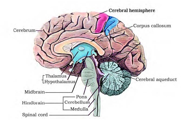
The structure is as follows:(a) Structure of the brain
 1. The brain serves as the body's "command and control system" and is the primary organ for processing information. In the skull, it is protected.
2. It is covered by three membranes called cranial meninges: the dura mater, which is robust and fibrous, is the outermost layer; the arachnoid, which is thin and sensitive; and the pia mater, which is an extension of the brain tissue, is the deepest layer. This layer has a high blood supply and is highly vascularised.
3. The three main regions of the brain are:
(i) Forebrain
(ii) Hindbrain
(iii) Midbrain
1. The brain serves as the body's "command and control system" and is the primary organ for processing information. In the skull, it is protected.
2. It is covered by three membranes called cranial meninges: the dura mater, which is robust and fibrous, is the outermost layer; the arachnoid, which is thin and sensitive; and the pia mater, which is an extension of the brain tissue, is the deepest layer. This layer has a high blood supply and is highly vascularised.
3. The three main regions of the brain are:
(i) Forebrain
(ii) Hindbrain
(iii) Midbrain
Forebrain – has three main parts – cerebrum, hypothalamus, thalamus
The cerebrum is the most significant and vital component of the brain. A deep fissure separates it longitudinally into two halves, each of which is referred to as the cerebral hemisphere. The corpus callosum, a network of nerve fibres, connects these two hemispheres. The walls of the cerebrum contain an outer cortex and an inner medulla, and the cerebral hemispheres are internally hollow. The cerebral cortex is known as grey matter because it is made up of neuronal cell bodies that give it a grey appearance. There are numerous folds (gyri) and grooves (sulci) in the grey matter. The intelligence increases with the number of convolutions. Sensory, motor, and association (neither motor nor sensory) areas make up the cerebral cortex. Complex processes including memory, communication, and intersensory associations are controlled by these particular regions. The axons of nerve fibres that make up the cerebral medulla give it its white look. That's why it's called white matter. The limbic system, also known as the limbic lobe, is a complex structure that is formed by a number of interconnected deep structures within the cerebral hemispheres, specifically the hippocampus and amygdala. Function: Memory, intelligence, consciousness, volition, and willpower are all centred in the cerebrum.-Thalamus
It is situated above the midbrain and is composed of grey matter. Its function is to transmit muscular and sensory signals to the brain, regulate emotional expression, and understand pain, heat, and cold.Hypothalamus
It is made up of the optic chiasma and is situated at the base of the thalamus. It is the intersection of the opposing sides of the optic nerve fibres. The infundibulum is a greyish projection of the hypothalamus that sits behind this structure. The pituitary gland is located there. Function: The hypothalamus contains centres that control blood pressure, homeostasis, body temperature, and appetite (hunger, sleep, exhaustion, thirst, pleasure, anger, and penance). The hypothalamic neurosecretory cells generate releasing factors or a number of hormones that are essential for controlling pituitary hormone activity. The hypothalamus contributes to the regulation of sexual conduct in addition to the limbic system.Midbrain
It consists of the cerebral peduncles and the corpora quadrigemina Cerebral Peduncles They are fibrous thick tracts which connect the cerebrum and the cerebellum. Role – Relay the sensory and motor impulses between the hindbrain and the forebrain Corpora quadrigemina Known as the corpora quadrigemina, the dorsal region of the brain has two pairs of solid lobes, one of which is called the superior colliculi and the other the inferior colliculi. Role: The corpora quadrigemina regulates eye and head movement as well as visual responses. In order to recognise and locate the source of sound, they also control head movement and auditory reflexes.2. Compare the following:
(a) Central neural system (CNS) and Peripheral neural system (PNS)
(b) Resting potential and action potential
(c) Choroid and retina
Solution:
The comparison is as shown below: (a) Central neural system (CNS) and Peripheral neural system (PNS)| Central neural system (CNS) | Peripheral neural system (PNS) |
| Consists of the spinal cord and the brain | It consists of the spinal nerves and the cranial nerves |
| The spinal column is protected by the vertebral column, whereas the brain is protected by the skull | No protective structures |
| No subdivisions | It is divided into the autonomic nervous system and the somatic nervous system |
| Processes information and regulates the responses to impulses. | Nerves of PNS pass impulses to the CNS and responses from the CNS to various structures of the body |
| Group of neurons known as nuclei | Group of neurons known as ganglia |
(b) Resting potential and action potential
| Resting potential | Action potential |
| When the neuron is at the resting phase, it is the potential difference across membrane | When the neuron is triggered, it is the potential difference across the membrane |
| The exterior side of the neuron is positively charged, while the interior side is negatively charged | The exterior side of the neuron is negatively charged, and the interior side of the neuron is positively charged |
| Permeability of K + ions is observed to be more by the plasma membrane of neurons | Permeability of Na + ions is observed to be more by the plasma membrane of the neurons |
| To maintain the resting potential, the sodium-potassium ATPase pump is activated, sending Na + ions outside the neuron | It functions in a reverse pattern wherein the sodium-potassium ATPase pump sends Na + ions to the neuron. |
| Choroid | Retina |
| Forms the mid-coat of the eyeball | Forms the inner coat of the eyeball |
| Forms the vascular layer of the eyeball | Forms the neurosensory layer of the eyeball |
| Has no photoreceptor cells | Has two kinds of photoreceptors – rods and cones |
| Prevents reflection of light in the eye and nourishes the retina | Imparts vision |
3. Explain the following processes:
(a) Polarisation of the membrane of a nerve fibre
(b) Depolarisation of the membrane of a nerve fibre
(c) Conduction of a nerve impulse along a nerve fibre
(d) Transmission of a nerve impulse across a chemical synapse
Solution:
(a) Polarisation of the membrane of a nerve fibre:

Impulse conduction through an axon
During its resting phase, a nerve fibre is referred to as being polarised. The nerve fibre membrane experiences a resting potential in this polarised state. The following are the stages that take place when a nerve fiber's membrane polarises: 1. The number of K+ ions outside the nerve fibres increases when a depolarised area of a nerve fibre gets polarised, whereas the axon membrane has an excessive amount of Na+ ions. 2. The membrane becomes more permeable to K+ ions and impenetrable to negatively charged proteins and Na+ ions as it begins to polarise. 3. A sodium-potassium pump uses active transport to transfer the two K+ ions to the axon, whilst the three Na+ ions are sent outside. 4. The passage of potassium and sodium ions leads the membrane's outer side to become electropositive and its inner side to become electronegative, polarising the nerve fibre.(b) Depolarisation of the membrane of a nerve fibre
 1. A nerve fibre is said to be in a depolarized state when it is triggered
2. In this condition, the nerve fiber's membrane experiences an action potential.
3. The following actions occur when the nerve fiber's membrane depolarises:
(i) The concentration of K+ ions is higher in polarised axons, while the concentration of Na+ ions is higher outside of axons.
(ii) When the stimulus triggers the nerve fibre, the permeability of the Na+ and K+ ion membranes is reversed.
(iii) The membrane becomes more permeable to Na+ ions.
(iv) There is a noticeable quick influx of Na+ ions into the axon.
(v) As a result, the membrane becomes positively charged on the inside and negatively charged on the outside.
(vi) As a result, the nerve fiber's membrane depolarises, causing an action potential to occur.
1. A nerve fibre is said to be in a depolarized state when it is triggered
2. In this condition, the nerve fiber's membrane experiences an action potential.
3. The following actions occur when the nerve fiber's membrane depolarises:
(i) The concentration of K+ ions is higher in polarised axons, while the concentration of Na+ ions is higher outside of axons.
(ii) When the stimulus triggers the nerve fibre, the permeability of the Na+ and K+ ion membranes is reversed.
(iii) The membrane becomes more permeable to Na+ ions.
(iv) There is a noticeable quick influx of Na+ ions into the axon.
(v) As a result, the membrane becomes positively charged on the inside and negatively charged on the outside.
(vi) As a result, the nerve fiber's membrane depolarises, causing an action potential to occur.
4. Write short notes on the following:
(a) Neural coordination
(b) Forebrain
(c) Midbrain
(d) Hindbrain
(e) Retina
(f) Ear ossicles
(g) Cochlea
(h) Organ of Corti
Solution:
(a) neurological coordination: This phenomenon occurs when two or more organs work together and enhance one another's functions via the body's neurological system. There are connections between the many physiological processes that occur in the body. Together, the neurological and endocrine systems are in charge of integrating and coordinating all of the organs' movements and activities so that they operate in unison. For quick coordination, this brain system provides a structured and methodical network for a point-to-point link. Through the hormones, the endocrine system provides chemical integration.(b) Forebrain –
Forebrain – has three main parts – cerebrum, hypothalamus, thalamus
The forebrain is the largest and most complex part of the brain, responsible for higher cognitive functions, sensory perception, and voluntary actions. It consists of three main parts: the cerebrum , thalamus , and hypothalamus . The cerebrum is divided into hemispheres and lobes, managing thought, memory, emotions, and sensory processing. The thalamus acts as a relay station, transmitting sensory and motor signals to the cerebral cortex. The hypothalamus regulates homeostasis, controlling hunger, thirst, body temperature, and hormonal secretions through the pituitary gland. Together, these structures coordinate complex behaviors, decision-making, and the body’s internal balance, playing a vital role in human physiology.C) Midbrain -
The midbrain , or mesencephalon, is a small yet vital part of the brainstem that connects the forebrain and hindbrain. It plays a crucial role in motor control , vision , hearing , and reflexes . The midbrain contains structures like the tectum and tegmentum , which manage auditory and visual reflexes. It also houses the substantia nigra , involved in regulating voluntary movements and producing dopamine. The midbrain ensures efficient sensory and motor signal transmission while contributing to maintaining alertness, arousal, and coordination of movements.d) The hindbrain is a crucial part of the brain responsible for coordinating basic life functions and motor activities. It consists of the cerebellum , pons , and medulla oblongata . The cerebellum controls balance, posture, and fine motor coordination. The pons acts as a bridge, relaying signals between different brain regions and aiding in sleep regulation. The medulla oblongata manages vital functions like heartbeat, breathing, and blood pressure. Together, these structures ensure smooth physiological processes and essential reflexive actions for survival.
e) The retina is a thin, light-sensitive layer of tissue located at the back of the eye, essential for vision. It contains specialized cells called photoreceptors — rods and cones —that detect light and color. Rods function in low-light conditions and enable black-and-white vision, while cones are active in bright light and perceive colors. The retina converts light signals into electrical impulses through a process called phototransduction, which are then transmitted to the brain via the optic nerve . Key areas of the retina include the macula , responsible for sharp central vision, and the fovea , specialized for detailed vision. It is vital for visual perception.
f) Ear ossicles
The ear ossicles are three tiny bones located in the middle ear: the malleus (hammer) , incus (anvil) , and stapes (stirrup) . These bones are the smallest in the human body and play a crucial role in hearing. They transmit and amplify sound vibrations from the eardrum to the inner ear's oval window. The stapes, the smallest bone, transfers these vibrations to the cochlea for further processing. Together, the ossicles ensure efficient sound conduction, enabling auditory perception.(g) Cochlea
The cochlea is a spiral-shaped, fluid-filled structure in the inner ear essential for hearing. It contains the organ of Corti , which houses sensory hair cells that convert sound vibrations into electrical signals. These signals are transmitted to the brain via the auditory nerve. The cochlea's structure allows it to detect a wide range of sound frequencies, making it vital for sound perception and distinguishing different tones in auditory processing.(h) Organ of Corti
The Organ of Corti is the primary sensory organ of hearing, located within the cochlea of the inner ear. It rests on the basilar membrane and contains sensory hair cells with tiny projections called stereocilia . These hair cells convert sound vibrations into electrical signals through a process called mechanotransduction . The signals are then transmitted to the brain via the auditory nerve. The Organ of Corti plays a critical role in detecting sound frequencies, enabling the perception of pitch and volume in hearing.5. Answer briefly:
(a) How do you perceive the colour of an object?
(b) Which part of our body helps us in maintaining the body balance?
(c) How does the eye regulate the amount of light that falls on the retina?
Solution:
Cone cells in the retina of the eye are responsible for colour vision. They are of three different types and react to red, green, and blue light, respectively. Different cone cells are triggered by different wavelengths of light, and when multiple types of cone cells are stimulated simultaneously, the other colours are detected. When all three types of cone cells are triggered simultaneously, a sensation of white light is observed. (b) The crista ampullaris, which is found in the three semicircular canals, the macula sacculi, which is found in the inner ear saccule, and the macular utriculi, which is found in the utricle, are the parts of the body that aid in preserving body balance. (c) The pupil is a hole in the middle of the iris. This opening allows light to enter the eye. The radial smooth muscles and the circular smooth muscles are the two muscle types in the iris that regulate the quantity of light that reaches the retina. In bright light, the smooth circular muscles contract, causing the pupil to shrink. As a result, the retina receives less light. When the light is dim, the radial smooth muscles contract, causing the pupil to enlarge so that the retina receives enough light.Benefits of Using NCERT Solutions for Class 11 Biology Chapter 18
Comprehensive Understanding : The solutions provide clear explanations of complex topics like nerve impulses, synaptic transmission, and hormonal regulation, making concepts easier to grasp.
Exam Preparation : These solutions are aligned with the NCERT syllabus, ensuring students are well-prepared for board exams and competitive tests.
Concept Clarity : Step-by-step answers help in breaking down challenging questions, improving problem-solving skills and conceptual clarity.
Time Management : Organized solutions save time during revisions by focusing on key topics and frequently asked questions.
Error-Free Answers : Authored by subject experts, they offer accurate and reliable explanations, reducing confusion.
Confidence Building : Regular practice with these solutions boosts confidence and helps students score better in exams.
NCERT Solutions for Class 11 Biology Chapter 18 FAQs
Why is neural coordination important?
What is the role of the brain in control and coordination?
How does neural control work?
What is the purpose of the neural pathways?


Get Free Counselling Today
and Clear up all your Doubts