NCERT Solutions for Class 11 Biology Chapter 7: Chapter 7 of NCERT Class 11 Biology, "Structural Organisation in Animals," explores the structural details and functional organization of animal tissues and organs. It begins by explaining animal tissues—epithelial, connective, muscular, and nervous tissues—their types, structure, and functions.
The chapter also discusses the anatomy and physiology of select organisms like earthworms, cockroaches, and frogs, providing insights into their systems, such as circulatory, digestive, and nervous systems. This chapter emphasizes the relationship between structure and function, offering foundational knowledge crucial for understanding advanced biological concepts in zoology. It is key for students preparing for board exams and competitive tests.NCERT Solutions for Class 11 Biology Chapter 7 Overview
Chapter 7 of NCERT Class 11 Biology, "Structural Organisation in Animals," delves into the study of tissues epithelial, connective, muscular, and nervous and their role in forming organs and organ systems. It explains the anatomy and physiology of animals like earthworms, cockroaches, and frogs, providing a deeper understanding of their structural and functional adaptations. This chapter is crucial as it lays the foundation for understanding the interdependence of structure and function in biology, a concept vital for advanced studies in zoology and medicine. Mastering this topic is essential for excelling in board exams and competitive exams.NCERT Solutions for Class 11 Biology Chapter 7 PDF
Chapter 7 of NCERT Class 11 Biology, "Structural Organisation in Animals," focuses on the structure and function of animal tissues and the anatomy of organisms like earthworms, cockroaches, and frogs. Understanding this chapter is essential for building a strong foundation in zoology. Below, we have provided a comprehensive PDF containing detailed NCERT solutions to help students understand key concepts and excel in their exams. Download the PDF for free now!NCERT Solutions for Class 11 Biology Chapter 7 PDF
Class 11 Biology Chapter 7 Structural Organisation in Animals Question Answers
Below is the NCERT Solutions for Class 11 Biology Chapter 7 Structural Organisation in Animals -1. Answer in one word or one line.
(i) Give the common name of Periplanata americana.
(ii) How many spermathecae are found in earthworms?
(iii) What is the position of ovaries in cockroaches?
(iv) How many segments are present in the abdomen of cockroaches?
(v) Where do you find Malpighian tubules?
Solution:
i) American cockroach ii) 4 pairs of spermathecae are found in earthworms. iii) Two ovaries are found lying laterally around the 2 nd to the 6 th abdominal segments. iv) 10 segments v) Malpighian tubules are found at the junction of the midgut and the hindgut of the alimentary canals of insects.2. Answer the following.
(i) What is the function of nephridia?
(ii) How many types of nephridia are found in earthworms based on their location?
Solution:
i) Nephridia in earthworms carry out the roles of osmoregulation and excretion. ii) The earthworm contains three different kinds of nephridia depending on where they are found, and they are Both sides of the intersegmental septa from segment 15 to the final one that exits into the gut include septal nephridia. From segment 3 to the final opening on the body surface, integumentary nephridia are connected to the lining of the body wall. In the fourth, fifth, and sixth segments, there are three paired tufts of pharyngeal nephridia.3. Draw a labelled diagram of the reproductive organs of an earthworm.
Solution:
The diagram of the reproductive organs of an earthworm is as follows:
4. Draw a labelled diagram of the alimentary canal of a cockroach.
Solution:
The diagram of the alimentary canal of a cockroach is as follows:
5. Distinguish between the following.
(a) Prostomium and peristomium
(b) Septal nephridium and pharyngeal nephridium
Solution:
a) Prostomium and peristomium The differences are as follows:| Prostomium | Peristomium |
| The small, fleshy lobe serves as a covering for the mouth and as a wedge to force open cracks in the soil in the earthworm crawls. | It is the crescentic aperture at the anterior end of the first segment of the earthworm comprising the mouth |
| Septal nephridium | Pharyngeal nephridium |
| Found at the anterior and posterior surface of septa occurring after segment 15 in earthworm | Found in three pairs in the 4 th , 5 th and 6 th segments located on either side of the alimentary canal |
| The excretory matter is discharged into the lumen of the alimentary canal | The excretory matter is discharged into the gut, in the pharynx or buccal cavity |
6. What are the cellular components of blood?
Solution:
The cellular components of blood are Red blood cells (RBC), white blood cells (WBC) and platelets.7. What are the following, and where do you find them in an animal body?
(a) Chondrocytes
(b) Axons
(c) Ciliated epithelium
Solution:
a) The cartilage cells are called chondrocytes. In adults, cartilage can be found between neighbouring bones of the hands, limbs, and vertebral column, as well as in the tip of the nose and outer ear joints. They are huge, mature, rounded cells that are located in clusters within the cartilage matrix. b) A long, thin projection of a neurone or nerve cell is called an axon. They can be found all over the body. They are in charge of carrying nerve impulses out of the cell body after emerging from the cyton. They come to an end in terminal arborisations, which are collections of branches. c) Columnar or cuboidal cells are referred to as ciliated epithelium if they have cilia on their free surface. They are found on the inside of hollow organs such as the fallopian tubes and bronchioles. Its free surface is made up of tiny, vibratile cytoplasmic structures known as cilia. The purpose of this cilium is to capture dust and other foreign objects.8. Describe various types of epithelial tissues with the help of labelled diagrams.
Solution:
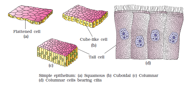
Epithelial tissues are found lining the body surface forming a protective surface. These cells are densely packed with a very little intercellular matrix.Various types of epithelial tissues are
i) Simple epithelium:
It is a single layer of cells which functions as a lining for body cavities, ducts, and tubes. Based on the structural modifications of the cells, Simple epithelial cells are further divided into 4 types.-
- Squamous epithelium
-
- Cuboidal epithelium
-
- Columnar epithelium
-
- Ciliated epithelium
ii) Compound epithelium
As in human skin, the compound epithelium is a layer of two or more cells that serves a protective purpose. Because they are made up of two or more cell layers, they are stronger and thicker than the ordinary epithelium; they provide protection. They cover the inner lining of the pancreatic and salivary ducts, the moist surface of the buccal cavity, and the dry skin surface.
9. Distinguish between
(a) Simple epithelium and compound epithelium
(b) Cardiac muscle and striated muscle
(c) Dense regular and dense irregular connective tissues
(d) Adipose and blood tissue
(e) Simple gland and compound gland
Solution:
a. Simple epithelium and compound epithelium| Simple epithelium | Compound epithelium |
| Composed of one layer of cells | Consist of many layers of cells |
| They are involved in the function of absorption and secretion | They are involved in the protection |
| Present in the stomach lining and intestine | Present in the lining of the buccal cavity and pharynx. |
| Cells rest on the basement membrane | Cells of the lowermost layer rest on the basement membrane |
| Cardiac muscle | Striated muscle |
| It is involuntary in function and never gets fatigued | It is voluntary in function, hence gets fatigued sooner |
| It is found in the heart | Found in the triceps, limbs and biceps |
| Branched fibres | Unbranched fibres |
| Uninucleated | Multinucleated |
| Dense regular connective | Dense irregular connective tissue |
| Collagen fibres are present in rows between parallel boundless fibres | Consists of Fibroblasts having several fibres that are differently oriented |
| Regular patterns of fibres observed | Irregular patterns of fibres observed |
| They are present in tendons and ligaments | They are present in the skin |
| Adipose tissue | Blood tissue |
| It is made of collagen fibres, fibroblasts, macrophages and adipocytes | It consists of RBC, WBC, platelets and plasma |
| It is a loose connective tissue | It is a fluid connective tissue |
| Its function is to synthesise, store and metabolise the fats | Its function is to transport food, gases, hormones and waste. |
| Present beneath the skin | Present in the blood vessels |
| Simple gland | Compound gland |
| It contains isolated glandular cells | Contains cluster of secretory cells |
| It is unicellular | It is multicellular |
| Ex: Goblet cells of the alimentary canal | Ex: salivary glands |
10. Mark the odd one in each series.
(a) Areolar tissue; blood; neuron; tendon
(b) RBC; WBC; platelets; cartilage
(c) Exocrine; endocrine; salivary gland; ligament
(d) Maxilla; mandible; labrum; antennae
(e) Protonema; mesothorax; metathorax; coxa
Solution:
- The answer is neuron because it is not a connective tissue.
- The answer is cartilage because it is not part of blood.
- The answer is ligament because it is connective tissue, whereas the rest are glands.
- The answer is antennae because the rest are the parts of a cockroach’s stomach.
- The answer is Protenema because it is a thread-like chain of cells found in the life cycle of moss, whereas others are the parts of segments of a cockroach’s leg.
11. Match the terms in column I with those in column II.
| Column I | Column II |
| (a) Compound epithelium | (i) Alimentary canal |
| (b) Compound eye | (ii) Cockroach |
| (c) Septal nephridia | (iii) Skin |
| (d) Open circulatory system | (iv) Mosaic vision |
| (e) Typhlosole | (v) Earthworm |
| (f) Osteocytes | (vi) Phallomere |
| (g) Genitalia | (vii) Bone |
Solution:
| Column I | Column II |
| (a) Compound epithelium | (iii) Skin |
| (b) Compound eye | (iv) Mosaic vision |
| (c) Septal nephridia | (v) Earthworm |
| (d) Open circulatory system | (ii) Cockroach |
| (e) Typhlosole | (i) Alimentary canal |
| (f) Osteocytes | (vii) Bone |
| (g) Genitalia | (vi) Phallomere |
12. Mention the circulatory system of earthworms briefly.
Solution:
- The earthworm's heart, capillaries, and blood arteries make up its closed, circular system.
- Because earthworms have a closed circulatory system, blood is limited to the heart and blood arteries.
- Blood flows in a single path during contraction.
- The fourth, fifth, and sixth segments have blood glands. They create haemoglobin, which is a type of blood cell that dissolves in blood plasma.
- The cells in the blood are phagocytic.
- Because they lack a specific respiratory system, their wet body surface facilitates respiratory exchange with their circulation.
13. Draw a neat diagram of the digestive system of a frog.
Solution:
The diagram is as below.
14. Mention the function of the following (a) Ureters in frogs (b) Malpighian tubules (c) Body wall in earthworms
Solution:
- Ureters in frog – Acts as a urinogenital duct which carries urine and sperm in the male frog.
- Malpighian tubules – Malpighian tubules are excretory organs in cockroaches.
- Body wall in earthworm – Helps in movement and burrowing
Benefits of Using NCERT Solutions for Class 11 Biology Chapter 7
Clear Conceptual Understanding : Simplifies complex topics like animal tissues and organ systems for easy comprehension.
Exam-Oriented Preparation : Solutions align with the NCERT syllabus, ensuring relevance for board exams and competitive tests.
Step-by-Step Explanations : Detailed answers enhance problem-solving skills and analytical thinking.
Saves Time : Pre-prepared solutions reduce time spent searching for accurate answers.
Boosts Confidence : Comprehensive coverage of topics ensures students are well-prepared.
Supports Self-Study : Ideal for independent learning and doubt resolution.
Foundation for Advanced Studies : Builds a strong base for topics in zoology, NEET, and medical entrance exams.
NCERT Solutions for Class 11 Biology Chapter 7 FAQs
What are the main points of structural organization in animals?
What is the structural unit of an animal?
What are the bits on structural organization in animals?
What do animals use for structure?


