






Renal Physiology & Special Senses Physiology MBBS 1st Year
 Aarti .18 Jul, 2026
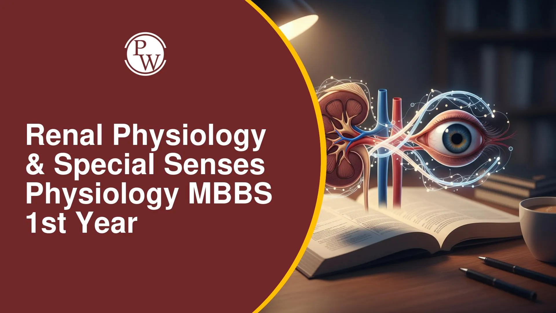
Renal Physiology and Special Senses are two of the most important topics in MBBS 1st Year Physiology. They help you understand how the kidneys maintain the body's internal balance and how the eyes and ears enable us to see and hear. These concepts form the foundation for understanding many clinical conditions and are frequently tested in university and competitive medical examinations.
This topic covers important concepts such as refractive errors, accommodation, photochemistry of vision, the visual pathway, hearing physiology, glomerular filtration rate (GFR), urine formation, acid-base balance, and concentrated urine formation. A clear understanding of these high-yield topics will strengthen your fundamentals and help you prepare effectively for both university exams and competitive medical entrance tests.
Special Senses and Renal Physiology
This content explores fundamental aspects of special senses, primarily vision and hearing, alongside vital concepts in renal physiology. It covers critical mechanisms, common pathologies, and their clinical relevance, aligning with key topics for MBBS 1st year examinations.
Vision
Vision is one of the most important special senses, allowing us to perceive and interpret the world around us. Understanding the basic structure and optical principles of the eye is essential before learning about refractive errors and visual physiology.
Reduced Eye (Schematic Eye)
The Reduced Eye is a simplified eye model. Light forms an image on the retina, transduced to electrical signals for the visual cortex. Refraction occurs at four planes, with greatest refraction at the anterior surface of the cornea due to the largest refractive index difference (air vs. cornea). These planes are simplified to a single plane just behind the cornea, with a total dioptric power of +59 Diopters.
Errors of Refraction (Ametropia)
Emmetropia is normal vision. Ametropia (refractive errors) means image formation doesn't occur on the retina and is most important for exams. Types include Index Ametropia and Curvature Ametropia. Newborns often have index hypermetropia. Main errors: Myopia, Hypermetropia, Presbyopia, Astigmatism.
1. Myopia (Short-Sightedness)
Myopia affects far vision, while near vision is normal (Memory Tip: "Myopia" -> "my piya" (my beloved) is near, so near objects are seen clearly). Far vision is for objects > 6 meters. The defect is an increased anteroposterior (AP) diameter of the eyeball, causing the image to form in front of the retina. Corrected with a concave lens or LASIK.
2. Hypermetropia (Long-Sightedness / Far-Sightedness)
Hypermetropia affects near vision, while distant vision is normal (Memory Tip: "Hypermetropia" -> "Hi piya" (greeting beloved) who is far, so far objects are seen clearly). The defect is a decreased anteroposterior (AP) diameter of the eyeball, causing the image to form behind the retina. Corrected with a convex lens.
3. Presbyopia
Loss of the power of accommodation with age, typically after 40 years, causing the near point of vision to recede (Memory Tip: "Presbyopia" -> "Bye-bye piya" or "Bye-bye Accommodation" after age 40).
For near vision, the eye undergoes accommodation. This reflex involves the second cranial nerve (afferent) to the visual cortex, then to the Edinger-Westphal nucleus via the third cranial nerve (efferent). Key changes: contraction of the ciliary body (most important), increased lens curvature and dioptric power, pupillary constriction, and medial rotation of the eyes (convergence). The Near Point of Vision (normal: 25 centimeters) is the nearest clear viewing distance.
4. Astigmatism
Astigmatism is caused by an irregular curvature of the cornea or lens (Memory Tip: "Egg-shaped" cornea/lens). Light rays do not converge at a single focal point. Corrected with spherical and cylindrical lenses.
Photochemistry of Vision
Photochemistry of vision converts light (electromagnetic energy) into electrical signals in the retina. Rods are for dim light vision, and cones for color and bright light vision.
Rod Photoreceptor and Light Stimulation
Rod outer segments contain rhodopsin. In darkness, cyclic GMP keeps sodium channels open, maintaining a sodium current. Light bleaches rhodopsin to activated rhodopsin, which activates transducin. Transducin activates PDE6, breaking down cyclic GMP, closing sodium channels. This leads to hyperpolarization of the rod membrane, which is considered stimulation of the rod.
Wald's Visual Cycle
This cycle describes rhodopsin regeneration. Rhodopsin (opsin + 11-cis retinal) absorbs light, causing 11-cis retinal to isomerize to all-trans retinal. This forms intermediates, including Metarhodopsin II (activated rhodopsin). Opsin and all-trans retinal separate. In darkness, all-trans retinal converts back to 11-cis retinal (by isomerase) and recombines with opsin to regenerate rhodopsin. An alternative pathway uses Vitamin A to regenerate 11-cis retinal for extra rhodopsin production, crucial for dark sensitivity.
Night Blindness (Nyctalopia)
Vitamin A deficiency causes Night Blindness by hindering extra rhodopsin generation via the alternative pathway, impairing low light vision.
Dark Adaptation
Dark adaptation is the improvement of vision in darkness, occurring due to:
-
Increased Retinal Sensitivity: Primarily by generating extra rhodopsin. The Dark Adaptation Curve shows initial rapid cone adaptation, then slower but significant rod adaptation (up to 25,000 times sensitivity).
-
Pupillary Dilation.
-
Neural Adjustments in the visual pathway.
Pilots wear red light goggles before dark to preserve dark adaptation (Memory Tip: Red light does not interfere with dark adaptation).
Color Vision and Color Blindness
Color vision enables us to distinguish different colors based on the response of specialized cone cells in the retina. Any defect in these cone cells or their pigments can lead to color vision deficiency, commonly known as color blindness.
Color Vision
The Young-Helmholtz Tri-chromatic Theory posits three cone types, sensitive to Red (Erythrolabe), Green (Chlorolabe), and Blue (Cyanolabe). Color perception results from their differential stimulation.
Color Blindness
Tested with Ishihara charts.
-
Dichromats: Lack one cone type (Protanopia - Red; Deuteranopia - Green; Tritanopia - Blue).
-
Anomalous Trichromacy: Weakness in a cone type (e.g., Protanomaly).
Red-Green Blindness is the most common type.
Visual Pathway and Effects of Lesions
This is a very important long question. The visual pathway includes the optic nerve, optic chiasm (where nasal fibers cross), optic tract, lateral geniculate nucleus (LGN) (an obligate relay station), optic radiations, and visual cortex. Associated structures: Pretectal Nucleus (Pupillary Light Reflex; lesion causes Argyll Robertson Pupil), Suprachiasmatic Nucleus (circadian rhythm), and Superior Colliculus (eye-hand coordination).
Retinal Projection: Nasal vision falls on temporal retina (temporal fibers); temporal vision falls on nasal retina (nasal fibers). Light rays invert.
|
Visual Pathway and Effects of Lesions |
||||
|---|---|---|---|---|
|
Lesion Location |
Fibers Affected |
Visual Field Loss |
Name of Condition |
Type of Hemianopia |
|
Optic Nerve (CRN II) Lesion |
All fibers of one optic nerve. |
Blindness of the corresponding eye. |
Monocular Blindness |
None |
|
Optic Tract Lesion |
Nasal fibers (contralateral eye) & Temporal fibers (ipsilateral eye) |
Homonymous hemianopia (e.g., right optic tract lesion causes loss of the right visual field in both eyes). |
Homonymous Hemianopia |
Homonymous |
|
Optic Chiasm Lesion (Crossed Fibers) |
Both nasal fibers (crossing) from both eyes. |
Loss of temporal half of vision in both eyes. |
Bitemporal Hemianopia |
Heteronymous |
|
Optic Chiasm Lesion (Uncrossed Fibers) |
Temporal fibers (uncrossed) from both eyes. |
Binasal hemianopia (loss of nasal half of vision in both eyes). |
Binasal Hemianopia |
Heteronymous |
Hearing
Middle Ear Functions are a highly probable exam question.
Functions of the Middle Ear
The Middle Ear transmits, enhances, and depresses sound transmission. (Memory Tip: The middle ear performs seemingly opposite functions – transmitting, enhancing, and depressing sound.)
1. Transmission of Sound
Sound vibrates the tympanic membrane, then malleus, incus, and stapes. The stapes footplate moves inward at the oval window, transmitting sound to the inner ear.
2. Enhanced Transmission of Sound (Impedance Matching)
The middle ear overcomes impedance from inner ear fluids by enhancing sound pressure by approximately 22 times (known as impedance matching). This occurs via:
-
Concentration of Vibrations (Area Ratio): Vibrations from the 55 mm² tympanic membrane concentrated onto the 3.2 mm² stapes footplate, increasing pressure 17-fold.
-
Ossicular Leverage: The ossicular lever system increases force by 1.3 times.
Total enhancement: 17 × 1.3 ≈ 22 times.
3. Depressed Transmission of Sound (Attenuation Reflex / Tympanic Reflex)
This reflex reduces sound transmission to protect the cochlea from loud sounds, filter low-frequency sounds, and improve speech. Loud sounds trigger reflex contraction of the stapedius and tensor tympani muscles. This makes the ossicular chain rigid and damps sound pressure by moving the malleus inward and stapes outward.
Organ of Corti and Mechanism of Hearing
The process of hearing begins when sound waves enter the ear and are converted into electrical signals that the brain can interpret. The Organ of Corti, located within the cochlea, is the sensory organ responsible for detecting sound and initiating this process.
Structure of Cochlea and Organ of Corti
The cochlea contains three fluid-filled compartments: scala vestibuli, scala media (endolymph, high K+), and scala tympani (perilymph). The Organ of Corti, on the basilar membrane within the scala media, is the sound transduction organ. It houses inner hair cells (single row) and outer hair cells (multiple rows) with stereocilia and a kinocilium. These hair cells synapse with the 8th cranial nerve.
Mechanism of Hearing (Sound Transduction)
-
Sound vibrates the basilar membrane.
-
This creates a shearing force between basilar and tectorial membranes.
-
Stereocilia bend, opening potassium channels.
-
Potassium influx from endolymph causes depolarization of the hair cell (unique as K+ influx leads to depolarization).
-
Depolarization opens voltage-gated calcium channels.
-
Calcium influx triggers glutamate release.
-
Glutamate excites 8th cranial nerve fibers, sending signals to the auditory cortex for sound perception.
Renal Physiology: Important Topics for Self-Study
The following kidney topics are very, very important for exams:
-
Long Questions:
-
GFR and Factors Influencing GFR (full chance).
-
Mechanism of Urine Formation.
-
Concentrated Urine Formation.
-
Acid-Base Balance (full chance).
-
Short Note/Reasoning:
-
JG Apparatus.
-
Renal Circulation Peculiarities (very, very high chance).
-
Transport Maximum for Glucose (TMG) (very, very high chance).
-
Concept of Clearance (very important).
-
Renal Handling of Glucose (TMG, Renal Threshold).
Renal Physiology & Special Senses Physiology MBBS 1st Year FAQs
What are the four main errors of refraction and how are they generally corrected?
Explain the key changes occurring during the accommodation reaction for near vision.
Describe the process of photochemistry of vision in rods when light strikes.
How does the middle ear enhance sound transmission to the inner ear?
What is unique about potassium influx in hair cells during the mechanism of hearing?
Which renal physiology topics are most important for MBBS 1st Year exams?




 Live & recorded classes available at easeDashboard for progress trackingMillions of practice questions at your fingertips