Cell Junctions, Diagram, Types and Functions in Animal Cells
Cell Junctions: Cell junctions, or junctional complexes, are important structures found in cells that help them stick together. They act like glue, holding neighbouring cells or a cell and its surroundings together. In animals, cell junctions are crucial for maintaining the barrier between cells and controlling the movement of substances between them. They are found abundantly in tissues that cover surfaces or line organs, such as the skin or intestines.
Cell junctions also play a key role in allowing cells to communicate with each other. They contain special parts called gap junctions that allow molecules and signals to pass between cells. Overall, cell junctions are essential for keeping cells healthy and enabling them to work together effectively. Students studying for the NEET exam can learn more about the types and functions of cell junctions in the chapter on the structural organisation in animals .
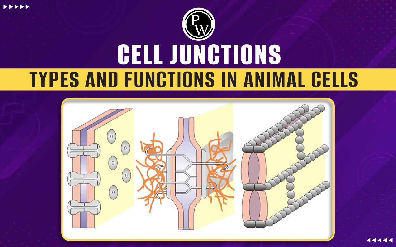
Cell Junctions Diagram
The Cell junction diagram is provided below:

Structural organisation in Animals MCQ for NEET
Cell Junctions Types
There are various types of connecting junctions that play a role in binding cells together:
- Tight Junctions (also known as occluding junctions or zonula occludens)
- 2 . Adhering Junctions (also known as zonula adherens)
- Desmosomes (also known as macula adherens)/ or hemidesmosomes
- Gap Junctions (also known as nexus or septate junctions)
These cell junctions are commonly found between epithelial cells but can also be found between other types of cells.
Gap Junctions
Gap junctions in animal cells, like plasmodesmata in plant cells, act as channels that connect adjacent cells, allowing ions, nutrients, and other substances required for cell communication to move more easily.
Gap junctions, also called macula communicans or communicating junctions, are protein complexes that form channels between two animal cells. These channels enable the passage of cytoplasm, water, nutrients, and signaling molecules.
Structure of Gap Junctions
Gap junctions in animal cells are protein complexes made up of connexins. These connexins assemble to form a structure known as a connexon. Six connexins come together to create a connexon, and two connexons combine to form a full channel that spans both the cell membranes and the extracellular space between them.
Tissues, Definition, Types of Animal Tissues and Functions
Functions of Gap Junctions
Gap junctions serve as channels that connect adjacent cells, facilitating the exchange of ions, nutrients, and other essential substances required for cell communication. They allow for the passage of cytoplasm, water, nutrients, and signaling molecules between cells. Gap junctions play an important role in transferring ions, second messengers, and small metabolites between neighbouring cells. They are involved in various physiological processes, such as:
- Cardiac Muscle: Gap junctions help transmit the action potential from cell to cell in cardiac muscle, contributing to the rhythmic contraction of the heart.
- Brain: Certain electrical synapses in the brain use gap junctions to transmit action potentials from synaptic terminals to postsynaptic cells without the delay required for neurotransmitter release.
- Uterus: As childbirth approaches, smooth muscle cells in the uterus form gap junctions, allowing for coordinated and powerful contractions to begin.
Mutations in connexin genes can lead to genetic disorders, highlighting the crucial role of gap junctions in maintaining tissue homeostasis.
Adhesion Junctions
Adherens junctions, also known as zonula adherens or adhering junctions, are connecting structures found in animal cells. They consist of specialized proteins known as cadherins and play an important role in anchoring cells to one another
Adherens junctions exhibit significant morphological diversity. Some appear as isolated streaks or spots, while others form bands that encircle the cell. Actin filament bundles located directly beneath the cell membrane are linked to band-type junctions.
Structure of Adhesion Junctions
Epithelial cells contain adhesion junctions, which aid in joining two cells together. They are made up of various anchoring proteins in the cell membrane, including:
- Catenins
- Cadherins
- Alpha-actinin
- Vinculin
Cadherins, transmembrane proteins, are used to construct adherens junctions. The extracellular segments of cadherins bind to one another, while the intracellular segments bind to catenin proteins. Catenins are linked to actin filaments, which are part of the cell's cytoskeleton.
Inside the cell, actin filaments connect with adhesion junctions to form a marginal band around the cell. This band can contract, allowing cells to transform shape. The formation of ducts and other structures in epithelial tissues depends on this contraction.
Functions of Adhesion Junctions
Adherens junctions play a role in tissue folding, bending, and cell adhesion. Complex tissue structures can be formed by the contraction of actin filaments within adherens junctions, which can distort epithelial cell sheets.
Desmosome Junctions
Desmosome junctions, also known as macula adherens or anchoring junctions, are specialized cell connections that play an important role in tissue integrity, especially in tissues subjected to mechanical stress.
Mutations in the genes that control desmosome formation can cause a variety of skin and heart diseases, emphasizing the importance of these junctions in maintaining tissue integrity.
Structure of Desmosome Junctions
Desmosomes are spot-like structures made up of proteins that span adjacent cell membranes. These proteins are linked to intermediate filaments within the cells, which vary depending on the cell type (desmin in cardiac cells and keratin in epithelial cells).
Functions of Desmosome Junctions
Desmosomes help tissues resist mechanical stress by evenly distributing the stress throughout the tissue. This is accomplished by connecting the intermediate filaments to form a nearly continuous fibrous network.
Tight Junctions
Epithelia are cell layers that form a barrier between cell groups and a hollow space. The apical surface is the part of the cell that faces the lumen, while the basolateral surface is made up of the cell's sides and base. Tight junctions tightly seal neighbouring epithelial cells just below the apical surface.
Structure of Tight Junctions
Tight junctions are formed by various proteins that help cells adhere to one another, forming a barrier that is difficult for substances to pass through while allowing for selective permeability. Tight junctions are made up of proteins like claudins, occludins, junctional adhesion molecules (JAMs), and zonula occludens (ZO).
Functions of Tight Junctions
- They restrict the movement of molecules and ions between cells, requiring most substances to enter cells via diffusion or active transport before passing through the tissue. This control allows precise regulation of which substances can pass through the barrier.
- They prevent the movement of integral membrane proteins (represented by red and green ovals) between the cell's apical and basolateral surfaces. This allows each surface to maintain distinct functions, such as receptor-mediated endocytosis at the apical surface and exocytosis at the basolateral surface.
The permeability of tight junctions varies with location and can be selectively permeable. For example, in the gut, tight junctions act as a selective barrier, preventing water-soluble molecules from diffusing. They also play a role in controlling the localization of membrane-bound proteins.
Cell Junctions Functions
Cell junctions serve various functions, including:
- Forming a seal: They create a seal between polarized epithelial cells. For example, tight junctions in the urinary bladder's epithelial cells prevent urine from leaking into the extracellular space.
- Regulating tissue homeostasis: Cell junctions help regulate tissue homeostasis by influencing critical cell processes such as tissue barrier function, cell proliferation, and migration.
- Enabling communication: Specialized protein complexes called communicating junctions facilitate communication between neighboring cells through cell junctions.
- Reducing stress: Cell junctions play a crucial role in reducing stress on cells.
- Maintaining tissue structure: Other junctions mainly function as adherence sites that help maintain tissue structure and integrity.
Physics Wallah provides excellent, cost-effective NEET Online Coaching in India . PW NEET courses cover the entire syllabus for class 11 and NEET exams, which includes Chemistry, Physics, Botany, and Zoology. Classes are held over six days and include two live sessions per day.
| NEET Exam Important Links | |
|---|---|
| NEET Biology Syllabus | NEET Biology Diagrams |
| NEET Biology MCQ | NEET Biology Chapter wise Weightage |
| NEET Exam Notes | NEET Previous Year Question papers |
Cell Junctions FAQs
How many types of cell junctions are found in vertebrates?
What are some examples of cell junctions?
What is the definition of cell junctions in Class 11?
Where are gap junctions typically found?