Structural Organisation in Animals, Tissues, Cockroach, Frog
Structural organisation in Animals: In a unicellular organism, such as a single-celled organism, all vital functions like digestion, respiration, and reproduction are carried out by the individual cell itself. On the other hand, a multicellular organism exhibits a hierarchy of structural organisation, in which various specialized structures cooperate to ensure the survival of the organism.
For example, the body of a relatively simple multicellular organism such as the Hydra is made up of thousands of different types of cells. The human body, on the other hand, is made up of billions of cells, each of which performs a different function that is essential to life.
This article provides comprehensive details into Structural organisation in Animals for Class 11 students, aligned with the updated NEET syllabus to aid in NEET preparation. NEET aspirants should use Structural Organisation in Animals NCERT PDF Notes to gain a better understanding of the chapter.
Structural Organisation in Animals Levels
A multicellular organism displays various levels of structural organisation in animals that collaborate to ensure its survival.
Cellular Level of Organisation
Cells are the fundamental units of structure and function at the cellular level. In multicellular animals, cells specialize and differentiate, resulting in a significant division of labor.
Structural organisation in Animals MCQ for NEET
Tissue Level of Organisation
Moving to the tissue level, tissues are comprised of one or more types of cells, originating from a common source and specialized to carry out specific functions. These cells are held together by extracellular materials, forming tissues. Across complex animals, there are only four basic types of tissues present.
Organ Level of Organisation
Further organisation occurs at the organ level, where animal tissues are arranged in various ways to form organs. Organs perform specific functions by coordinating the activities of their constituent tissues, which include the stomach, liver, and urinary bladder.
Organ System Level of Organisation
At the organ-system level, multiple organs interact physically or chemically to perform major life processes in synchrony. For example, the digestive system contributes to the digestion and absorption of nutrients from food.
Organism
Finally, an individual is made up of several organ systems that work together to ensure its survival. This level represents the highest level of structural organisation in the organism.
NOTE: Structural Organisation in Animals is an important chapter of the Class 11 as well as the NEET exam. Structural Organisation in Animals Class 11 NCERT revision notes for NEET cover all topics that are important.
Animal Tissues
Animal tissues are categorized into four primary classes based on the specialized functions carried out by their constituent cells. These categories include epithelial, connective, muscular, and neural tissue. 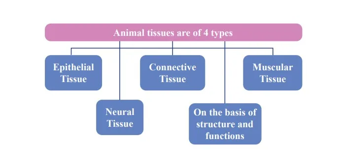 Epithelial Tissue
Epithelial tissue, commonly referred to as epithelium (plural: epithelia), was termed by Ruysch as 'epithelium' (derived from Greek epiupon, thelio meaning "grows upon"), denoting a tissue growing upon other tissues.
Epithelial tissue typically comprises one or more layers of closely packed similar cells, forming a protective covering or lining on both external and internal surfaces of various body structures. Notably, epithelial tissue is the earliest to develop in the embryo and emerge in the animal kingdom.
Functions of Epithelial Tissue
Epithelial tissue serves several vital functions including protection, filtration, diffusion, secretion, and excretion.
Simple Epithelium
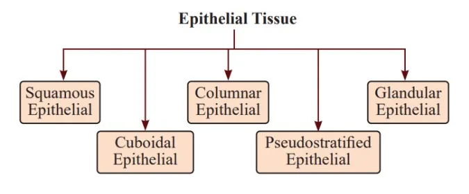
Simple epithelium consists of a single layer of cells directly resting on the basement membrane, serving as linings for body cavities, tubes, and ducts. This type of epithelium is further classified into three types based on the structural modifications of the cells: squamous epithelium, cuboidal epithelium, and columnar epithelium
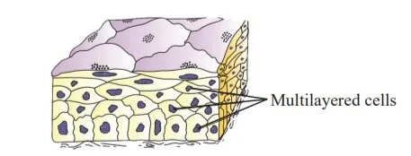
Compound Epithelium
Compound epithelium comprises multiple layers of cells (multilayered), with cells of the deepest layer resting on the basement membrane. It is typically found in areas requiring protection against mechanical, chemical, or osmotic stresses, with a limited role in secretion and absorption. Compound epithelium is divided into two main types: stratified epithelium (Non-Strechtable) and transitional (Strechtable) epithelium.
Cell Junctions
Cell junctions are structural and functional connections between neighboring cells, particularly prominent in tissues with minimal intercellular material. Three types of cell junctions are observed in epithelial and other tissues.
Biotechnology and its applications
Connective Tissue
Connective Tissue serves as a essential link and supportive framework for various tissues and organs within complex organisms. It is the most prevalent and widely distributed tissue type in such organisms, characterized by loosely arranged cells with significant intercellular space. This tissue's adaptability and functionality stem from a combination of cells, fibers, and extracellular matrix components.
Types of Connective Tissue
Below is the flow chart of Types and subtypes of Connective Tissue: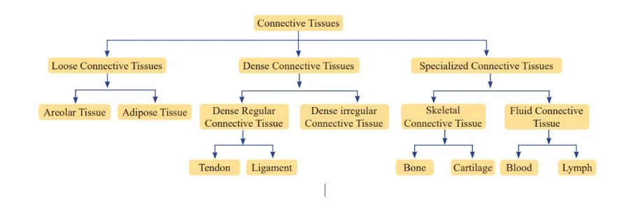
Features of Connective Tissue
- Origin: Derived from mesoderm during embryonic development.
- Function: Connects and supports various tissues and organs in the body.
- Composition: Rich intercellular matrix with loosely arranged cells.
- Variability: Exists in various forms, from soft to specialized types like cartilage, bone, adipose tissue, and blood.
- Fiber Production: Cells like fibroblasts produce structural proteins like collagen, enhancing tissue strength and flexibility.
- Ground Substance: Modified substances between cells and fibers contribute to the extracellular matrix.
- Vascularization: Most connective tissues, except cartilage, are penetrated by blood vessels and nerves.
Functions of Connective Tissue
Connective tissue fulfills several essential functions:
- Support: It provides a structural framework for organs and tissues.
- Protection: Connective tissue shields and cushions delicate organs, enhancing their resilience.
- Transport: As exemplified by blood, a fluid connective tissue, it facilitates the transport of vital nutrients, gases, and waste products throughout the body.
- Repair and Healing: Connective tissue plays a pivotal role in wound healing and tissue regeneration processes.
sexual reproduction in flowering plants
Loose Connective Tissue
Composition: Comprised of loosely arranged cells and fibers within a semi-fluid matrix.
Distribution: Widely distributed throughout the body.
Cellular Components: Includes fibroblasts responsible for fiber production, mast cells secreting inflammatory mediators, macrophages for phagocytosis, adipocytes, and plasma cells.
Function: Serves as a supportive framework for epithelial tissues, typically found beneath the skin.
Types:
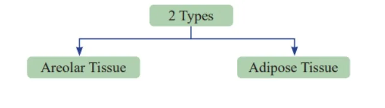
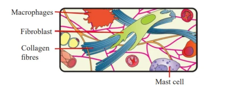
Areolar Tissue
Characteristics: Exhibits maximum intercellular space or matrix.
Cell Types: Features fibroblasts, mast cells, macrophages, adipocytes, and plasma cells.
Location: Predominantly found beneath the skin.
Function: Provides support for epithelium.
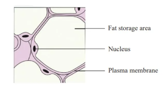
Adipose Tissue
Composition: Comprised of adipocytes storing fat.
Storage Function: Stores excess nutrients converted into fat.
Location: Mainly situated beneath the skin and around organs like the eyes, heart, and kidneys.
Types: Differentiates into white fat and brown fat based on the characteristics of adipocytes.
Dense Connective Tissue
Dense Connective Tissue is characterized by the tightly packed arrangement of fibers and fibroblasts.
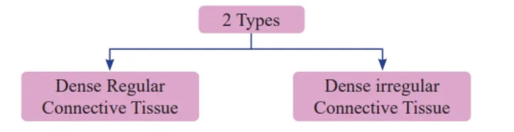
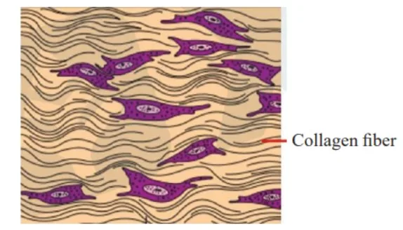
Dense Regular Connective Tissue: exhibits a consistent orientation of fibers, typically arranged in parallel bundles. Tendons, for instance, connect skeletal muscles to bones, while ligaments connect one bone to another. Ligaments are notably rich in elastin fibers and contain relatively fewer collagen bundles. Examples of Dense Regular Connective Tissue include tendons and ligaments.
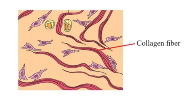
Dense Irregular Connective Tissue: On the other hand, Dense Irregular Connective Tissue displays fibers oriented in an irregular pattern, with fibroblasts and various fibers, predominantly collagen, oriented differently. This type of tissue is commonly found in the skin.
Specialized Connective Tissue
The three types of specialized connective tissue is given below: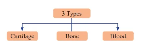
Cartilage
Cartilage, a type of specialized connective tissue, exhibits a solid and pliable intercellular material known as the matrix, reinforced by chondroitin salts. Collagen fibers are crucial in providing structural support and stability to cartilage. Cartilage cells, or chondrocytes, maintain the extracellular cartilage matrix.
There are three main types of cartilage: hyaline, fibrous, and calcified cartilage, each with distinct properties and functions.
Bones
Bones, another form of connective tissue, possess a hard and non-pliable matrix rich in calcium salts and collagen fibers, contributing to their strength and rigidity. Bone cells, or osteocytes, are housed within spaces called lacunae, where they reside and maintain bone tissue. The primary protein found in bones is ossein.
Blood
Blood is classified as a fluid connective tissue composed of plasma, red blood cells (RBCs), white blood cells (WBCs), and platelets. It is the main circulating body fluid, transporting various substances throughout the body.
Muscle Tissues
Muscle tissue originates from the mesodermal layer during embryonic development. Comprising multiple muscle fibers or cells, each muscle fiber consists of numerous delicate myofibrils. These muscle fibers possess the unique ability to contract and relax, crucial for various physiological functions.
Myology, also called Sarcology, is the dedicated field of muscle study. All muscles in the body derive from the mesoderm and exhibit distinct characteristics such as excitability, contractility, extensibility, and elasticity. Muscles contribute significantly to overall body weight, constituting approximately 40-50% of it.
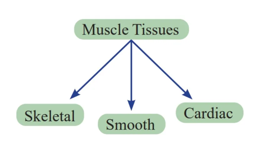
Skeletal Muscles
Skeletal muscle fibers attached to bones are elongated, cylindrical, and unbranched cells arranged parallelly in bundles enclosed by tough connective tissue. Being multinucleated, they act synergistically to facilitate bodily movement, adjustment to environmental changes, and the maintenance of posture. Examples include the biceps and triceps.
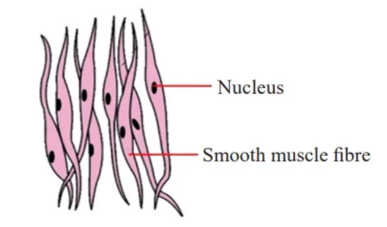
Smooth Muscles
Smooth muscles feature fusiform fibers that taper at both ends and lack striations. Held together by cell junctions within a connective tissue sheath, these muscles operate involuntarily under the autonomic nervous system's control. They predominantly constitute the walls of internal organs like blood vessels, stomach, and intestines.
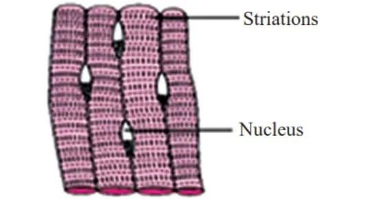
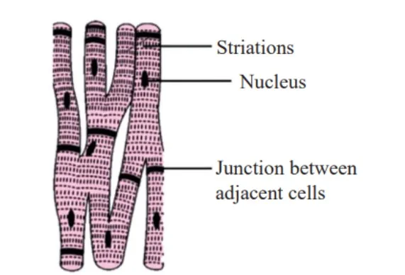
Cardiac Muscles
Found exclusively in the heart walls, cardiac muscles function involuntarily and possess communication junctions known as intercalated discs. These discs facilitate synchronized contraction, where a signal triggering contraction in one cell prompts adjacent cells to contract, ensuring effective pumping action throughout the circulatory system. The functions of cardiac muscles are:
- Cardiac muscles contract autonomously, generating the force necessary for blood circulation.
- These muscles maintain a rhythmic and continuous pumping action, ensuring the circulation of oxygenated blood throughout the body.
Neural Tissue
Neural tissues play a pivotal role in orchestrating the body's response to various stimuli. Comprising neurons and neuroglia, these tissues are fundamental in controlling and coordinating bodily functions.
Neurons: The Building Blocks of the Nervous System.Neurons, the primary units of the neural system, are excitable cells responsible for generating, conducting, and transmitting impulses.
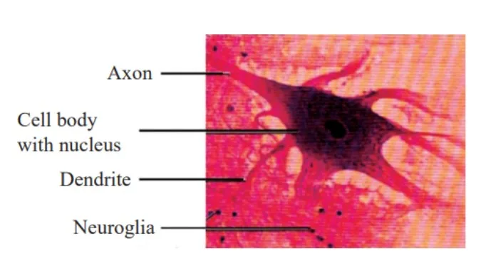
Structure of Neuron
Neurons consist of three main parts: the cell body, dendrites, and axon. Each component serves a specific function in the transmission of nerve impulses.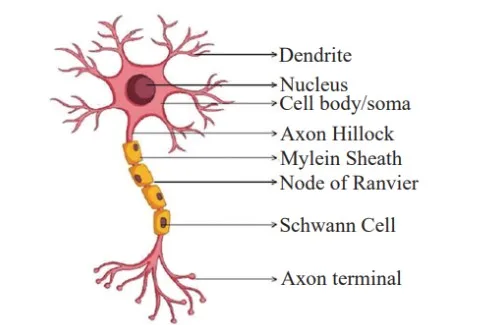
Cell Body
- Also known as cyton or soma, it houses uninucleated cytoplasm containing various organelles.
- Nissl's granules, composed of free ribosomes and rough endoplasmic reticulum (RER), facilitate protein synthesis.
- Neurofibrils aid in internal conduction within the cytoplasm.
Dendrites
- Short fibers branching from the cell body that contain Nissl's granules.
- Transmit impulses toward the cell body.
Axon
- A lengthy fiber terminating in synaptic knobs.
- Synaptic knobs contain synaptic vesicles filled with neurotransmitters.
- Responsible for transmitting nerve impulses away from the cell body.
- Myelinated and non-myelinated neurons are classified according to whether or not they have a myelin sheath.
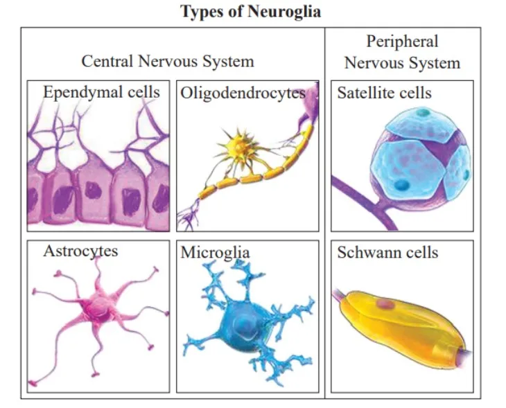
Neuroglia
Neuroglia are cells that protect and support neurons, comprising more than half the volume of neural tissue.
Neural tissues, consisting of neurons and neuroglia, are essential for maintaining the body's homeostasis and coordinating its responses to external stimuli. Understanding the structure and function of these components is crucial for comprehending the complexities of the nervous system.
Introduction to Cockroach
Cockroaches are nocturnal omnivorous creatures commonly found inhabiting moist environments. Typically, they exhibit brown or black coloration, although vibrant hues such as yellow, red, and green are observed in tropical areas. Their size varies from 0.25 to 3 inches (0.6-7.6 cm). Cockroaches serve as vectors for numerous diseases and are significant household pests.
Kingdom: Animalia
Phylum: Arthropoda
Class: Insecta
Order: Blattodea
Family: Blattidae
Genus: Periplaneta
Species: americana
Their habitat is widespread, with cockroaches being cosmopolitan in distribution, favoring dark and damp habitats globally, often residing within human dwellings, posing serious pest problems and transmitting various diseases. Regarding their coloration, while typically brown or black, cockroaches may display vivid yellow, green, or red tones, particularly in tropical regions.
Morphology of Cockroach
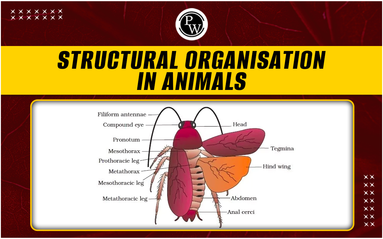
Adult cockroaches typically measure 34-53 mm in length. Its body is covered in a strong, brown chitinous exoskeleton. Males' wings extend past the abdomen. The segmented body is divided into three sections: the head, thorax, and abdomen. Each segment is made up of hardened plates called sclerites, which include dorsal tergites and ventral sternites. They are connected by a thin, flexible articular membrane called the arthrodial membrane.Head
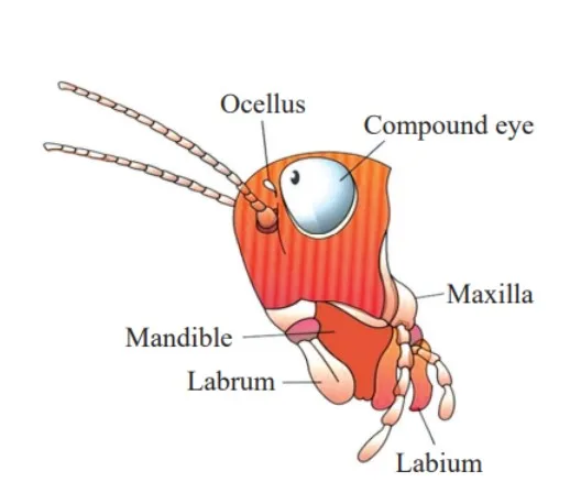
The triangular head results from the fusion of six segments and exhibits considerable mobility in all directions due to its flexible neck. Positioned anteriorly at right angles to the body's longitudinal axis, the head is equipped with thread-like antennae, compound eyes, and mouthparts specialized for biting and chewing.
Mouth Parts
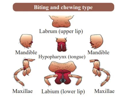
Thorax
The thorax is composed of three parts: the prothorax, mesothorax, and metathorax. The head is linked to the thorax by a neck, a short extension of the prothorax. Each thoracic segment bears a pair of walking legs.
There are two pairs of wings: the forewings (mesothoracic), known as tegmina, are opaque, dark, and leathery, covering the hind wings when at rest. The hind wings (metathoracic) are transparent, membranous, and utilized for flight.
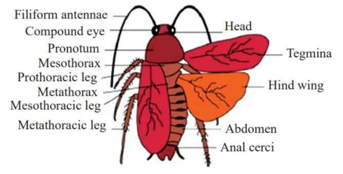
Abdomen
The abdomen consists of ten segments. In females, the 7th, 8th, and 9th sterna form a brood (genital) pouch, housing the female gonopore, spermathecal pores, and collateral glands. In males, the genital pouch is located at the abdomen's hind end, dorsally bounded by the 9th and 10th tergites and ventrally by the 9th sternum.
It contains the dorsal anus, ventral male genital pore (gonopore), and gonapophysis. Both sexes feature the 10th segment (tergum), which bears a pair of jointed anal cerci. Males also possess a pair of short, thread-like anal styles attached to the 9th sternum, absent in females.
Anatomy of Cockroach
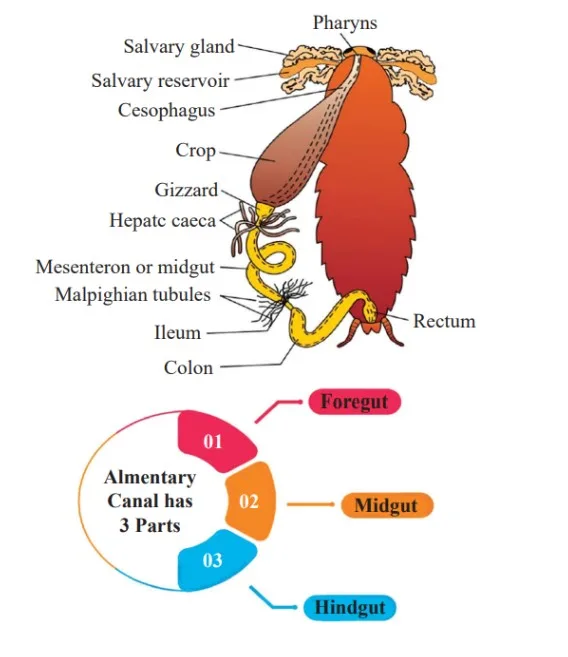
The anatomy of a cockroach consists of various organ systems including the digestive system, excretory system, respiratory system, and reproductive system.Digestive System of Cockroach
The cockroach digestive system consists of foregut, hindgut, midgut.Foregut
A protective cuticle lines the foregut and comprises the pharynx, esophagus, crop (for temporary food storage), and gizzard (proventriculus). The gizzard facilitates food grinding with its outer layer of thick circular muscles and inner chitinous teeth plates.
Midgut
Also known as the mesenteron, the midgut lacks a cuticular lining and features hepatic or gastric caeca at the foregut-midgut junction, which secrete digestive juices. Additionally, numerous malpighian tubules at the midgut-hindgut junction serve excretory functions.
Hindgut
Broader than the midgut, the hindgut includes the ileum, colon, and rectum, which opens through the anus.
Circulatory System of Cockroach
An open blood vascular system characterizes the circulatory system of cockroaches. Blood vessels, poorly developed, open into the haemocoel, bathing visceral organs in haemolymph, a mixture of colorless plasma and haemocytes. The heart, an elongated muscular tube along the mid-dorsal line of the thorax and abdomen, pumps blood anteriorly to sinuses and receives blood through ostia.
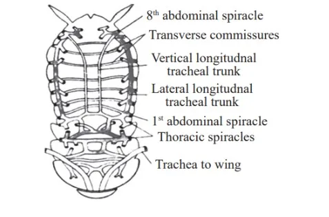
Respiratory System of Cockroach
Cockroaches possess a tracheal system consisting of a network of tracheal tubes opening through spiracles on the lateral sides of the body. Oxygen is transported to tissues through branching tracheal tubes, with gas exchange occurring at tracheoles via diffusion. Sphincters regulate the opening of spiracles.
Nervous System of Cockroach
The nervous system comprises fused and segmentally arranged ganglia connected by longitudinal connectives. The double, solid, and ganglionated ventral nerve cord extends along the thorax and abdomen. The supra-oesophageal ganglion (brain) supplies nerves to antennae and compound eyes. Even if the head is severed, cockroaches can survive for a week due to the distribution of the nervous system throughout the body.
Sense Organs of Cockroach
Sense organs include antennae, eyes, maxillary palps, labial palps, and anal cerci. Antennal sensory receptors monitor the environment, while compound eyes provide mosaic vision, particularly useful during nocturnal activities.
Reproductive System of Cockroach
The reproductive system of a male cockroach consists of the ejaculatory duct, testes, utricular gland, vas deferens, and phallic gland. The reproductive system of a female cockroach consists of genital chamber, two large ovaries, vagina, colleterial gland, spermatheca, external genitalia and oviduct.
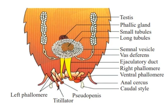
Male Reproductive System
The male reproductive system includes testes, seminal vesicles, mushroom gland, and phallic gland. External genitalia are represented by asymmetrical chitinous structures surrounding the male gonopore. Sperms are stored in spermatophores and discharged during copulation.
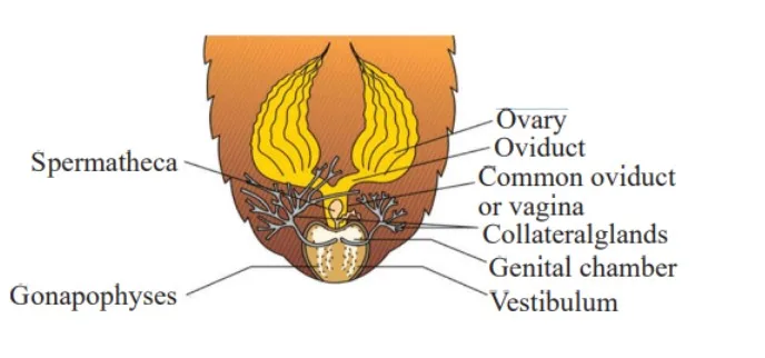
Female Reproductive System
The female reproductive system comprises ovaries, oviducts, spermathecae for sperm storage, and colleterial glands. These glands aid in forming ootheca, capsules containing eggs. Females typically produce multiple oothecae, each containing numerous eggs.
Frog
These organisms adapt to terrestrial and freshwater environments, exhibiting a dual habitat preference. As ectothermic creatures, they lack a constant internal body temperature, a trait commonly known as being cold-blooded or poikilotherms. They possess the remarkable ability to change their skin coloration for camouflage, aiding in protection from predators. During extreme environmental conditions such as summer and winter, they exhibit behavioral adaptations like aestivation and hibernation, seeking refuge in deep burrows.

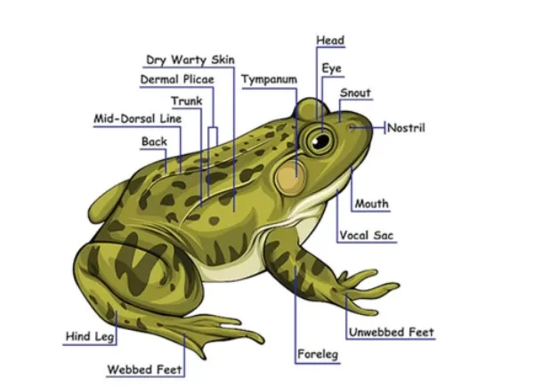
Morphology of Frog
Integumentary System : Their skin is characterized by a moist, smooth, and slippery texture due to mucus. Dorsally, they typically display an olive-green hue with irregular dark spots, while the skin appears uniformly pale yellow ventrally.
Anatomical Structure: They lack a distinct neck and tail and possess a pair of nostrils above the mouth. Bulged eyes are covered by a nictitating membrane, offering protection while submerged. Forelimbs and hind limbs facilitate various locomotive functions, aided by webbed digits for swimming.
Taxonomic Classification of frog:
Phylum: Chordata
Subphylum: Vertebrata
Superclass: Tetrapoda
Class: Amphibia
Order: Anura
Genus: Rana
Species: tigrina
Division: Gnathostomata
Anatomy of Frog
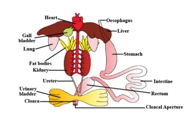
The anatomy of a frog consists of various organ systems, including the digestive, excretory, respiratory, and reproductive systems.Digestive System of Frog
Their digestive system comprises an alimentary canal along with associated digestive glands. Due to their carnivorous diet, the alimentary canal is relatively short, featuring a reduced length of intestine. The mouth leads to the buccal cavity, which in turn connects to the esophagus via the pharynx.
From the stomach, partially digested food known as chyme transitions into the duodenum, where it encounters bile from the gall bladder and pancreatic juices from the pancreas. Subsequent digestion of carbohydrates and proteins occurs within the intestine, with absorption facilitated by numerous fingerlike folds known as villi and microvilli. Undigested waste is expelled through the rectum and cloacal aperture.
Respiratory System of Frog
Respiration in these organisms occurs via two distinct methods, adapted for both aquatic and terrestrial environments. In water, cutaneous respiration takes place through the skin, allowing for the exchange of dissolved oxygen. On land, respiration is facilitated by a combination of buccal cavity, skin, and lungs, with gaseous exchange primarily occurring through the skin.
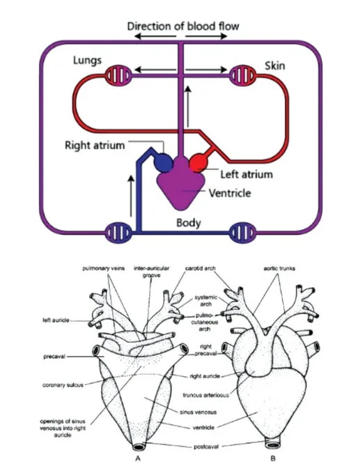
Circulatory System of Frog
Their circulatory system comprises a blood vascular system and a lymphatic system. The heart, situated in the upper part of the body cavity, consists of three chambers: two atria and one ventricle. Deoxygenated blood is received by the sinus venosus before being pumped to various body parts via arteries. The veins collect oxygenated blood and return it to the heart. Specialized portal circulations, such as hepatic and renal, further regulate blood flow.
Excretory System of Frog
The excretory system eliminates nitrogenous wastes and comprises a pair of kidneys, ureters, cloaca, and urinary bladder. Each kidney contains numerous uriniferous tubules or nephrons, through which wastes are filtered and excreted. Urea is the primary excretory product, expelled through the cloaca.
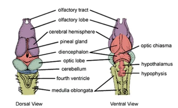
Endocrine and Neural Systems of Frog
Control and coordination within these organisms are achieved through a well-developed endocrine system, which secretes hormones for chemical coordination. Major endocrine glands include the pituitary, thyroid, parathyroid, thymus, pineal body, pancreatic islets, adrenals, and gonads. The nervous system, organized into central, peripheral, and autonomic components, also plays a crucial role in coordinating various physiological processes.
Reproductive System of Frog
The reproductive systems of males and females exhibit distinct structures and functions. In males, testes are paired structures near the kidneys, while ovaries occupy a similar position in females. Fertilization occurs externally in freshwater environments, leading to the development of larvae known as tadpoles, which undergo metamorphosis into adult frogs. This reproductive strategy contributes to population maintenance and ecological balance by controlling insect populations and supporting agricultural ecosystems.
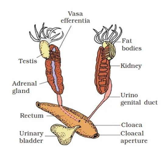
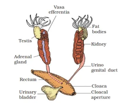
Male Reproductive System
In the male reproductive system, testes are yellowish oval structures attached to the upper part of the kidneys via the mesorchium, a double fold of peritoneum. Approximately 10 to 12 vasa efferentia emerge from the testes, entering the kidneys and opening into Bidder’s canal, which connects to the urinogenital duct. This duct leads to the cloaca, a small chamber for excreting feces, urine, and sperm.
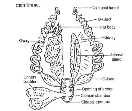
Female Reproductive System
In the female frog reproductive system, ovaries are located near the kidneys on their ventral side. Eggs released by the ovaries are collected by a pair of oviducts, each opening separately into the cloaca. A mature female frog can lay 2500 to 3000 eggs at once. Fertilization occurs externally in freshwater environments.
| NEET Exam Important Links | |
|---|---|
| NEET Biology Syllabus | NEET Biology Diagrams |
| NEET Biology MCQ | NEET Biology Chapter wise Weightage |
| NEET Biology Notes | NEET Previous Year Question papers |
Structural Organisation in Animals FAQs
What is the structural organisation of an animal cell?
Is the structural organisation in animals important for exams like NEET?
What are the four levels of structural organisation in animals?
What does structural organisation in animals refer to in Class 11?