

Excretory System In Mammals
Excretory System of Class 11
Consists of a pair of kidneys, a pair of ureters, a urinary bladder and urethra.
In birds, cyclostomes, cartilaginous fishes, snake and crocodile urinary bladder is absent.

Fig. Excretory system of human
Kidney
Kidney is perfectly bean shaped structure with inner concavity called hilum.
Ureter, blood vessels, nerve etc. enter or leave the kidney through hilus (hilum)
In human the right kidney is at lower level than the left one along the sides of the lumber vertebrae.
It measures about 10 cm long, 5 cm wide and 4cm thick, weighing about 135 to 150 gms.
Each kidney covered by three protective layer which are as follows:
(i) Renal capsule – Renal capsule forms innermost protective layer. It consists of white fibrous connective tissue with a few yellow elastic fibres and few muscles.
(ii) Adipose capsule – It is middle covering which is made of adipose tissue. It acts as a shock absorber.
(iii) Renal Fascia – It is outermost fibrous covering with which kidney is anchored to the abdominal wall.
Kidney is also covered by double fold of peritoneum only on the ventral side. Therefore, kidney is posterior to peritoneum and is called retro-peritoneal.
Internally divided into outer granulated cortex and inner striated medulla.
The medulla is distinct by the presence of 15-16 conical pyramids with broader end towards cortex. Each pyramid ends as renal papilla where many ducts of Bellini converge to open.
The renal papilla fits over small funnel-like minor calyx, 2 to 3 minor calyces together open in still wider funnel called major calyx.
In man each kidney has 2 - 3 major calyces which open at a common place pelvis, that forms the wide mouth of ureter.
Between the renal pyramids and the minor and major calyces the cortical parts remain extended into medullary part and are called as renal columns of Bertini.
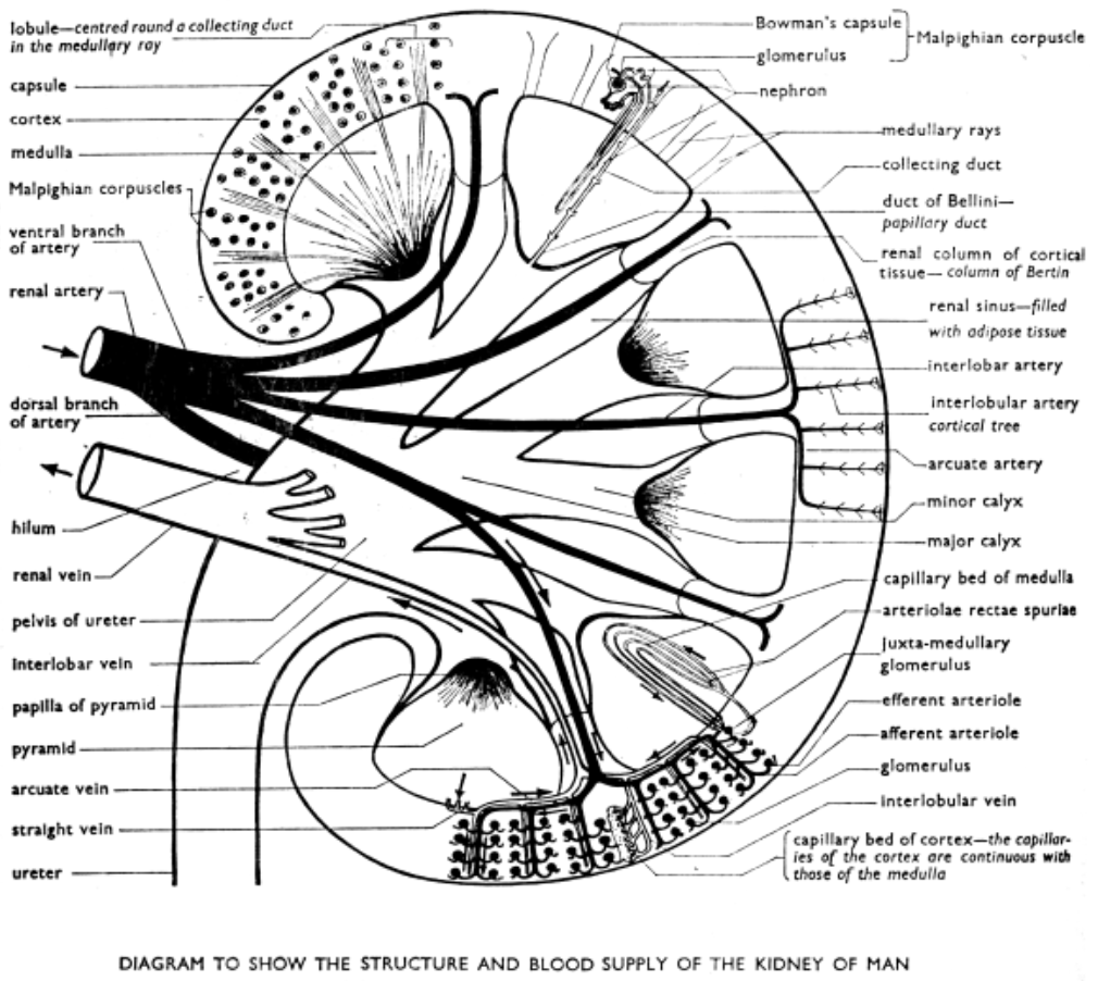
Fig. Structure and blood supply of the kedney of man
Nephron (Uriniferous tubules) :
In man each kidney has 1.2 million nephrons. It is a thin, long (-3 cm), twisted tubule extended from cortical to medullary part; has four parts : Bowmann’s capsule, proximal convoluted
tubule (PCT), loop of Henle and distal convoluted tubule (DCT). The latter opens into the collecting duct through collecting tubule. Many collecting ducts open in a common duct of
Bellini which converge at renal pyramid.
Two types of nephrons : Cortical and Juxtamedullary.
The cortical nephrons (85%) have short loop of Henle extended little in medulla.
The juxtamedullary nephrons (rest 15%) have long loop of Henle extended deep into medulla and their Bowman’s capsule lie near the junction of cortex and medulla.
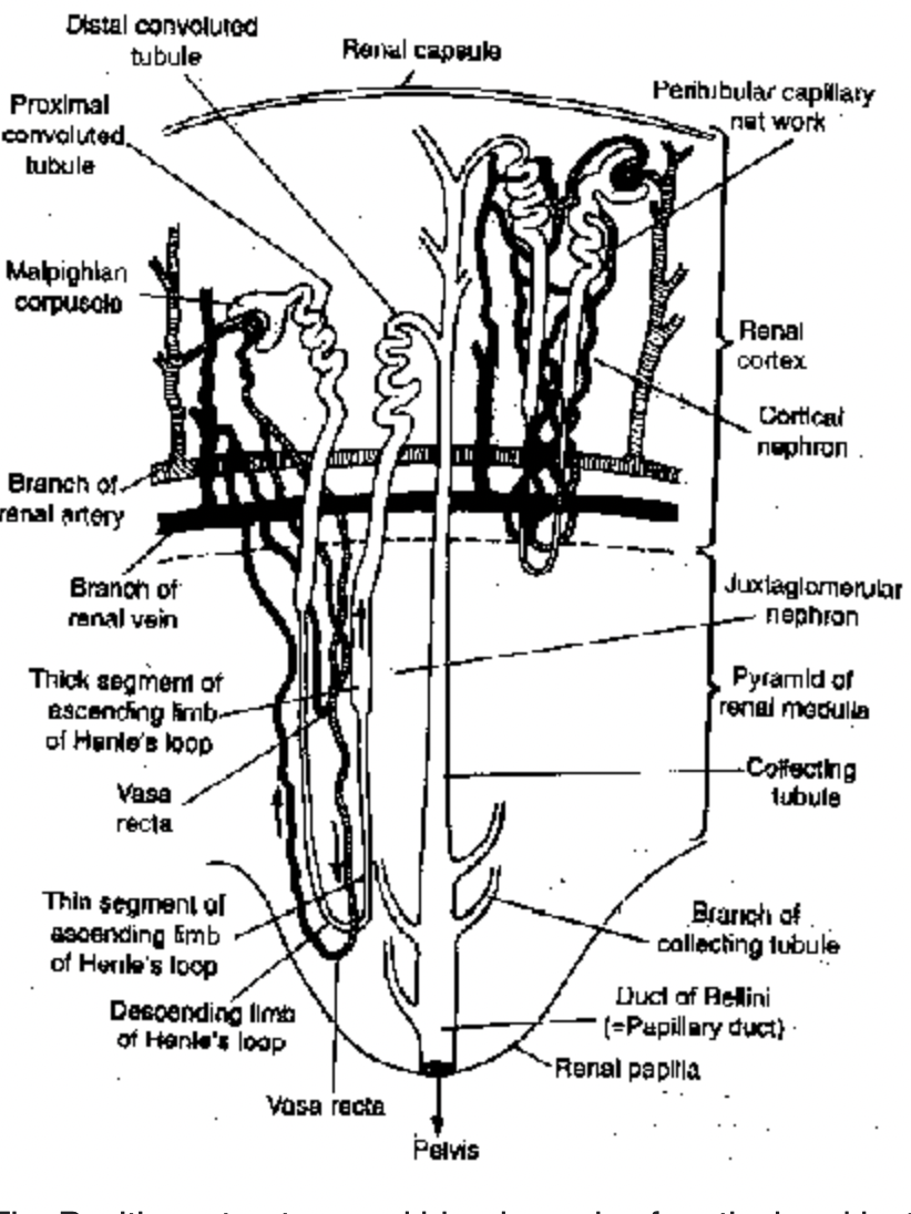
Fig. Position, structure and blood supply of cortical and juxtamedullary nephrons in a mammalian kidney
Bowman’s capsule is made of two layered squamous epithelium glomerulus and together called as Malpighian body or renal corpuscle. Podocytes are the cells of inner membrane with long process closely applied to the walls of capillaries.
Blood enters the glomerulus through afferent arteriole (larger diameter)and leaves through efferent arteriole (smaller diameter).
PCT is the highly coiled part of the tubule behind the Bowman’s capsule in cortical part, lined with cone-shaped brush border cells.
Loop of Henle (Pars recta) is U-shaped loop extended in medulla, divided as (i) thin walled straight descending limb, (ii) thin-walled ascending limb and (iii) thick walled ascending
limb. It is longer in birds and mammals but short or absent in other vertebrates.
DCT also a coiled part lies at the level of PCT, lined by cuboidal epithelium but without any true brush border.
Jaxtaglomerular Apparatus
This includes 3 types of cells in the cortical part of the kidney surrounding the renal corpuscles, PCT and DCT. These are :
(i) The cells around the afferent arteriole called juxtaglomerular cells (JGC)
(ii) The cells around DCT called macula densa and
(iii) the Lacis cells or Polkissen situated in the angle between afferent and efferent arteriole of each glomerulus.
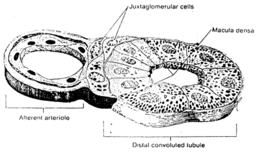
Fig. Juxtaglomerular apparatus
Collecting tubule is about 20 mm long straight tubule, many of these join to form duct of Bellini.
Vasa recta :
The efferent renal arterioles break up into capillaries, around the loop of Henle, these are called vasa recta.
Ureter :
Narrow distensible tube, emerging from the hilus is basically a metanephric duct, lined with transitional epithelium. Being muscular it also undergoes peristalsis to pass urine from kidney to urinary bladder.
Urinary bladder :
Single, median, pyriform bag (300-800 ml) in the pelvic area has upper body with two openings of ureter and lower trigone leading to urethra and guarded by sphincter.
Wall is stretchable, muscular (detrusor) and lined with stratified transitional epithelium.
Urethra :
The tube emerging from urinary bladder to release urine having sphincters at both ends.
In women it is a short (- 4 cm) tube opening separately below clitoris in the vulva.
In man it is long (- 20 cm) tube also serving as reproductive tract; divided into 3 parts : the upper prostatic part; the middle membranous part and the lower penile part.