
Earthworm Classification and Body wall
Anatomy of Earthworm,Cockroach & Frog of Class 11
Classification
Phylum – Annelida; Class – Oligochaeta; Order : Opisthopora; Family – Megascolecidae; Genus – Pheretima; Species – posthuma.
Salient Features
- Detailed anatomy by Prof. K.N. Bahl (1926)
- Earthworms has cosmopolitan distribution, with around 1800 species placed under several genera. About 40 species of earthworms are found in India. Largest genus is Pheretima that includes 500 species, widespread in tropical and temperate regions of the world. Thirteen of these species found in India.
- Pheretima posthuma is the most common species.
- Earthworm is nocturnal and lives in burrows in damp soil rich in humus. Burrows are usually 20-30 cm deep but may reach 2-3 mtrs. during dry season in search of water.
- Earthworm eats its way into the soil to excavate a burrow. Organic matter in the soil serves as its food. Unused organic food and residual soil is egested out through anus in the form of small spherical worm castings.
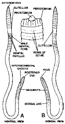
Fig. A. Ventral view of P.posthuma;
B. Dorsal view of its anterior end
External Features
- Body elongated, rounded, narrow, bilaterally symmetrical and cylindrical. The anterior end pointed while the posterior end blunt. A mature worm is
- around 15-20 cms in length and 3 to 5 mm in width.
- It is quite slimy to touch and appears brown due to the presence of porphyrin pigment in its body wall. Porphyrin protects the body from injurious
- effect of bright light.
- The body divided into about 100-120, small, ring-like segments, also called metameres or somites by distinct annular grooves or furrows in the body
- wall. External segmentation extends internally into the body cavity.
- The dorsal surface relatively darker and bears a dark coloured median line of dorsal blood vessel visible through semi-transparent integument.
- It lacks a distinct head. The first segment at anterior end is called the buccal segment or peristomium bearing the terminal crescentic mouth. It is prolonged anteriorly into a fleshy lobe, the prostomium which overhangs the mouth.
- In mature worms a thick collar or girdle-like glandular thickening of body wall called clitellum is present around segments 14, 15 and 16. During breeding season, it forms cocoon in which ova are laid and fertilised.
- With the presence of clitellum body can be differentiated into a pre-clitellar, clitellar and post-clitellar regions.
- In each segment except the first, last and clitellar segments, there is a ring of tiny, curved, S-shaped, yellowish chitinous setae. Setae are half embedded in body wall and half projected backwards upon body surface. Arrangement of setae in Pheretima is perichaetine, in Lumbricus and Eutypheus is Lumbricin or Octochaetine (2 × 4)
Note : If we put body wall in 20% KOH, then setae remain unaffected.
Following apertures occur upon the body surface :
Mouth : Relatively larger, transversely crescentic and terminal aperture at anterior end.
Anus : Small and vertical slit-like aperture at the end of last anal segment.
Dorsal pores : Minute apertures of the coelomic chambers located mid-dorsally, one in each intersegmental groove, behind the segment 13 upto the last but one groove.
Nephridiopores : Each segment except the first two have about 200-250 minute pores of integumentary nephridia, scattered upon the surface of each segment except the first two. In the clitellar region, the number is ten times more and this region is known as ‘forest of nephridia’.
Spermathecal pores : In each of the four intersegmental grooves between segments 5/6, 6/7, 7/8 and 8/9, there is a pair of ventro-lateral elliptical spermathecal pores.
Male genital pores : These are a pair of crescentic apertures located ventro-laterally upon 18th segment.
Female genital pores : Single minute female genital pore is located in the mid-ventral line of 14th segment.
Just behind the clitellum, each of the 17th and 19th segments bears a pair of small and conical ventro-lateral genital or copulatory papillae. These papillae help in copulation.
Body Wall
Body wall is thin, soft and slimy. It consists of the following layers from the surface inwards :
Cuticle : is thin and elastic, non-cellular protective membrane, formed by two super-imposed layers of collagen fibres secreted by underlying epidermis.
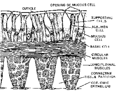
Fig. V.S. of skin of earthworm
Epidermis : is composed of supporting cells, glandular cells, basal cells (replacement cells) and sensory cells.
Muscular layers : has outer thin layer of circular muscles and inner thick layer of longitudinal muscle.
Coelomic epithelium : is outer envelope of coelomic cavity, made up of squamous cells.
Each segment bears a ring of setae lodged in setigerous sacs. The setigerous sac has two sets of muscles, the protractor muscles which attach the sac with outer layer of circular muscles and one pair of retractor muscles which join the sac with a thin layer of circular muscles which forms an additional ring below the longitudinal muscle layer.
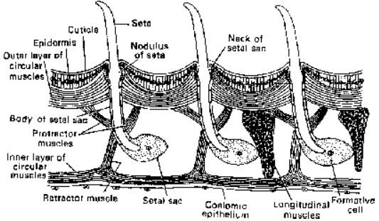
Fig. Setae in situ
Body wall maintains shape, provides protection, perceive stimuli, help in locomotion and respiration.
Note : Body wall of Microscolex phosphoreus is phosphorescent in nature.
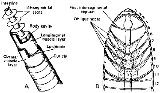
Fig. A. Diagram of “tube within a tube” type of body in earthworms.
B. Arrangement of septa in anterior part of body
Coelom
Schizocoel divided by septa from 4/5 onwards.
Each septum consists of a thin layer of muscle fibres lined by coelomic epithelium on both surfaces.
The first six septa are cone-like and run obliquely backwards from the body wall to the gut wall.
The first nine septa i.e. upto septum 13/14 are without perforations. The remaining septa beginning from septum 14/15 are perforated by numerous sphinctered apertures.
Coelomic fluid is milky, white and alkaline. It has fluid matrix of watery plasma containing proteins, salts and numerous minute nucleated corpuscles. Corpuscles are of following four types:
Granulocytes (Phagocytes) : Most numerous and largest corpuscles. These serve to store nutrients.
Leucocytes : These are smaller and fewer. Their function is uncertain.
Mucocytes : These are elongated, vase-like corpuscles, each with an expanded fan-like part and a narrow nucleated stalk-like part. Function of these is also uncertain.
Chloragogen cells/Yellow cells : These are also numerous but much smaller and star-shaped corpuscles, each with finger-like pseudopodia. They are supposed to be excretory in function.
Originate from visceral peritoneum of stomach and intestine. Analogous to liver as store food in the form of glycogen. (Storage of reserve food, deamination of proteins, urea formation)
Note : Other corpuscles are formed by lymph glands, present on dorsal side of intestine from 26th segment to last segment. Each gland is having 10 lobes.
Coelomic fluid serves as a hydrostatic skeleton to assist the musculature of body wall in bringing about the locomotion of body. It oozes out upon body surface through dorsal pores, keeping the body wall moist to facilitate respiration and to destroy bacteria and other harmful micro-organisms.
Locomotion
- Movement in earthworm involves the musculature of the body wall and setae.
- A wave of contraction affecting the circular muscles, begins at the anterior end and travels posteriorly, causing the body to become thinner and longer.
- After the wave of contraction has passed over the front half of the animal, a wave of contraction affecting the longitudinal muscles sets in at the anterior end, causing the thickening and
- thus shortening of the body. This is again followed by the wave of thinning and the process is repeated.
- Each wave of circular contraction causes the segments affected to move forward, but the segments in a state of longitudinal contraction, do not move as they are anchored to the ground by
- the protruded setae.
- Setae always protrude during longitudinal contraction and retract during circular contraction.
- The earthworm travels a distance of about 25 cms in one minute.
- When the direction of the waves is reversed, the worm crawls backward.
Madreporite is a thick, rounded sieve-like calcareous plate, situated on the aboral surface of the central disc. The pores of the madreporite allow water into the system.
Tiedemann’s bodies are also known as racemose glands. In Asterias they are nine in number. Exact function is not known. They are considered to be lymphatic glands and filtering device.
Types of larvae:
Name of organism Name of larva
Holothuria Auricularia
Asterias Bipinnaria; Brachiolaria
Antedon Doliolaria
Echinus Echinopluteus
Ophioderma Ophiopluteus
