Pelvic Girdle, Anatomy, Bone, Functions and Pectoral Girdle
Pelvic girdle is a ring like bony structure that connects the axial skeleton to the lower limbs. In this article, pelvic girdle functions, anatomy, bones, muscles, and pctoral girdle are provided.
Share
Pelvic Girdle: The pelvic girdle, also known as the hip girdle, is comprised of a single bone called the hip bone or coxal bone (coxal meaning "hip"). It acts as the point of attachment for each lower limb. Each hip bone is firmly connected to the axial skeleton through its attachment to the sacrum of the vertebral column. Additionally, the right and left hip bones meet at the front to attach to each other. Together, these structures form the bony pelvis, which includes the two hip bones, the sacrum, and the coccyx attached below the sacrum.
Unlike the bones of the shoulder girdle, which are highly mobile to facilitate a wide range of upper limb movements, the bones of the pelvis are strongly fused to form a mostly immobile, weight-bearing structure. This immobility is important for stability, as it allows the weight of the body to be easily transferred from the vertebral column, through the pelvic girdle and hip joints, to either lower limb when the other limb is not bearing weight. Therefore, the pelvis' lack of mobility provides a stable foundation for the upper body, which rests atop the mobile lower limbs. The article below details the pelvic girls and their difference from the pectoral girdle.Pre Fertilisation: Structures and Events
What is Pelvic Girdle?
The pelvic girdle, also known as the bony pelvis, is the structure in the lower part of the trunk that connects the legs to the torso. It forms a basin-like shape and is a crucial part of the appendicular skeleton, which attaches the lower limbs to the axial skeleton. This girdle, along with the sacrum and coccyx of the vertebral column, comprises the bony framework of the pelvis. Its main functions include providing support and stability to the body, and transferring.What is Pelvic Bone?
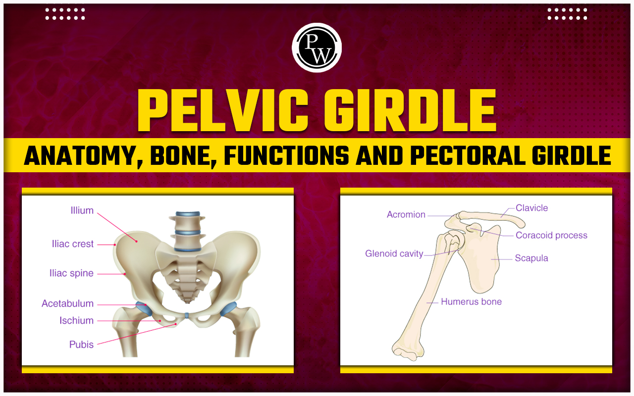
The pelvic bone, or hip bone, is a large structure located in the lower part of the body. It consists of two hip bones, each made up of three sections: the pubis, ischium, and ilium, which fuse together during puberty to form a single bone. The sacrum and coccyx form the posterior part of the pelvic skeleton.Pelvic Girdle Anatomy
The pelvic region, located between the abdomen and the thighs, contains several important structures that contribute to its overall function:Bony Pelvis (Pelvic Skeleton)
- The sacrum, coccyx, and two hip bones (ilium, ischium, and pubis) fused to form this structure.
- It serves to support the weight of the upper body and transmit forces to the legs.
Pelvic Cavity
This space is enclosed by the pelvic bones. It is divided into two parts:- Greater (false) pelvis: Located superior to the pelvic brim.
- Lesser (true) pelvis: Situated inferior to the pelvic brim, containing most of the pelvic organs.
Pelvic Floor (Pelvic Diaphragm)
- It supports the pelvic organs by acting as a sling made up of muscular and connective tissue.
- It is made up of several muscle layers, including the levator ani and coccygeus muscles, which support organs and close the rectum/urogenital openings, the urogenital diaphragm, which adds support to the urogenital openings, and the external anal and urethral sphincters, which regulate urination and defecation.
Perineum
- This is the area between the anus and the scrotum/labia majora.
- It contains muscles and structures that support the pelvic organs.
Pelvic Girdle Variations Between the Sexes
The male and female pelvises exhibit distinct differences in size, shape, and structure, reflecting their respective physiological functions. These differences include:- Overall Size and Shape: The female pelvis is generally wider and more rounded, while the male pelvis is narrower and more compact.
- Pelvic Inlet: The female pelvic inlet is wider and oval-shaped, whereas the male pelvic inlet is narrower and heart-shaped.
- Pelvic Sides: The female pelvic sides are wider apart, allowing for childbirth, while the male pelvic sides converge towards the pelvic outlet.
- Subpubic Angle: The angle formed by the pubic bones is wider in females (90-100 degrees) and narrower in males (70 degrees).
- Ischial Bones: The ischial bones, which form part of the pelvis, are wider apart in females, creating a larger pelvic outlet.
- Iliac Crests: The iliac crests are more prominent in males, making the false pelvis deeper and narrower.
- Sacrum: The female sacrum is shorter, wider, and more curved than the male sacrum, with a less pronounced promontory.
- Acetabula: The acetabula, which are the sockets of the hip joints, are wider apart and more anteriorly facing in females compared to males.
The Pelvic Girdle Consists of
The pelvic girdle, also known as the pelvic bone or hip bone, is a complex structure that forms the base of the vertebral column and supports the trunk of the body. Pelvic girdle consists of two hip bones, a sacrum, and a coccyx.- Hip Bones (Coxal Bones): Each hip bone is made up of three fused bones ilium, ischium, and pubis. These bones fuse together during adolescence to form a single bone. The ilium is the largest and uppermost bone of the hip bone, contributing to the acetabulum, the socket of the hip joint. The ischium forms the lower and back part of the hip bone and supports the body when sitting. The pubis forms the front part of the hip bone and joins the opposite pubic bone at the pubic symphysis.
- Sacrum: The sacrum is a triangular bone located at the base of the spine, between the two hip bones. It consists of five fused vertebrae and forms the back part of the pelvic girdle. The sacrum connects the spine to the hip bones and helps transmit the weight of the body to the lower limbs.
- Coccyx: The coccyx, also known as the tailbone, is a small triangular bone located at the base of the sacrum. It consists of three to five fused vertebrae and serves as an attachment point for ligaments and muscles of the pelvic floor.
| Other NEET Biology Topics | ||
|---|---|---|
| Ribosomes | Pollination | Apomixis |
| Centrosome | Embryo | Tissues |
Pelvic Girdle Bones
The pelvic girdle, also known as the hip girdle, is a bony ring in the lower body that connects the torso to the legs. It is formed by two large hip bones (coxal bones), each made up of three fused bones:1. Ilium
Location: The uppermost and largest part of the hip bone. Function: Forms the flaring portion of the hip and attaches to the sacrum in the back.2. Ischium
Location: The lower and back portion of the hip bone. Function : Forms the inferior and posterior parts of the hip bone and is the part you sit on.3. Pubis
Location: The front and lower portion of the hip bone. Function: Meets the pubis of the other hip bone at the pubic symphysis in the midline. Although not technically part of the pelvic girdle, the sacrum (the lower five fused vertebrae of the vertebral column) and the coccyx (the tailbone) are also part of the bony pelvis.Pelvic Girdle Muscles
The pelvic girdle, also known as the bony pelvis, is a ring-shaped structure located in the lower part of the torso. It serves as a connection between the spine and the legs, consisting of three bones: the sacrum, the two innominate bones (formed by the fusion of the ilium, ischium, and pubis), and the coccyx. The following are the muscles Associated with the Pelvic Girdle1. Pelvic Floor Muscles
Function: Support the organs within the pelvis, including the bladder, rectum, and uterus (in females). They also play a role in urinary and fecal continence, as well as sexual function. Key Muscles: Levator ani, Coccygeus, Iliococcygeus, Pubococcygeus, Puborectalis.2. Hip Muscles
Function: Surround the hip joint and facilitate leg movement at the hip, essential for activities like walking and running. Key Muscles: Gluteus maximus, Gluteus medius, Gluteus minimus, Piriformis, Obturator internus, Superior gemellus, Inferior gemellus, Quadratus femoris, IliopsoasPelvic Girdle Functions
The pelvic girdle performs several important functions:- Support: It provides support for the organs of the abdomen and pelvis, such as the intestines, bladder, uterus (in females), and rectum. Additionally, the muscles of the pelvic floor help support these organs.
- Stability: The pelvic girdle helps maintain the stability and balance of the body during various activities like standing, walking, and running. The structure of the pelvis bones and their alignment contribute significantly to this stability.
- Weight Transfer: It transfers weight from the upper body to the legs. When standing, the weight of the upper body is transmitted through the sacrum to the innominate bones and then to the legs.
- Muscle Attachment: The pelvic girdle provides attachment points for many important muscles, including the hip muscles. These muscles are essential for leg movement in various directions, such as flexion, extension, abduction, adduction, rotation, and circumduction.
- Childbirth (in females): For females, the pelvic girdle is specifically adapted for childbirth. The sacrum and coccyx are slightly movable to facilitate the passage of the baby's head during birth. During pregnancy, hormones relax the ligaments in the pelvis, allowing for slight movement of the bones.
Pelvic Girdle and Pectoral Girdle
The pectoral and pelvic girdles are two important skeletal structures with different functions and characteristics. Here's a comparison of their key differences:| Pelvic Girdle and Pectoral Girdle | ||
|---|---|---|
| Feature | Pectoral Girdle | Pelvic Girdle |
| Function | Connects upper limbs (arms) to thorax | Connects lower limbs (legs) to lower axial skeleton |
| Bones | Clavicle (collarbone), scapula (shoulder blade) | Ilium, ischium, pubis (fused to form os coxa in adults) |
| Attachment to axial skeleton | Indirect attachment to sternum | Attaches to the sacrum (lower part of vertebral column) |
| Movement | Allows for a wide range of motion at the shoulder joint | Provides stability and weight transfer |
| NEET Exam Important Links | |
|---|---|
| NEET Syllabus | NEET Biology Diagrams |
| NEET Biology MCQ | NEET Biology Chapter wise Weightage |
| NEET Biology Notes | NEET Previous Year Question papers |
Pelvic Girdle FAQs
How many bones make up the pelvis?
The pelvis consists of three bones: the hip bone, sacrum, and coccyx. These bones connect the axial skeleton to the lower limbs and help bear the weight of the upper body. They also serve as attachment points for numerous muscles and ligaments within the pelvis and lower limbs.
What is pelvic girdle pain during pregnancy?
Pelvic girdle pain in pregnancy, also known as pregnancy-related pelvic girdle pain (PGP) or symphysis pubis dysfunction (SPD), is a condition characterized by uncomfortable symptoms caused by stiffness in the pelvic joints or uneven movement of the joints at the back or front of the pelvis.
Where is the pelvic girdle located?
The pelvic girdle is a ring of bones located around the base of the spine in the body.
What are the four types of pelvis?
In obstetric practice, pelvises are often classified into four main types based on their shape: gynecoid, android, anthropoid, and platypelloid. This classification is primarily based on the shape of the pelvic inlet.
What is the function of the pelvic girdle?
The pelvic girdle's primary function is to support the weight of the upper body when sitting and to transfer this weight to the lower limbs when standing. It also serves as an attachment point for muscles of the trunk and lower limbs and protects the internal pelvic organs.
Talk to a counsellorHave doubts? Our support team will be happy to assist you!

Join 15 Million students on the app today!

Free Learning Resources
PW Books
Notes (Class 10-12)
PW Study Materials
Notes (Class 6-9)
Ncert Solutions
Govt Exams
Our Other Websites
Class 6th to 12th Online Courses
Govt Job Exams Courses
UPSC Coaching
Defence Exam Coaching
Gate Exam Coaching
Other Exams
Know about Physics Wallah
Physics Wallah is an Indian edtech platform that provides accessible & comprehensive learning experiences to students from Class 6th to postgraduate level. We also provide extensive NCERT solutions, sample paper, NEET, JEE Mains, BITSAT previous year papers & more such resources to students. Physics Wallah also caters to over 3.5 million registered students and over 78 lakh+ Youtube subscribers with 4.8 rating on its app.
We Stand Out because
We provide students with intensive courses with India’s qualified & experienced faculties & mentors. PW strives to make the learning experience comprehensive and accessible for students of all sections of society. We believe in empowering every single student who couldn't dream of a good career in engineering and medical field earlier.
Our Key Focus Areas
Physics Wallah's main focus is to make the learning experience as economical as possible for all students. With our affordable courses like Lakshya, Udaan and Arjuna and many others, we have been able to provide a platform for lakhs of aspirants. From providing Chemistry, Maths, Physics formula to giving e-books of eminent authors like RD Sharma, RS Aggarwal and Lakhmir Singh, PW focuses on every single student's need for preparation.
What Makes Us Different
Physics Wallah strives to develop a comprehensive pedagogical structure for students, where they get a state-of-the-art learning experience with study material and resources. Apart from catering students preparing for JEE Mains and NEET, PW also provides study material for each state board like Uttar Pradesh, Bihar, and others
Copyright © 2026 Physicswallah Limited All rights reserved.