Difference Between Cardiac Muscle And Skeletal Muscle with Characteristics
Difference Between Cardiac Muscle And Skeletal Muscle: Muscles are extremely important in living things, accounting for roughly 40% of our body weight. They help us do essential things like moving, chewing, and speaking. Muscles also help control important body functions like heart rate, digestion, and breathing. Our bodies have three types of muscles: skeletal, smooth, and cardiac.
They differ in how they're built, where, and what they do, all working together to make our bodies function well. Skeletal muscles are for moves we control, smooth muscles work involuntarily inside our organs and blood vessels, and cardiac muscles pump blood in our hearts. Knowing the differences between these muscles gives us a good understanding of how they work and keep our bodies healthy. The article below goes into more detail about the specific difference between cardiac muscle and skeletal muscle.| NEET Biology Syllabus | NEET Biology Diagrams |
| NEET Biology MCQ | NEET Biology Chapter wise Weightage |
| NEET Biology Notes | NEET Previous Year Question papers |
Difference Between Cardiac Muscle And Skeletal Muscle Overview
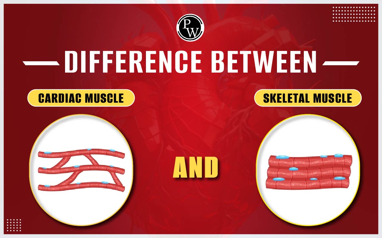
Cardiac and skeletal muscles exhibit distinct features. Cardiac muscles are autonomously controlled, while the somatic nervous system governs skeletal muscles. Cardiac muscles have a semi-spindle shape and are smaller than cylindrical skeletal muscles. Tendons connect skeletal muscles to bones, and myoblasts generate muscle fibers. Cardiac muscles are located in the heart's myocardium, connected by intercalated discs and T-tubules to form a syncytium.
Cardiac and skeletal muscles differ in several aspects. Skeletal muscles are more interconnected, lack gap junctions, and rely on syncytium for contraction. They are multinucleated, with fewer mitochondria and smaller T-tubules. Cardiac muscles have one or two nuclei, more mitochondria, larger T-tubules, and gap junctions. They also have a higher density of endomysium and mitochondria associated with larger blood vessels. See the article below to learn more about the difference between cardiac muscle and skeletal muscle.
Difference Between Cardiac Muscle And Skeletal Muscle
Human muscles are divided into three types: skeletal, smooth, and cardiac. Among these, cardiac muscles, which are located solely in the heart, play an important role in pumping blood throughout the body. These muscles made up of branched and striated fibers and a single nucleus, have a limited regenerative capacity following injury. Skeletal muscles, found all over the body, are in charge of movement and posture maintenance. They function voluntarily and under conscious control, responding to nerve impulses to contract or relax. Notably, skeletal muscles have a greater capacity for regeneration after injury or damage. The table below shows the detailed difference between cardiac muscle and skeletal muscle:
|
Difference Between Cardiac Muscle And Skeletal Muscle |
||
|---|---|---|
| Characteristic | Cardiac Muscle | Skeletal Muscle |
| Structure | Exclusive to the heart | Distributed throughout the body |
| Appearance | Branched and striated fibers with a single nucleus | Long, cylindrical, and striated fibers with multiple nuclei |
| Function | Pumps blood throughout the body | Responsible for movement and maintaining posture |
| Control | Involuntary, not under conscious control | Voluntary, under conscious control |
| Contraction | Rhythmically and continuously to pump blood | In response to nerve impulses, can be prompted to contract or relax |
| Fatigue | Low rate of fatigue due to continuous rhythmic contractions | Can become fatigued with prolonged use |
| Regeneration | Limited capacity for regeneration after injury or damage | Higher capacity for regeneration after injury or damage |
Cardiac Muscle
Cardiac muscle, found in the heart's walls, shares similarities with skeletal muscle as it is striated but functions involuntarily. Cardiac muscle cells, called cardiomyocytes, contain one to four nuclei and require a rich blood supply for oxygen and nutrients. Their coordinated contraction plays a vital role in blood circulation. Cardiac muscles have many mitochondria, myoglobins, and a well-established blood supply to support the heart's function. Cross striations, resulting from alternating thick and thin filaments, are distinctive features of cardiac muscles. Actin filaments appear thin in an electron microscope, whereas myosin filaments are thick, darker bands. The branching pattern is a common characteristic of cardiac muscle fibers. T-tubules in cardiac muscle are particularly large, extending across the Z-Discs. Intercalated discs connect cardiac myocytes, creating an electrochemical syncytium that aids in force transmission during muscle contraction. T-tubules play an important role in the excitation-contraction-coupling (ECG) process.
Skeletal Muscle
Tendons are collagen fiber bundles that connect skeletal, striated muscle, to bones. It is entirely under the voluntary control of the somatic nervous system, allowing for functions such as locomotion, facial expressions, posture, and other deliberate movements. Myocytes, or muscle cells, are the basic structural units of skeletal muscle. They are organized into long, cylindrical, multinucleated structures known as muscle fibers. These fibers result from the fusion of myoblasts. Myofibrils, which consist of thick and thin myofilaments, can be found within muscle fibers. Thick filaments are made up of myosin filaments, while thin filaments are made up of actin filaments. Under a microscope, these filaments in skeletal muscle show distinct banding patterns. Troponin and tropomyosin are required for muscle contraction and are also found in muscle fibers. Actin and myosin are arranged in a repeating unit known as a sarcomere, the basic functional unit of muscle fibers, contributing to their striated appearance. The interaction between actin and myosin is what causes muscle contraction.
Difference Between Cardiac Muscle And Skeletal Muscle FAQs
What are the primary difference between cardiac muscle and skeletal muscle?
What sets skeletal and cardiac muscles' action potentials (AP) apart?
Which muscle is considered the strongest in the human body?
How does the heart differ from cardiac muscle?
What is the smallest muscle in our body?