Eukaryotic Cells, Structure, Characteristics, Diagram, Examples
Eukaryotic cells have an organized nucleus and organelles that are surrounded by membrane-bound organelles. The article below discusses the structure, diagram, characteristics, and examples of eukaryotic cells.
Eukaryotic Cells: Most living things are classified as either unicellular or multicellular organisms. Unicellular organisms, such as bacteria, are made up of a single cell that performs all of the vital functions. Multicellular organisms, such as plants, animals, and humans, are made up of many cells, some of which specialize to perform specific functions. It is important to note that fungi can be both unicellular and multicellular.
All organisms, unicellular or multicellular, may have a distinct nucleus surrounded by a nuclear membrane. These organisms are termed eukaryotes. Thus, eukaryotes refer to all life forms that have a well-defined nucleus surrounded by a nuclear membrane. They can be unicellular eukaryotes, such as fungi, or multicellular, such as plants, animals, and humans. These organisms are made up of individual units known as eukaryotic cells. The provided article contains more information on the characteristics, structure, and examples of eukaryotic cells.What are Eukaryotic Cells?
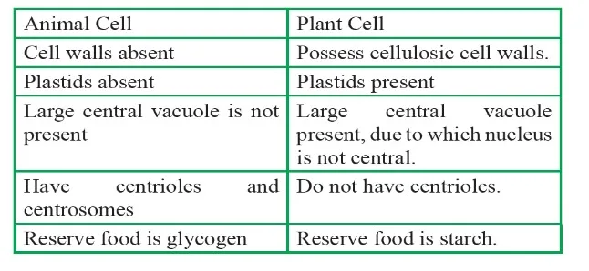
A eukaryote is a cell or organism that has a distinct nucleus enclosed within a nuclear membrane. This nucleus contains well-organized chromosomes containing genetic material. Eukaryotic cells also contain a variety of membrane-bound organelles, including mitochondria for cellular energy production, the Golgi apparatus for secretion, the endoplasmic reticulum for cellular transport, and lysosomes for cellular digestion. Even so, there are some exceptions, such as red blood cells' lack of mitochondria and nucleus and some species' lack of mitochondria, such as the oxymonad. Eukaryotic cells, or monocercomonoides, are usually larger and contain other membrane-bound organelles in addition to a clearly defined nucleus. Although there are some unicellular exceptions, most of them are multicellular organisms. A wide variety of organisms, including protists, plants, fungi, and animals, are classified as eukaryotes. The first microfossils that resembled eukaryotic organisms date back approximately 1.8 billion years, meaning that the evolution of eukaryotes is thought to have taken place between 1.7 and 1.9 billion years ago.Table: Comparison of Animal and Plant Cells
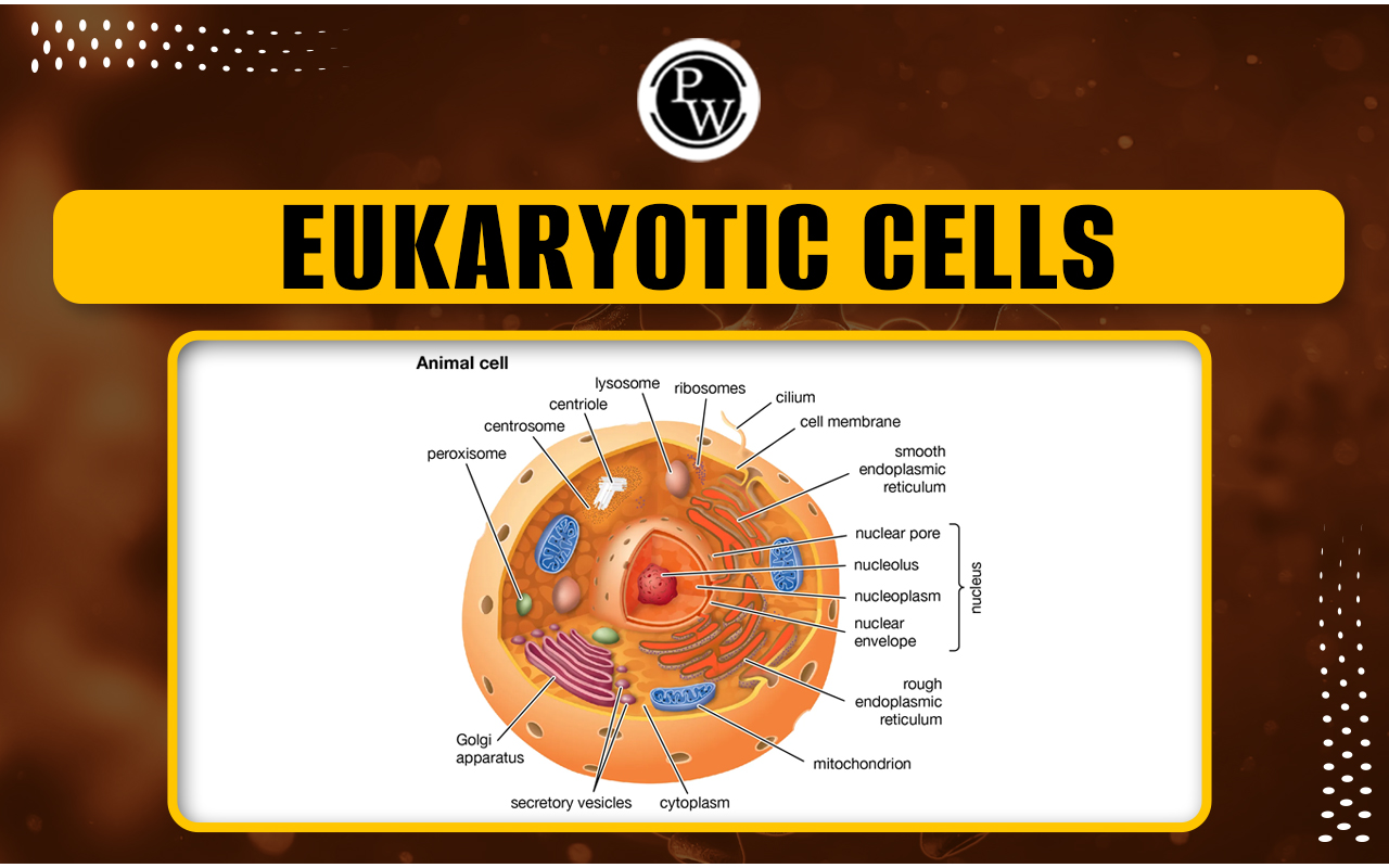
Eukaryotic Cells Diagram
The image below shows a diagram of a eukaryotic cell of both plant cell and animal cell: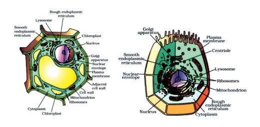
Eukaryotic Cells Characteristics
Eukaryotic cells have the following unique characteristics:- Nucleus Enclosure: One characteristic that sets eukaryotic cells apart is the presence of a nucleus, a specialized chamber surrounded by a membrane for protection. The nucleus functions as the central nervous system of the cell, storing its genetic material and directing all cellular processes.
- Organelle Complexity: Membrane-bound organelles, each with a distinct function in cellular function, are abundant in eukaryotic cells. These organelles, which include mitochondria, the Golgi apparatus, the endoplasmic reticulum, and lysosomes, add to the cell's complexity and efficiency in performing various functions.
- Linear DNA Configuration: Eukaryotic cells' genetic material is organized as double-stranded linear DNA molecules, which are neatly packaged within the nucleus. This configuration enables precise regulation of gene expression and facilitates complex cellular processes.
- Dimensional Advantage: Eukaryotic cells are typically larger in size than their prokaryotic counterparts. This increased size allows for the intricate structures and organelles required to carry out various cellular functions efficiently.
- Multicellular Complexity: While multicellularity is a common feature of eukaryotic organisms, there are some exceptions, such as unicellular organisms. Multicellular eukaryotes have a complex organization of specialized cell types that form tissues, organs, and organ systems, all of which contribute to the organism's survival and function.
- Taxonomic Diversity: Eukaryotic cells represent a wide range of organisms, including protists, plants, fungi, and animals. Despite their diversity, eukaryotic cells have fundamental similarities that highlight their evolutionary relatedness and common ancestry.
Eukaryotic Cells Structure
Eukaryotic cells exhibit a higher level of complexity compared to prokaryotic cells. They contain a wide range of subcellular structures that are essential for maintaining energy balance, metabolic processes, and gene expression. In the sections that follow, we will look at the distinct structures found in eukaryotic plant and animal cells, as well as their roles and significance in cellular life. A eukaryotic cell's structure consists of the following components:Plasma Membrane
The electron microscope, introduced in the 1950s, revealed the detailed structure of the plasma membrane. Prior to this, chemical studies, particularly on human red blood cells (RBCs), provided information about its composition. The cell membrane is made up primarily of lipids and proteins, with phospholipids arranged in a bilayer. The lipids are oriented with polar heads facing outward and hydrophobic tails inward, protecting nonpolar tails from the aqueous environment. Membrane proteins, classified as integral or peripheral based on how easily they can be extracted, contribute to its structure. Integral proteins are partially or completely embedded in the membrane, whereas peripheral proteins rest on its surface. In addition, cholesterol can be found in the plasma membrane, and further biochemical analysis has revealed the presence of proteins and carbohydrates. The ratio of protein to lipid varies between cell types. Human erythrocyte membranes, for example, are made up of about 52% protein and 40% lipid.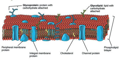
Fluid Mosaic Model of Plasma Membrane
Singer and Nicolson proposed the fluid mosaic model in 1972 to describe the structure of cellular membranes, such as those found in the nucleus, mitochondria, and vacuoles. This model portrays proteins as icebergs floating within a sea of lipids. It emphasizes the quasi-fluid nature of the lipid bilayer, enabling lateral movement of proteins. This fluidity is vital for membrane function.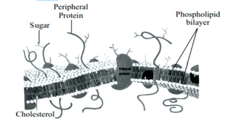
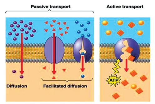
Transport of Molecules Across Cell Membrane
A fundamental role of the plasma membrane is the selective transport of molecules across it.- Passive transport allows molecules to move across the membrane without expending energy. This includes simple diffusion, where molecules move along a concentration gradient, and osmosis, the movement of water across a membrane.
- Diffusion: For polar molecules unable to pass through the lipid bilayer, carrier proteins facilitate their transport via facilitated diffusion.
- Active transport moves ions or molecules against their concentration gradient, requiring energy typically derived from ATP, as seen in the Na+/K+ pump.
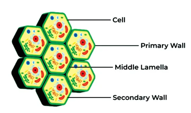
Cell Wall
The cell wall, which is found in plant and fungal cells, is a rigid structure that extends beyond the plasma membrane.- Primary walls , present in young plant cells, allow for growth and are gradually replaced by secondary walls.
- Secondary walls primarily consist of cellulose, along with other polysaccharides, lignin, and suberin.
- Middle lamella , rich in calcium pectate, acts as a glue, holding adjacent cells together.
Endomembrane System
The endomembrane system, which includes a variety of membrane-bound organelles, coordinates cellular functions. Organelles such as the endoplasmic reticulum (ER), Golgi complex, lysosomes, and vacuoles collaborate to carry out various cellular processes, excluding mitochondria, chloroplasts, and peroxisomes.Endoplasmic Reticulum (ER)
The ER is a network of tubular structures in the cytoplasm, dividing it into luminal and extra luminal compartments. Smooth ER lacks ribosomes and synthesizes lipids, while Rough ER has ribosomes on its surface and is involved in protein synthesis.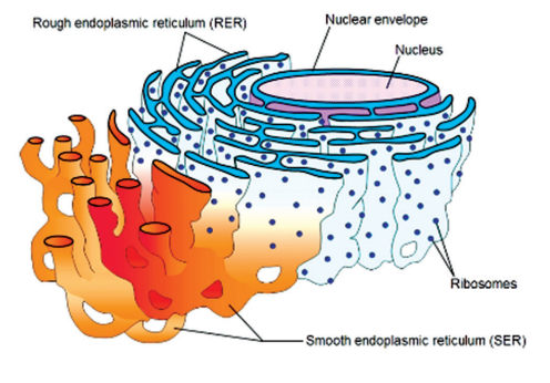
Golgi Apparatus
The Golgi apparatus, named after its discoverer, Camillo Golgi, packages and modifies proteins synthesized in the ER before transporting them to their final destination. It consists of flattened sacs or cisternae with distinct forming and maturing faces. Functions include protein modification, packaging into vesicles, and synthesis of glycoproteins and glycolipids.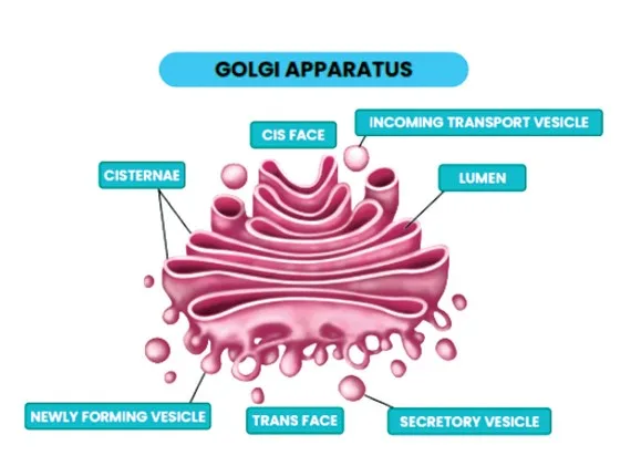
Lysosomes
Lysosomes are membrane-bound vesicles that contain hydrolytic enzymes for cellular digestion and originate in the Golgi. They participate in intracellular and extracellular digestion, and autolysis, breaking down cellular components.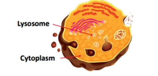
Vacuoles
Vacuoles are membrane-bound in plant and fungal cells store water, nutrients, and waste products. They help maintain turgor pressure and can have roles in osmoregulation and digestion.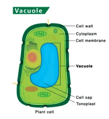
Mitochondria
Mitochondria, also known as the cell's powerhouse, are organelles that produce energy through aerobic respiration. While not easily visible under a microscope, their numbers vary depending on cell activity, and they exhibit diverse shapes and sizes. Mitochondria have a double membrane structure, with outer and inner membranes dividing the organelle's lumen into two compartments. The inner membrane contains infoldings called cristae, which increase its surface area for energy production. Functionally, mitochondria synthesize ATP, the cell's primary energy currency, through aerobic respiration. They possess their own DNA, RNA, and ribosomes, allowing semi-autonomous control over their replication and protein synthesis.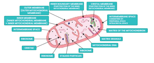
Plastids
Plastids are found exclusively in plant cells and euglenoids, which are membrane-bound organelles that play various roles, including photosynthesis and storage. Plastids vary in size, shape, and pigmentation, with different types classified based on their color and function. Leucoplasts, for example, are colorless and serve as storage organelles for starch, lipids, or proteins, while chloroplasts contain chlorophyll and are involved in photosynthesis.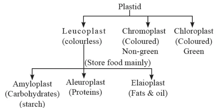
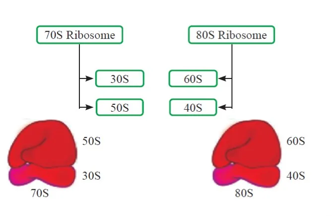
Ribosomes
Ribosomes are non-membranous, dense granular structures responsible for protein synthesis in the cell. Composed of RNA and proteins, ribosomes assemble amino acids into proteins based on instructions from messenger RNA (mRNA). These structures come in two subunits—larger and smaller—and exist in both prokaryotic and eukaryotic cells. In eukaryotes, ribosomes can be free in the cytoplasm or bound to the endoplasmic reticulum.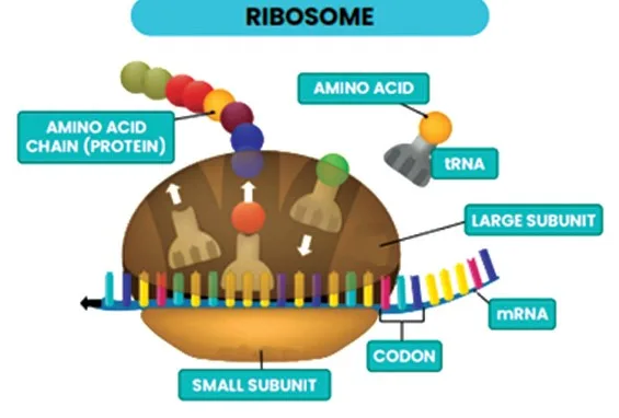
Cytoskeleton
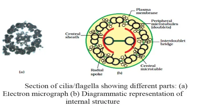
The cytoskeleton is a network of protein filaments that provides structural support, facilitates cell motility, and maintains cell shape. It comprises three main types of filaments: microtubules, microfilaments, and intermediate filaments. Microtubules are hollow tubes made of tubulin protein and participate in cell division and intracellular transport. Microfilaments, made of actin protein, enable cell movement and contraction. Intermediate filaments provide mechanical strength and stability to cells.Cilia and Flagella
Cilia and flagella are hair-like structures extending from the cell membrane, involved in cell movement and sensory functions. Cilia are shorter and numerous, while flagella are longer and fewer in number. Both structures consist of a core called the axoneme, composed of microtubules arranged in a 9 + 2 array. Centrioles, found in the centrosome, play a crucial role in their formation.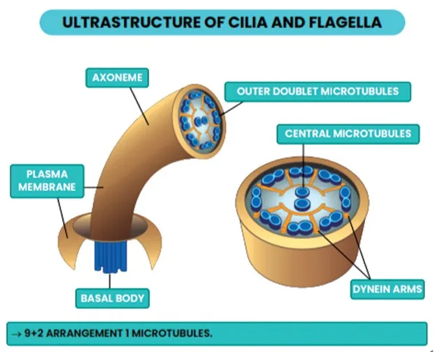
Centrosome and Centrioles
The centrosome contains two cylindrical structures called centrioles, positioned perpendicular to each other. Centrioles consist of nine evenly spaced fibrils of tubulin protein and play key roles in cell division and the formation of cilia and flagella.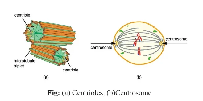
Nucleus
The nucleus was discovered by Robert Brown in 1831, the nucleus is often referred to as the brain of the cell due to its role in genetic regulation and cellular activities. A double membrane binds it, called the nuclear envelope, which is perforated by nuclear pores for molecular exchange. The nucleus contains chromatin, a complex of DNA and proteins, and a nucleolus, where ribosomal RNA synthesis occurs. Chromosomes, condensed structures of chromatin, are visible during cell division and contain genetic information essential for cell function.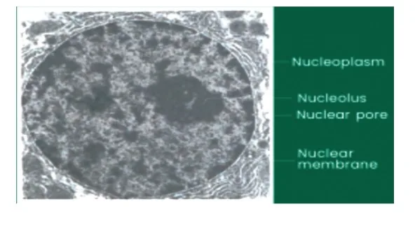
Chromosomes
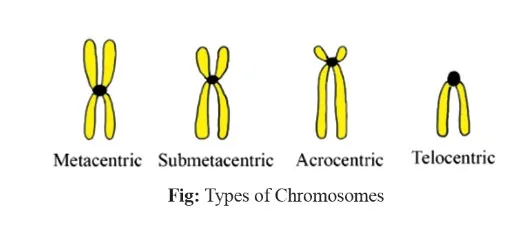
Chromosomes are dense structures visible during cell division, composed of condensed chromatin fibers. Each chromosome has a centromere, with disc-shaped kinetochores on its sides. Human cells typically have 46 chromosomes, carrying genetic information essential for cellular functions. Chromosomes are classified based on the position of the centromere into four types: metacentric, submetacentric, acrocentric, and telocentric. Each type exhibits distinct centromere placement, affecting chromosome shape.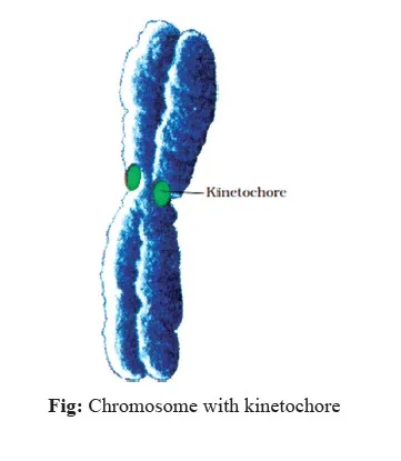
 Satellite regions, seen in some chromosomes, indicate secondary constrictions, providing insights into genetic structure and function. These cellular components collectively contribute to the organization, function, and dynamics of eukaryotic cells, highlighting the intricate and coordinated nature of cellular processes.
Eukaryotic Cells Examples
Eukaryotic cells are found in plants, animals, fungi, protozoa, and other complex organisms. Examples include:- Plant Cells: These have cellulose cell walls, a large central vacuole for turgor pressure, and chloroplasts for photosynthesis.
- Fungal Cells: Featuring chitin cell walls, some fungi have septa allowing organelle passage.
- Animal Cells: Lacking cell walls, they have membranes offering diverse shapes and phagocytic abilities.
- Protozoa: Unicellular, they may use cilia for movement and possess a pellicle for support.
| NEET Exam Important Links | |
|---|---|
| NEET Biology Syllabus | NEET Biology Diagrams |
| NEET Biology MCQ | NEET Biology Chapter wise Weightage |
| NEET Biology Notes | NEET Previous Year Question papers |
Eukaryotic Cells FAQs
What is a eukaryotic cell?
Eukaryotic cells feature a nucleus enclosed within a nuclear membrane and are found in large and complex organisms like protozoa, fungi, plants, and animals. They belong to the kingdom Eukaryota.
What are the two types of eukaryotes?
The two primary types of eukaryotic cells are plant cells, which have a cell wall, large vacuole, and chloroplasts for photosynthesis, and animal cells, which lack a cell wall and chloroplasts.
What defines eukaryotic cells at a class 11?
Eukaryotic cells are characterized by having a nucleus and organelles organized within the cell, all enclosed by membrane-bound organelles.
What are the four main types of eukaryotes?
The four main types of eukaryotes are animals, fungi, plants, and protists.
What are the differences between eukaryotic and prokaryotic cells?
Prokaryotes lack a nucleus and membrane-bound organelles, and they are unicellular. Eukaryotes, on the other hand, possess a nucleus and membrane-bound organelles and can be either unicellular or multicellular. Bacteria serve as an example of prokaryotes.


🔥 Trending Blogs
Talk to a counsellorHave doubts? Our support team will be happy to assist you!

Join 15 Million students on the app today!

Free Learning Resources
PW Books
Notes (Class 10-12)
PW Study Materials
Notes (Class 6-9)
Ncert Solutions
Govt Exams
Our Other Websites
Class 6th to 12th Online Courses
Govt Job Exams Courses
UPSC Coaching
Defence Exam Coaching
Gate Exam Coaching
Other Exams
Know about Physics Wallah
Physics Wallah is an Indian edtech platform that provides accessible & comprehensive learning experiences to students from Class 6th to postgraduate level. We also provide extensive NCERT solutions, sample paper, NEET, JEE Mains, BITSAT previous year papers & more such resources to students. Physics Wallah also caters to over 3.5 million registered students and over 78 lakh+ Youtube subscribers with 4.8 rating on its app.
We Stand Out because
We provide students with intensive courses with India’s qualified & experienced faculties & mentors. PW strives to make the learning experience comprehensive and accessible for students of all sections of society. We believe in empowering every single student who couldn't dream of a good career in engineering and medical field earlier.
Our Key Focus Areas
Physics Wallah's main focus is to make the learning experience as economical as possible for all students. With our affordable courses like Lakshya, Udaan and Arjuna and many others, we have been able to provide a platform for lakhs of aspirants. From providing Chemistry, Maths, Physics formula to giving e-books of eminent authors like RD Sharma, RS Aggarwal and Lakhmir Singh, PW focuses on every single student's need for preparation.
What Makes Us Different
Physics Wallah strives to develop a comprehensive pedagogical structure for students, where they get a state-of-the-art learning experience with study material and resources. Apart from catering students preparing for JEE Mains and NEET, PW also provides study material for each state board like Uttar Pradesh, Bihar, and others
Copyright © 2026 Physicswallah Limited All rights reserved.