Cell The Unit of Life, Theory, Fundamentals, Types for NEET Exam
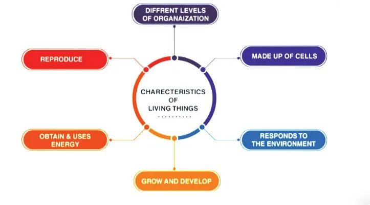
Cell The Unit of Life: As you explore your surroundings, you will encounter a wide range of living and non-living beings. Indeed, you have considered the complexities of their existence. In-depth examinations of their characteristics merely highlight the rich tapestry of diversity. However, the fundamental principles of cell theory highlight the essential unity amidst this diversity: the cellular organization, which serves as the foundation of all life forms.
This chapter provides a comprehensive explanation of cell structure and function, as cells are the fundamental building blocks of life. Indeed, when we dissect any organism down to the cellular level, the cell emerges as the smallest, self-contained entity. Explore the accompanying cell notes to thoroughly understand what constitutes a cell, its structural intricacies, its various types, and the pivotal roles they fulfil.
Cell the Unit of Life Overview
Cell The Unit of Life notes comprehensively overview all cell-related concepts. The article deeply explores Chapter 11 of the NCERT, which focuses on Cell The Unit of Life. Class 11 notes Cell The Unit of Life has been prepared by Physics Wallah experts per the updated NEET syllabus to assist students in properly preparing for their exams.
Notes: "Cell the unit of life", is an important chapter in Class 11 Biology. The revision notes for cell the unit of life chapter covers all the essential topics that are important for understanding the concept of cells as the basic structural and functional units of life.
What is Cell?
A cell, the fundamental unit of life, consists of all living organisms and tissues of the body and functions autonomously. It is made up of three key components: the cell membrane, the nucleus, and the cytoplasm, all of which are necessary for cellular activity. The cell membrane functions as both a protective barrier and a selective gatekeeper, controlling the influx and efflux of substances.
The nucleus, which contains the nucleolus and most of the cell's DNA, is where critical cellular processes like ribonucleic acid (RNA) production occur. Simultaneously, the cytoplasm, a ubiquitous fluid within cells, creates an environment for biochemical reactions and protein synthesis. The human body is made up of approximately 30 trillion cells, demonstrating the extent of cellular organization.
Within their membranes, which are equipped with surface receptors, cells contain a variety of organelles, including the Golgi complex, mitochondria, and endoplasmic reticulum. These organelles serve specific functions that are vital to the functioning of cells. The study of these structures and functions is the focus of cell biology, highlighted by the complex cellular architecture.
Organisms are classified according to the type of cells they contain:
- Unicellular organisms comprise a single cell and can carry out essential life functions independently.
- Multicellular organisms, which consist of multiple cells, exhibit complex cellular interactions that support the organism's overall survival and functionality.
Sexual Reproduction in Flowering Plants
Discovery Of Cell
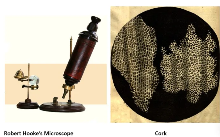
Robert Hooke first discovered cells in 1665 using a compound microscope. He discovered many tiny structures called "cellula" while studying thin cork sections. Hooke's important observation was documented in his book Micrographia.
Following Hooke's discovery, Anton Van Leeuwenhoek investigated these cellular entities using improved microscope lenses. His improved microscope allowed for superior magnification of specimens. Van Leeuwenhoek used his single-lens microscope to observe organisms known as "animalcules," which included bacteria and protozoa.
Cell Theory
Scientists were already observing cells from various organisms in the early 1800s. Several German scientists, including Theodor Schwann and Matthias Jakob Schleiden, recognized cells as the fundamental building blocks of all living organisms. Rudolf Virchow, a German physician, made a significant breakthrough around 1850 when he discovered that cells divide and give rise to new cells, as revealed by microscopic examination. Virchow confirmed the theory that living cells divide to reproduce through research, which also highlighted that new cells can only arise from pre-existing living cells.
Virchow, Schwann, and Schleiden made significant contributions to the development of cell theory knowledge, which is essential to biological research.
The cell theory includes the following principles:
- All organisms are composed of cells.
- The life processes of an organism occur within its cells.
- Every cell originates from pre-existing cells.
Despite their diversity, cells share common characteristics. Cells have plasma membranes, cytoplasm, ribosomes, and DNA. These components are found in all cells, including bacteria and humans.
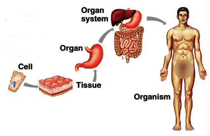
Cells perform various functions within a living organism, including vital processes like digestion, respiration, and reproduction. For example, in the human body, an amalgamation of cells results in the formation of tissues, which then assemble to form organs. Multiple organs work together to form organ systems, and the integration of multiple organ systems results in the functioning of the human body.
Formulations Of Cell Theory
- Theodore Schwann and Matthias Schleiden collaborated to develop cell theory in the 19th century, a fundamental concept in contemporary biology.
- Plant tissues are made up of different cell types, as demonstrated by Schleiden's 1838 botanical investigations.
- Theodore Schwann's zoological investigations 1839 identified the 'plasma membrane' as an important component of animal cells and the distinct cell walls found in plant cells.
- Schwann's observations led to the theory that plant and animal bodies comprise cells and their byproducts.
- In 1855, Rudolf Virchow expanded on the initial cell theory by proposing that new cells arise from dividing existing cells, introducing the concept of " Omnis cellula-e cellula ."
- Virchow is discovery improved cell theory and provided a mechanism for cell formation, highlighting the continuity of cellular life processes.
- The contributions of Schleiden, Schwann, and Virchow formed the foundation of cells as the basic units of life and the components of every living thing.
An Overview Of Cell
A microscope was used to examine specimens of onion peel and human cheek cells. The onion cell, a prototypical plant cell, has a distinct cell wall as an outer boundary adjacent to the cell membrane. On the other hand, human cheek cells are distinguished by an outer membrane rather than the rigid cell wall found in plant cells.
Each cell contains a dense, membrane-bound organelle known as the nucleus, which houses chromosomes containing genetic material, specifically DNA. Eukaryotic cells have membrane-bound nuclei, while prokaryotic cells do not. The cellular volume is occupied by cytoplasm, a viscous, semi-fluid substance.
Cytoplasm, a semi-fluid medium, fills the intracellular space and is the primary site of many cellular activities in plant and animal cells. Various biochemical reactions within the cytoplasm support the cell's metabolic processes and facilitate its ongoing functions.
Types Of Cell - According to Size & Shape
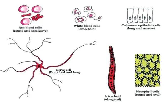
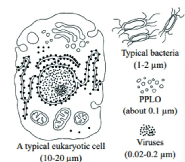
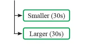
Cells vary greatly in size, shape, and functionality. Their morphology varies from disc-like, polygonal, columnar, cuboid, thread-like, and irregular, with their shapes frequently corresponding to the specific functions they perform. The table below highlights some important cell types, along with their distinct features and dimensions:
| Types of Cell | |
|---|---|
| Cell Type | Feature/Size/Shape |
| Smallest cell | 0.3 μm in length |
| Bacteria | 3-5 μm |
| Ostrich egg | Largest isolated single cell |
| Human red blood cell (erythrocyte) | 7.0 μm in diameter |
| Nerve cells (neurons) | Longest cells in humans |
Types Of Cell- According to the Nuclear Organisation
Cells can be likened to factories, each with diverse workers and departments collaborating toward a shared goal. Different types of cells undertake specific functions. Based on their structure, cells are categorized into two types:
- Prokaryotic cells
- Eukaryotic cells
Prokaryotic Cell
Prokaryotic cells, unicellular microorganisms, are among the oldest life forms on Earth. Among these organisms, there are various prokaryotes, some of which are known as extremophiles due to their ability to thrive in harsh environments. In addition, some prokaryotes are photoautotrophic, meaning they can synthesize nutrients using solar energy.
These cells are represented by various organisms such as bacteria, blue-green algae, Mycoplasma, or PPLO (Pleuro Pneumonia Like Organisms). They are typically smaller in size and reproduce at a faster rate compared to eukaryotic cells.
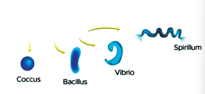
Prokaryotic cells exhibit considerable diversity in both shape and size. The four primary shapes of bacteria are:
- Bacillus (rod-like): These bacteria are elongated and appear individually or in chains of two or three connected cells. An example is Bacillus coagulans.
- Coccus (spherical): These bacteria are spherical or oval-shaped. An example is Streptococcus pyogenes .
- Vibrio (comma-shaped): These bacteria have a curved, comma-like shape. An example is Vibrio cholerae .
- Spirillum (spiral): These bacteria display a spiral or helical shape. An example is Spirillum minus .
Structure Of Prokaryotic Cell
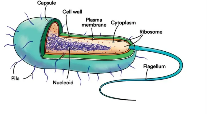
Prokaryotic cells have a relatively uniform structural organization and exhibit various shapes and functions. A cell wall supports the plasma, or cell membrane that encloses all prokaryotes. However, exceptions, such as Mycoplasma, lack a cell wall entirely.
Unlike eukaryotic cells, prokaryotic cells lack a distinct nucleus and a nuclear membrane to protect their genetic material. In addition to the primary genomic DNA, which usually forms a single chromosome or circular DNA, many bacteria contain additional circular DNA molecules known as plasmids.
Plasmids are important in conferring specific traits to bacteria, such as antibiotic resistance, and monitoring bacterial transformation by foreign DNA. Prokaryotic cells lack membrane-bound organelles like a nuclear envelope, with ribosomes being their sole organelle.
Cell Envelope and its Modifications
The cell envelope of most prokaryotic cells, especially bacterial cells, comprises three tightly bound layers: the outermost glycocalyx, followed by the middle cell wall, and finally the innermost plasma membrane. While functioning collectively as a protective unit, each layer of the envelope serves distinct roles.
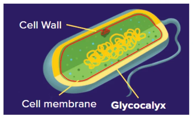
Glycocalyx
The composition and thickness of the glycocalyx vary among different bacteria. It can range from a loose sheath, the slime layer, to a thick and tough capsule.
Cell Wall
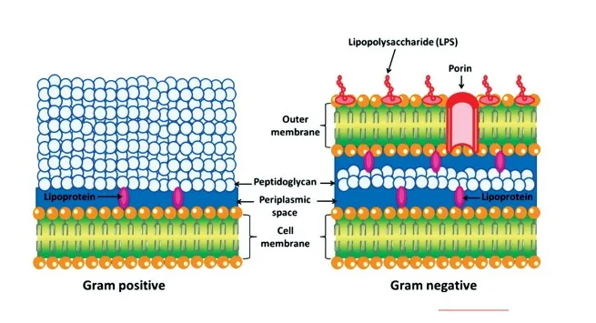
The cell wall determines the cell's shape and provides structural support to prevent rupture or collapse. In Gram-positive bacteria, the cell wall is thicker and composed of a single layer of peptidoglycan, while in Gram-negative bacteria, it consists of a double layer: a thin outer membrane of lipids and an inner wall of peptidoglycan.
Plasma Membrane
The selectively permeable plasma membrane interacts with the external environment and shares structural similarities with eukaryotic membranes.
Mesosome
Mesosomes are specialized membranous structures formed by extensions or infoldings of the plasma membrane into the cell. These features are characteristic of prokaryotes and play crucial roles in various cellular processes, including cell wall formation, DNA replication, and respiration.
Chromatophores
Chromatophores are pigment-containing membranous extensions of the plasma membrane found in some prokaryotes, such as cyanobacteria. They contribute to photosynthesis and other metabolic processes.
Cytoplasm
Enclosed within the cell membrane, the cytoplasm constitutes the internal environment of the cell. It contains a liquid matrix composed of water, proteins, and other biomolecules.
Nucleoid
The nucleoid is an irregularly shaped region within prokaryotic cells that houses the genetic material. Unlike the nucleus of eukaryotic cells, the nucleoid is not surrounded by a nuclear membrane. The prokaryotic genome typically consists of a circular, double-stranded DNA molecule.
DNA
Deoxyribonucleic acid (DNA) carries genetic information and is present within the cytoplasm of prokaryotic cells in a closed circular structure. In contrast, eukaryotic DNA is organized into chromosomes within the nucleus.
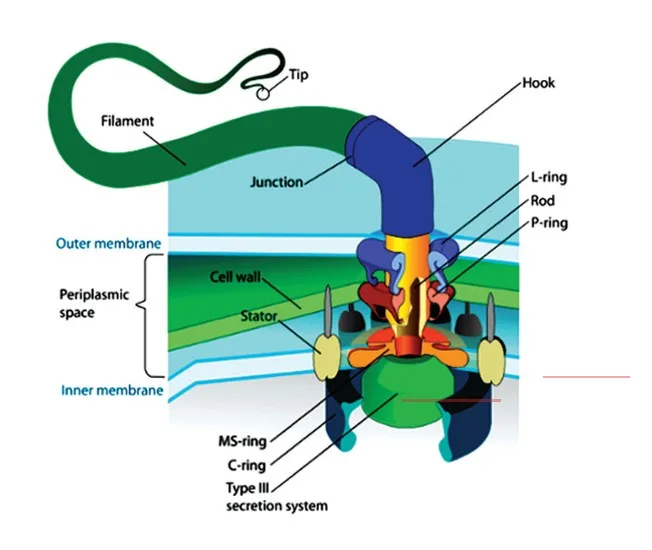
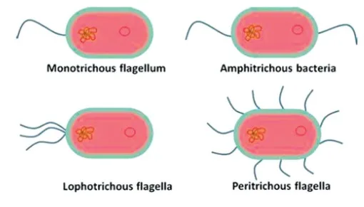
Motility
Bacterial cells may exhibit motility, facilitated by flagella, thin filamentous extensions of the cell wall. Flagella consist of three parts: the filament, hook, and basal body, which enable movement.
Types of Flagella
Ribosomes
Prokaryotic ribosomes, measuring about 15 nm by 20 nm, are associated with the plasma membrane and serve as the site of protein synthesis. Polyribosomes, or polysomes, may form when multiple ribosomes attach to a single mRNA molecule, facilitating efficient protein production.
Inclusion Bodies
Inclusion bodies store reserve materials within the cytoplasm of prokaryotic cells. These non-membrane-bound structures, which include phosphate granules, cyanophycean granules, and glycogen granules, are essential for cellular metabolism and energy storage. Gas vacuoles, found in certain photosynthetic bacteria, aid in buoyancy regulation.
Gram Staining
Gram staining, also known as Gram's method, is a staining technique utilized to differentiate and categorize bacterial species based on variations in cell wall composition and response to staining procedures. Developed by Danish bacteriologist Hans Christian Gram in 1884, this method divides bacteria into two main groups:
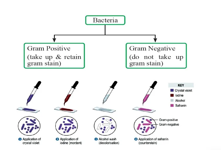
- Gram-positive bacteria:
Gram-positive bacteria retain the violet color of the crystal violet stain in the Gram stain. Staphylococci, streptococci, and pneumococci are some examples, as are pathogens that cause diseases such as diphtheria and anthrax. After being treated with iodine and ethanol, these bacteria develop a visible violet color and form a thick layer of peptidoglycan in their cell walls. Additionally, their cell walls contain teichoic acids and phosphate, which contribute to their rigid structure.
- Gram-negative bacteria:
In Gram's staining method, Gram-negative bacteria absorb the red color of the counterstain rather than the crystal violet stain. This is a distinguishing characteristic of bacteria possessing a cell wall composed of a thin layer of peptidoglycan and an outer lipid-rich layer. Gram-negative bacteria are stained with a counterstain such as safranin and are destained with an alcohol wash, resulting in their pink coloration under a microscope.
Eukaryotic Cell's
Eukaryotes include a wide variety of organisms, including protists, fungi, plants, and animals. The presence of membrane-bound organelles in eukaryotic cell's causes extensive compartmentalization of the cytoplasm. These cells have a well-organized nucleus enclosed by a nuclear envelope, as well as a number of complex locomotory and cytoskeletal structures. Chromosomes are structures that organize eukaryotic cell's genetic material.
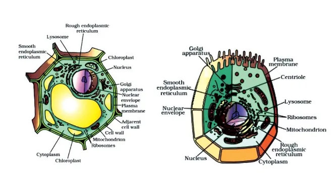
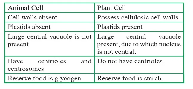
Table: Comparison of Animal and Plant Cells
Structure Of Eukaryotic Cell
Eukaryotic cells are known for their complexity, requiring numerous components to function simultaneously while carrying out various cellular processes. Despite the diverse structures found in eukaryotic cells, certain components are consistently present.
Plasma Membrane
The detailed understanding of the plasma membrane's structure began with the invention of the electron microscope in the 1950s. However, prior to this, chemical studies, especially on human red blood cells (RBCs), provided insights into its composition.
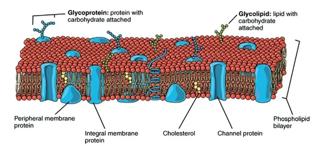
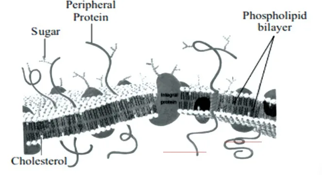
The plasma membrane consists mainly of lipids and proteins, with phospholipids being the most abundant lipid arranged in a bilayer. This arrangement ensures that the hydrophilic heads face outward towards the aqueous environment while the hydrophobic tails are shielded within the membrane.
Membrane proteins can be categorized as integral or peripheral, depending on their association with the lipid bilayer. Integral proteins are embedded within the membrane, while peripheral proteins are found on the membrane's surface.
Additionally, cholesterol is present in the plasma membrane, along with proteins and carbohydrates. The ratio of proteins to lipids varies among cell types, with human erythrocytes having approximately 52% protein and 40% lipids.
Fluid Mosaic Model of Plasma Membrane
The fluid mosaic model, proposed by Singer and Nicolson in 1972, is widely accepted. It describes the plasma membrane as a dynamic structure composed of a fluid lipid bilayer with embedded proteins. According to this model, proteins float within the lipid bilayer, resembling icebergs in a sea of lipids. The fluidity of the membrane allows lateral movement of proteins.
Transport of Molecules Across the Cell Membrane
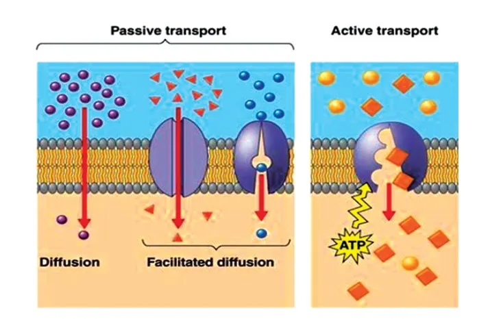
The plasma membrane's selective permeability enables the transport of molecules across it. This transport occurs through various mechanisms:
Passive Transport: Molecules move across the membrane without the need for energy input, driven by concentration gradients. This includes simple diffusion and osmosis.
Active Transport: Some molecules are transported against their concentration gradient, requiring energy, typically in the form of ATP. Examples include the Na+/K+ pump.
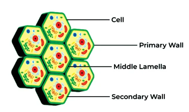
Cell Wall
The cell wall is a rigid, non-living structure that surrounds the plasma membrane in fungi and plant cells. It provides support, protection against mechanical damage and infection, facilitates cell-to-cell interactions, and acts as a barrier to unwanted macromolecules. The primary cell wall is flexible and capable of growth, while the secondary cell wall, formed later, consists mainly of cellulose, along with other polysaccharides, lignin, and suberin.
Endomembrane System
The endomembrane system coordinates the functions of various membranous organelles within the cell, including the endoplasmic reticulum (ER), Golgi apparatus, lysosomes, and vacuoles. Notably, mitochondria, chloroplasts, and peroxisomes are not part of this system.
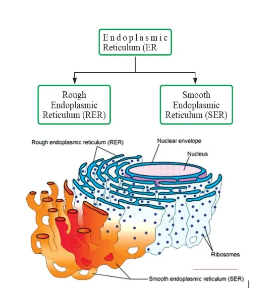
Endoplasmic Reticulum
The ER is a network of tubular structures that divides the intracellular space into distinct compartments. The Golgi apparatus consists of stacked cisternae and is involved in packaging and modifying cellular materials. Lysosomes are vesicles containing hydrolytic enzymes, while vacuoles are membrane-bound compartments containing various substances.
This comprehensive overview highlights the importance of these cellular structures and their functions in maintaining cell integrity and facilitating essential cellular processes.
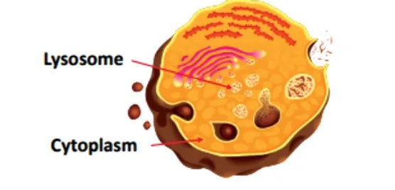
Lysosomes
Lysosomes are membrane-bound vesicles formed by the Golgi apparatus, containing a variety of hydrolytic enzymes that function optimally in an acidic pH environment. These enzymes are responsible for breaking down carbohydrates, proteins, lipids, and nucleic acids. Lysosomes play crucial roles in digesting large extracellular particles, breaking down intracellular substances, and facilitating autolysis.
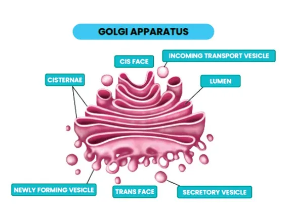
Golgi Apparatus
The Golgi apparatus, discovered by Camillo Golgi in 1898, is a cellular organelle involved in processing, modifying, and packaging proteins and lipids synthesized by the cell. It consists of a series of flattened, membrane-bound sacs called cisternae, which are stacked parallel to each other near the nucleus.
The main function of the Golgi apparatus is to receive proteins and lipids from the endoplasmic reticulum and modify them through processes such as glycosylation, phosphorylation, and sulfation. These modified molecules are then sorted, packaged into vesicles, and transported to their final destinations within the cell or outside the cell via exocytosis.
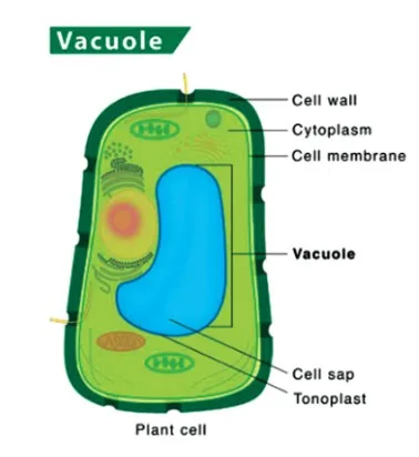
Vacuoles
Vacuoles are membrane-bound organelles found in the cytoplasm, containing water, sap, excretory products, and other materials not needed by the cell. In plant cells, vacuoles are particularly large and contribute to maintain turgor pressure. The tonoplast is the membrane surrounding the vacuole, regulating the movement of materials into and out of the vacuole.
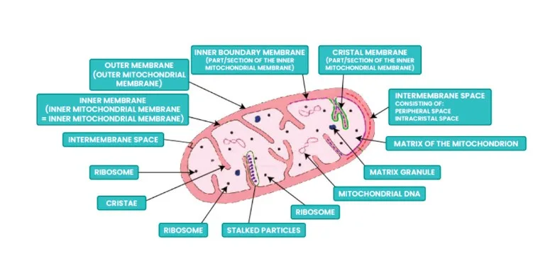
Mitochondria
Mitochondria, often referred to as the powerhouse of the cell, are double-membrane-bound organelles responsible for cellular respiration and energy production. They vary in shape and size and contain their own DNA, ribosomes, and enzymes. The inner membrane of mitochondria forms infoldings called cristae, increasing the surface area for metabolic reactions. Mitochondria divide through a process called fission.
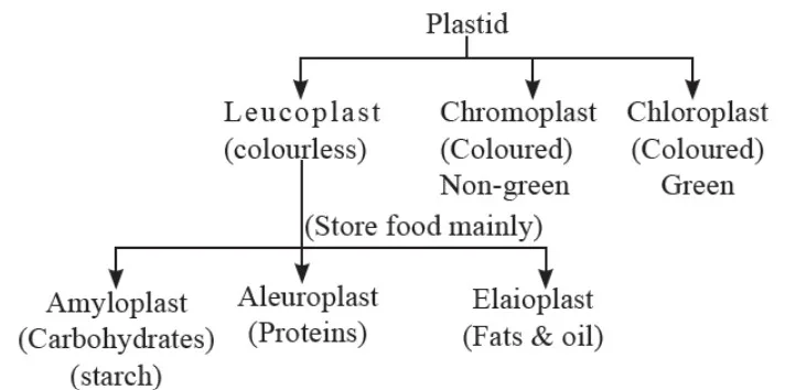
Plastids
Plastids are organelles found in plant cells and some protists, characterized by their ability to store pigments and perform photosynthesis. Chromoplasts contain pigments responsible for colors such as red, orange, and yellow, while chloroplasts contain chlorophyll and are essential for photosynthesis, giving plants their green color. Plastids exhibit various shapes and sizes and are crucial for plant growth and development.
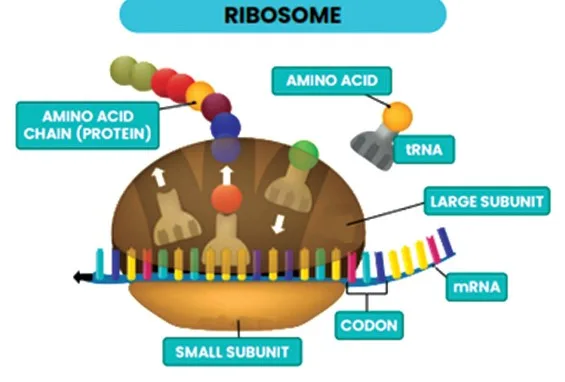
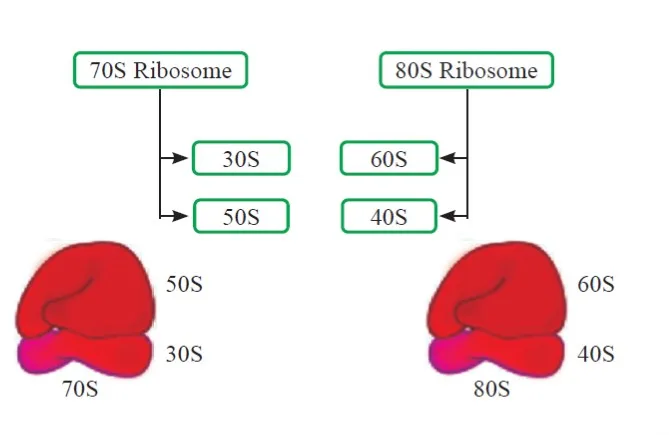
Ribosomes
Ribosomes are small, non-membranous organelles responsible for protein synthesis. They consist of two subunits and can be found free in the cytoplasm or attached to the endoplasmic reticulum. Ribosomes translate mRNA into proteins through a process called translation, playing a fundamental role in cellular function.
Cytoskeleton
The cytoskeleton is a network of protein filaments present in the cytoplasm, providing structural support, facilitating cell motility, and maintaining cell shape. It includes three main types of filaments: microtubules, microfilaments, and intermediate filaments. Cytoskeletal elements are dynamic and essential for various cellular processes, including cell division and intracellular transport.
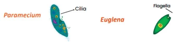
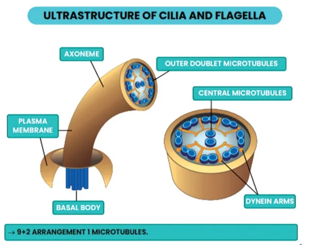
Cilia and Flagella
Cilia and flagella are hair-like structures protruding from the cell membrane, involved in cell movement and sensory functions. Cilia are shorter and more numerous, while flagella are longer and fewer in number. Both structures contain microtubules arranged in a 9+2 pattern and play crucial roles in cellular locomotion and signal transduction.
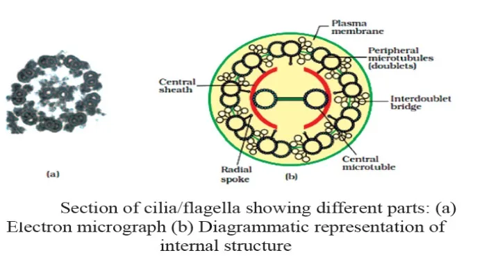
Structure of Cilia And Flagella
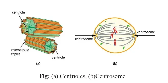
Centrosome and Centrioles
The centrosome is an organelle containing two centrioles oriented perpendicular to each other. Centrioles are composed of microtubules arranged in a specific pattern and play essential roles in organizing the cytoskeleton and facilitating cell division. They serve as the basal body for the formation of cilia and flagella and are vital for maintaining cell structure and function.
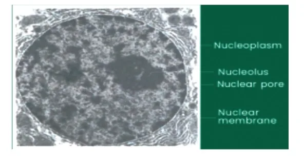
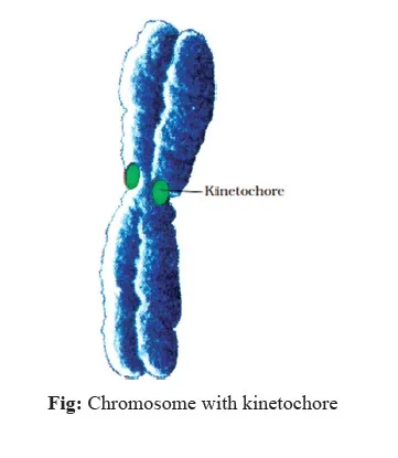
Nucleus and Chromosomes
The nucleus is the control center of the cell, containing genetic material in the form of chromatin, which condenses into chromosomes during cell division. Chromosomes consist of DNA wrapped around histone proteins and contain genes responsible for encoding proteins and regulating cellular processes. The nucleus plays crucial roles in gene expression, DNA replication, and cell division, ensuring the transmission of genetic information to daughter cells.
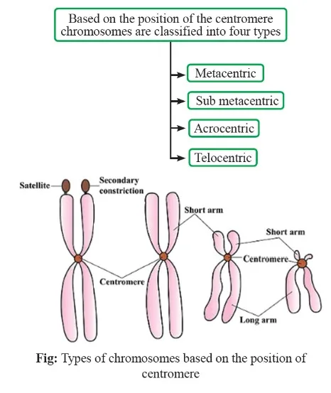
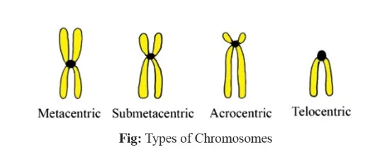
Types of Chromosome
Physics Wallah's NEET Online Coaching for Class 11 provides comprehensive preparation for the NEET UG Exam. We offer free and paid courses in multiple languages, with experienced faculty, recorded and live classes, doubt-clearing sessions, and study materials. Enroll now to begin your NEET preparation journey.
| NEET Exam Important Links | |
|---|---|
| NEET Biology Syllabus | NEET Biology Diagrams |
| NEET Biology MCQ | NEET Biology Chapter wise Weightage |
| NEET Biology Notes | NEET Previous Year Question papers |
Cell The Unit of Life FAQs
Why are cell the unit of life?
What is the significance of cell the unit of life in Class 11 Biology?
What is the concept of cells and the cell theory in Class 11?
What defines a cell in Class 11?
Which organism has the largest single cell?