Structure of Ear - Diagram, Functions and Mechanism of Hearing

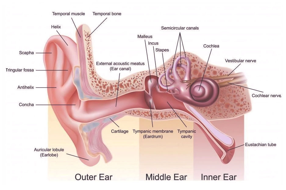
Structure of Ear: The ear, responsible for hearing and balance, is a complex organ housed within the skull. It comprises three main sections: the external ear, middle ear, and inner ear, each filled with either air or fluid. These sections contain the mechanisms for sound transmission and the sensory structures for hearing and balance.
Also known as the vestibulocochlear organ, the ear functions as a receptor and filter, transforming auditory signals into information that the brain can interpret. Its primary tasks are detecting, transmitting, and converting sounds into electrical impulses. The article below contains NEET Biology Notes on the structure of ear, physiology, and functions of the ear.
Structure of Ear Diagram
The following is a Structure of Ear:
Sexual Reproduction in Flowering Plants
Anatomy of Ear
Structure of ear is composed of three main sections:
- Outer ear
- Middle ear
- Inner ear
Outer Ear
The outer ear, also known as the external ear or auris externa, is essential for our ability to hear. Let's examine its structure and function:
1. Auricle (Pinna):The auricle is the visible part of the ear on the outside of the head. It is made of a curved framework of cartilage covered by skin. The auricle directs sound waves into the ear. The different parts of the auricle include:
- Helix: The outermost curved ridge of cartilage running from the top of the ear to the earlobe.
- Antihelix: A Y-shaped ridge inside the helix, following the outer curve of the ear.
- Crux helix: A C-shaped ridge extending from the top of the helix to the bottom of the antihelix.
- Tragus and antitragus: Smaller ridges of cartilage around the ear canal.
- Concha: Depressions between the ridges of cartilage leading to the ear canal.
- Lobule (Earlobe): The only part of the auricle not supported by cartilage. Its skin contains sebaceous glands that secrete sebum, an oily substance that moisturizes and protects the ear.
Structural Organisation in Animals
2. Ear Canal (External Acoustic Meatus):- The ear canal extends from the concha (part of the auricle) to the eardrum (tympanic membrane).
- It follows an S-shaped path, which helps prevent water and debris from entering the ear.
- The walls of the canal are made up of one-third cartilage and two-thirds bone.
- The skin lining the ear canal contains ceruminous glands that produce cerumen (earwax), which helps trap debris and insects.
- The tympanic membrane is at the inner end of the ear canal.
- Commonly known as the eardrum, it vibrates when sound waves reach it, initiating a chain reaction in the middle ear.
- The eardrum is crucial for transmitting sound energy to the middle ear and beyond.
Function of Outer Ear: The outer ear is crucial for hearing as it gathers sound waves and directs them to the eardrum. The folds and ridges of the auricle amplify these sound waves, while the ear canal protects the eardrum from damage.
Middle Ear
The middle ear consists of the following parts:
- Tympanic Cavity: The tympanic cavity is a narrow, air-filled space. It is separated from the outer ear by the tympanic membrane and from the inner ear by a bony wall. The anterior wall of the tympanic cavity contains the eustachian tube.
- Eustachian Tube: The eustachian tube is a 4 cm long passage that balances air pressure on both sides of the tympanic membrane. It connects the tympanic cavity to the nasopharynx.
- Ear Ossicles: The ear ossicles transmit sound waves from the eardrum to the middle ear. There are three ear ossicles in the human ear:
- Malleus: Shaped like a hammer, it is attached to the tympanic membrane via its handle and to the incus via its head. It is the largest of the ear ossicles.
- Incus: Shaped like an anvil, it connects with the stapes.
- Stapes: The smallest ossicle and the smallest bone in the human body.
Function of middle ear: The middle ear is essential for converting sound waves into vibrations. These vibrations are then transmitted to the inner ear for further processing.
| Important NEET Biology Notes | ||
|---|---|---|
| Selaginella | Embryo | Malvaceae |
| Pinus | Polyembryony | Volvox |
Inner Ear
The inner ear, also known as the labyrinth, is a complex part of our auditory and balance system. The following is the structure of inner ear:
- The inner ear is situated at the end of the ear canals, within the skull's temporal bone.
It consists of two main parts: the bony labyrinth and the membranous labyrinth.
- The bony labyrinth encloses the membranous labyrinth, which contains perilymph (a fluid similar to extracellular fluid).
- Within the membranous labyrinth, there is endolymph, a fluid similar to cytosol, and these two fluids do not mix.
- The inner ear is composed of three main sections:
- Cochlear component: Responsible for hearing. The cochlea, shaped like a snail's shell, converts sound waves into nerve signals the brain interprets as sound.
- Vestibular component: Includes the utricle and saccule, which manage stationary balance.
- Semi-circular component: Regulates balance during movement and is positioned in front of the vestibular component.
| Important NEET Biology Notes | ||
|---|---|---|
| Chara | Funaria | Apomixis |
| Rhizopus | Spirogyra | Bacteriophage |
Functions of the inner ear:
- Hearing: The cochlea transforms sound waves into nerve impulses that our brain perceives as sound.
- Balance and Equilibrium: The vestibular system (including the utricle, saccule, and semi-circular canals) helps maintain balance, detect head movements, and adjust posture.
- Gaze Stability: The inner ear aids in stabilizing vision when focusing on a single object.
Function of Ear
The human ear is an organ that combines the functions of hearing and maintaining balance. It enables us to perceive the rich auditory world around us and stay stable on our feet.
Hearing Function:
- The primary role of the ear is to hear. It captures sound waves from the environment and translates them into recognizable sounds.
- When sound reaches the outer ear, it travels through the ear canal and strikes the eardrum (tympanic membrane).
- The eardrum responds to these sound waves by vibrating, and these vibrations are transmitted to three small bones in the middle ear called ossicles (malleus, incus, and stapes).
- The ossicles amplify the sound, ensuring efficient transmission to the inner ear.
Equilibrium Maintenance:
In addition to hearing, the ear plays a crucial role in maintaining balance and coordinating head and eye movements. The ear is anatomically divided into three parts: the outer ear, middle ear, and inner ear. The specific structures responsible for balance are as follows:
- Maculae (Saccule and Utricle): These are specialized sensory areas located in the vestibule of the inner ear. They detect changes in head position and linear acceleration (such as in elevators or moving vehicles). Maculae help us maintain balance in static conditions.
- Cristae (Semicircular Canals): These are three fluid-filled loops within the inner ear. Each semicircular canal is oriented in a different plane (horizontal, anterior, and posterior). The cristae within these canals detect rotational movements of the head. When you turn your head, fluid movement inside the canals stimulates hair cells, providing information about dynamic equilibrium.
- Organ of Corti (Cochlea): The cochlea is a spiral-shaped structure within the inner ear, resembling a snail shell. Inside the cochlea lies the organ of Corti, which is responsible for hearing.
The organ of Corti contains specialized sensory cells called hair cells that respond to sound vibrations. When sound waves enter the cochlea, they cause the basilar membrane to vibrate.
These vibrations stimulate the hair cells, which then transmit nerve impulses to the brain via the auditory nerve. The brain processes these impulses, allowing us to perceive different sounds.
How Do We Hear?
Hearing is a fascinating process that allows us to perceive sounds from our environment. It starts with sound waves passing through the air and into our ears. The following is a step-by-step explanation of how sound travels to your brain:
- Outer Ear: The visible part of the ear, known as the pinna, collects sound waves and directs them into the ear canal.
- Middle Ear: Sound waves travel down the ear canal and cause the eardrum, a thin membrane, to vibrate. These vibrations are passed on to three small bones in the middle ear: the malleus, incus, and stapes. These bones amplify the vibrations.
- Inner Ear: The amplified vibrations reach the cochlea, a spiral-shaped structure filled with fluid in the inner ear. Inside the cochlea are thousands of tiny hair cells with projections at their tops. Vibrations in the cochlear fluid cause the hair cells to bend.
- Electrical Signals: When hair cells bend, they produce electrical signals that travel along the auditory nerve to the brain. The brain interprets these signals as sound, enabling us to perceive different pitches and loudness levels.
- Brain Processing: Different parts of the brain handle various aspects of sound, including pitch, loudness, and location. This enables us to understand the meaning of sounds, such as speech, music, and environmental noises.
Next time you listen to a song or hear someone speak, consider the incredible journey of sound waves through your ears, transforming into electrical signals that your brain interprets as sound.
Physics Wallah provides the best NEET online coaching for class 11. PW offers free classes in multiple languages, reasonable fees, experienced faculty, and study materials. Physics Wallah helps students build a strong foundation for NEET preparation. Enroll Now!
| NEET Exam Important Links | |
|---|---|
| NEET Biology Syllabus | NEET Biology Diagrams |
| NEET Biology MCQ | NEET Biology Chapter wise Weightage |
| NEET Biology Notes | NEET Previous Year Question papers |
Structure of Ear FAQs
What is the structure of the ear?
What are the main functions of the ear?
What are the muscles involved in the structure of the ear?
How many bones are in the ear?
What is the physiology of the ear?