
Extraembryonic Membranes
Embryonic Development of Class 12
Extraembryonic Membranes
Four extraembryonic membranes (or embryonic membranes or foetal membranes) are formed in amniotes (reptiles, birds and mammals) outside the body of embryo by the cells of presumptive ectoderm, mesoderm and endoderm. These are :
- Chorion
- Amnion
- Allantois
- Yolk sac.
Chorion (serosa) : The outermost covering, formed by ectoderm and mesoderm as a protective layer head fold and tail fold of ectoderm and mesoderm emerge from respective parts of embryo, start growing and folding upon the dorsal side where both fuse (= sero-amniotic connection) to form outer chorion and inner amnion.
Amnion : It forms private (closest) chamber of embryo filled with amniotic fluid, isotonic to the body fluid. The aquatic medium for embryo to float and grow, denotes the aquatic origin of life.
This fluid having cells of embryo is used (amniocentesis) to test its sex and genetic disorders.
Allantois : It develops from the inner endoderm and outer mesoderm. It comes out from posterior end of embryo within the chorio-amniotic tail fold.
In chick embryo this stores the metabolic (excretory) wastes provides surface for exchange of respiratory gases. Its mesodermal part after combining with chorionic mesoderm forms allantochorion that forms placenta in mammals.
Yolk sac : It also develops from the endoderm and mesoderm. The fold grows out from gut towards the anterior end of embryo. In chick it grows bigger to surround the yolk, develops villi like structure with vitelline blood vessels extending within the yolk mass. In mammals during early part of development it forms yolk sac placenta (e.g. in metatherians) but degenerates later on.
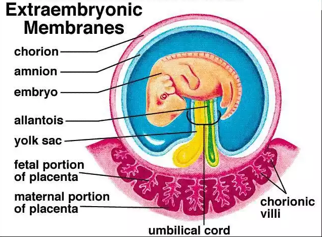
Fig.Extraembryonic membranes in a mammal