

Excretory System In Humans
Transportation in animals and plants of Class 7
Excretory System In Humans
Excretory system consists of the following organs
- A pair of kidneys
- Ureters
- Urinary bladder
- Urethra
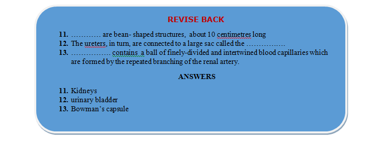
Kidneys are bean-
shaped structures, about 10 centimetres long. They are located just above the waist on either side of the backbone. The right kidney is placed slightly above the left. It measures about 10 cm long, 5 cm wide and 4cm thick, weighing about 135 to 150 gms., constituting 1% of body weight. Its inner concavity called hilum. Ureter, Blood vessels, nerve etc. enter or leave the kidney through hilus (hilum).Remains covered by peritoneium only on the ventral or front side hence termed as retroperitoneal. A narrow tube called the ureter runs from the inner side of each kidney.
The ureters, in turn, are connected to a large sac called the urinary bladder. Urine is collected and stored in the urinary bladder.
Leading from the bladder is another muscular tube called the urethra, which works as the outlet passage.
The end of the urethra is normally held closed by means of a ring of muscle (a sphincter), which controls the release of urine from the bladder.
Urine drains continuously out of the kidney into the ureters where it is forced downwards into the bladder by wave-like contractions of the ureter walls. The bladder stretches and expands in volume as it fills with urine, and when it is nearly full the stretching stimulates sensory nerve endings in its walls so that nerve impulses are sent to the brain. This is how a person knows when his bladder must be emptied. The sphincter muscle around the urethra is then voluntarily (i.e., consciously) relaxed to let urine drain from the bladder, through the urethra, and out of the body. This is called urination.
”The waste material is separated by the kidneys from the blood which is fed into it through the renal artery. The cleaner blood after the removal of wastes is sent back through the renal vein.
Structure of Kidneys
- A kidney in section shows a dark-coloured outer zone called the cortex, and a pale-coloured inner zone called the medulla. Each kidney is made of a large number of excretory units called nephrons.
- Each nephron consists of two parts - a rounded, cup-shaped body called the Bowman’s capsule, and the tubular part or the collecting tubule.
- Bowman’s capsule contains a ball of finely-divided and intertwined blood capillaries which are formed by the repeated branching of the renal artery. These structures are called glomeruli (singular glomerulus). The blood capillaries bring waste and excess water from body to the kidneys.
- The medulla is distinct by the presence of 15-16 conical pyramids with broader end towards cortex. Each pyramid ends as renal papilla where many ducts of Bellini converge to open. The renal papilla fits over small funnel-like minor calyx, 2 to 3 minor calyces together open in still wider funnel called major calyx.
Ureter
Narrow distensible tube, emerging from the hilus is basically a metanephric duct, lined with transitional epithelium. Being muscular it also undergoes peristalsis to pass urine from kidney to urinary bladder.
Urinary bladder :
- Single, median, pyriform bag (300-800 ml) in the pelvic area has upper body with two openings of ureter and lower trigone leading to urethra and guarded by sphincter.
- Wall is stretchable, muscular and lined with stratified transitional epithelium.
Urethra :
- The tube emerging from urinary bladder to release urine having sphincters at both ends.
- In women it is a short (4 cm) tube opening separately below clitoris in the vulva.
- In man it is long (20 cm) tube also serving as reproductive tract; divided into three parts : the upper prostatic part; the middle membranous part and the lower penile part.
Related Topics