Development of Diaphragm: Origin, Stages, and Clinical Importance
Development of diaphragm is a complex embryological process involving multiple structures such as the septum transversum, pleuroperitoneal folds, and cervical somites. Muscle cells migrate from the neck region, nerves grow alongside them, and connective tissue provides structural support. Any defect in this coordinated process can result in congenital diaphragmatic hernia, which affects lung development and breathing after birth.
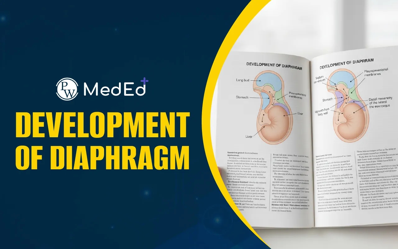

Development of Diaphragm: The diaphragm, crucial for mammalian respiration, forms from multiple embryonic sources. These include the septum transversum, pleuroperitoneal folds, and somites. Its complex development of diaphragm involves cell migration, muscle formation, nerve innervation, and vascularization. Defects during this process lead to Congenital Diaphragmatic Hernias (CDH), impacting lung development and causing severe health issues.
What is Development of Diaphragm?
The diaphragm is a vital skeletal muscle that separates the thoracic and abdominal cavities. It is essential for breathing. Understanding the development of diaphragm is important because defects in its formation cause serious birth defects. These notes explore the stages and components involved in its intricate embryological development. The diaphragm forms from different embryonic structures. Its muscle and connective tissues arise from three main sources.
Development of Diaphragm Embryology
The diaphragm develops from three primary components:
-
Septum Transversum: This is the first structure in the developing diaphragm. It is a thin, mesodermal sheet. The septum transversum separates the heart from the liver early in development. It likely forms a scaffold for later diaphragm morphogenesis. Its contribution to the adult diaphragm's central tendon is suggested.
-
Pleuroperitoneal Folds: These are transient, pyramid-shaped structures. They protrude from the body wall, separating the pleural and peritoneal cavities. They are important targets for migrating muscle progenitors and nerves. These folds appear to spread and fuse with the septum transversum.
-
Somites: These are the source of the diaphragm’s muscle cells. Muscle progenitors migrate from cervical somites (C3-C5) to form the diaphragmatic muscles. Genes like Pax3 and Met are vital for this migration.
Migration of Muscle and Nerve to Developing Diaphragm
Muscle progenitors and the phrenic nerve axons migrate to the developing diaphragm.
-
Muscle Progenitor Migration: Muscle cells move from the somites to the pleuroperitoneal folds. Hepatocyte Growth Factor (HGF) guides this migration. Loss of HGF can prevent muscle development in the diaphragm.
-
Phrenic Nerve Migration: The phrenic nerve originates from cervical nerves (C3-C5). It travels with muscle progenitors, eventually innervating the diaphragm. Neural cell adhesion molecules (NCAM) may guide nerve outgrowth.
Diaphragm Morphogenesis
After migration, muscle cells differentiate and form the diaphragm structure.
-
Myogenesis: Muscle progenitors become committed myoblasts. These then differentiate into myocytes and fuse to form multinucleate myofibers. These myofibers assemble into the costal and crural muscles of the diaphragm.
-
Nerve Branching and Innervation: Phrenic nerves branch into sternocostal, dorsocostal, and crural branches. They form neuromuscular junctions with myofibers. Signals from both nerve and muscle control this process.
-
Vascularization: Arteries such as the phrenic, internal thoracic, and intercostal arteries vascularize the diaphragm. This ensures proper blood supply for its function.
Development of Diaphragm Flowchart
Here is a Development of Diaphragm Flowchart:
Cervical somites C3–C5
↓
Migration of muscle progenitor cells
↓
Pleuroperitoneal folds receive migrating cells
↓
Fusion with septum transversum
↓
Muscle differentiation and formation of diaphragm
↓
Phrenic nerve innervation and vascularization
↓
Fully developed functional diaphragm
Congenital Diaphragmatic Hernias (CDH)
CDH is a common birth defect affecting the development of diaphragm. It occurs when the diaphragm fails to form correctly. This leads to abdominal organs moving into the chest cavity.
-
Impact of CDH: Herniated organs impede lung development, causing hypoplastic lungs. This results in severe respiratory problems and high mortality.
-
Types of CDH:
-
Bochdalek Hernias: These are the most common (90%) and occur in the posterior-lateral diaphragm, usually on the left side.
-
Morgagni Hernias: These form in the anterior diaphragm.
-
Central Hernias: These involve the central tendon.
-
Genetic Factors: Mutations in genes like Gata4, Zfpm2, and Nr2f2 link to CDH. Retinoic acid (RA) signaling is also critical; its disruption causes CDH. Defects in connective tissue formation, like in lysyl oxidase or Collagen 3a1 mutations, also contribute.
Congenital Diaphragmatic Hernia and Clinical Importance
Congenital diaphragmatic hernia occurs when the diaphragm fails to form completely. This allows abdominal organs such as the stomach and intestines to enter the chest cavity. As a result, lung development is severely restricted, leading to breathing difficulties after birth.
The most common type is Bochdalek hernia, which occurs in the posterolateral part of the diaphragm, usually on the left side. Other types include Morgagni hernia in the anterior region and central tendon defects. Genetic factors play an important role in this condition. Abnormal retinoic acid signaling and mutations in genes involved in connective tissue formation have been linked to diaphragm defects.
| Development of the Cardiovascular System | Types of Scabies |
| Parotid Gland | Necrotizing Enterocolitis |