CBSE Class 11 Biology Notes Chapter 21 Download PDF Here

CBSE Class 11 Biology Notes Chapter 21: Chapter 21, Neural Control and Coordination, in CBSE Class 11 Biology, focuses on how the human nervous system functions to regulate body activities. Key topics include the structure and function of neurons, central and peripheral nervous systems, reflex action, synaptic transmission, and sense organs. As per the CBSE class 11 syllabus, this chapter is an important part of Unit 5 – Human Physiology and carries significant weight in exams. The CBSE exam pattern includes a combination of objective, short-answer, and long-answer questions, with a focus on diagrams, explanations, and conceptual clarity. Studying this chapter thoroughly, along with practicing class 11 biology sample papers helps students perform well in both school exams and competitive exams like NEET.
CBSE Class 11 Biology Notes Chapter 21 Overview
This chapter provides information on the coordination and whole neurological system of all animals. This biology chapter is crucial and will support your further education. Our highly qualified instructors will help you grasp the material more fully. The neural control and coordination class 11 notes are available for download in PDF format. You can prepare for your exams with the help of the reference notes.- The body's communication is coordinated by the nervous system.
- Coordination is the process by which two or more organs work in tandem to enhance each other's functions. Our neurological system is set up with point-to-point connections for fast coordination.
- A neuron is a basic unit of brain organization that is capable of detecting, receiving, and transmitting various inputs.
CBSE Class 11 Biology Notes Chapter 21
Neural Control and Coordination
Coordination is the interaction of two or more organs to support and enhance one another's functions.Neural System
Neurons are the anatomical and functional building blocks of the nervous system. They can send, receive, and detect impulses.Human Nervous System
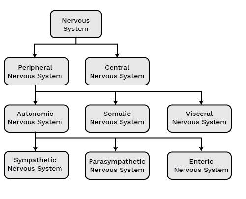
The classification of nervous system is divided into two types-
(i) central nervous system (CNS)
(ii) peripheral nervous system (PNS).
The peripheral nervous system is made up of all the nerves found in the brain and spinal cord, whereas the central nervous system is made up of the brain and spinal cord. The PNS contains two different types of nerve fibers:
(a) afferent nerve fibers or sensory nerve fiber
(b) efferent nerve fibers or motor nerve fiber
Impulses are carried from organs/tissues to the CNS in Afferent nerve fibers whereas, in efferent nerve fibers, impulses are transmitted from the CNS to the target tissue/organ.
PNS Is Classified Into Two Types-
(i) somatic nervous system
(ii) autonomic nervous system.
The somatic nervous system carries impulses from the central nervous system to skeletal muscles, while the autonomic nervous system innervates the smooth and cardiac muscles of the glands, which in turn carries the impulses from the central nervous system to the involuntary part of the body. The autonomic nerve system is divided into two categories:
(i) sympathetic nervous system
(ii) parasympathetic nervous system
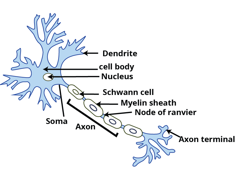
Structure and the Function of the Neuron
A neuron is the structural and functional unit of the nervous system. It comprises 3 parts-
(i) cell body
(ii) dendrites
(iii) axon.
The cytoplasm of the cell body is composed of granular entities known as Nissl's granules. Dendrites are short, many, finger-like projections that emerge from the cell body. Nerve impulses are transported by dendrites into or towards the cell body.
An axon is a long, single structure with branches at its distal end. Every axon terminates at the synaptic knob, a structure that resembles a bulb and is home to neurotransmitters. Schwann cells are the axon cells that generate and coat the myelin sheath. Nodes of Ranvier are the breaks in the myelin sheath that occur between two neighboring areas.
Types of Neurons
Multipolar Neurons- Several processes arise in them, having one axon and many dendrites. For example, in the cerebral cortex.
Bipolar Neurons- They are the ones with one axon and one dendrite. For example, the retina.
Unipolar Neurons- Unipolar neurons have only two fibers arising close together, i.e., one axon, no dendrite. For example, embryonic stage.
Generation and Conduction of Nerve Impulses
Neurons are the cells that are capable of being electrically activated and stimulated. The neuronal membrane exhibits polarisation. Ion channels that are selectively permeable and have differing permeabilities to various ions are present in the membrane. When there is no nerve impulse being conveyed, the membrane becomes more permeable to potassium ions and impermeable to sodium ions. The potential of the membrane at rest is this.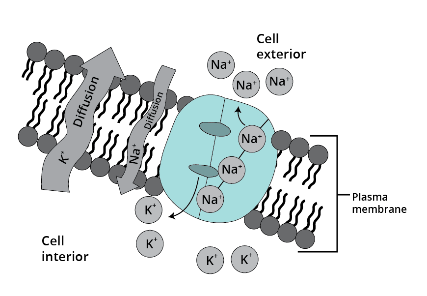 Over time, potassium ions become more permeable whereas sodium ions become less permeable. An outward-moving polarisation of sodium ion outflow takes place in the axonal membrane. The polarity is inverted once more. This series of actions is repeated along the axon, aiding in the nerve impulse's conduction.
Transmission of Impulses
the process by which a nerve impulse is transferred between neurons through connections known as synapses. A synaptic cleft, which separates the membranes of the pre-synaptic and postsynaptic neurons, forms the junction known as a synapse.Two Types of Synapses are There-
(i) Electrical synapse
(ii) Chemical synapse.
Pre- and post-synaptic neurons are close together in an electrical synapse. It is possible for electric current to travel directly between neurons. Nerve impulses are transmitted through this mechanism more quickly than through a chemical synapse.
Pre- and post-synaptic neurons in chemical synapses are divided by a fluid-filled area known as the synaptic cleft. Neurotransmitters play a role in these synapses.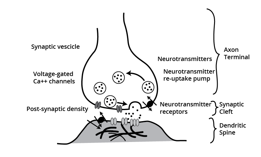
Central Nervous System
The brain controls and coordinates all the functions of the body thus, performing as a command and control system. The protection of the human brain is inside the skull. The covering of the brain is done in 3 layers known as meninges. They are-
(i) outermost dura mater,
(ii) middle arachnoid,
(iii) innermost pia mater.
The division of the brain is into:
(a) forebrain,
(b) midbrain,
(c) hindbrain.
The thalamus, hypothalamus, and cerebrum make up the three regions of the forebrain. The greatest portion of the brain is thought to be the cerebrum. The left and right cerebral hemispheres refer to the partition of the cerebrum into these two halves.
The corpus callosum, a large white central canal commissure, connects these hemispheres. The cerebral cortex is the layer of cells that covers the cerebral hemispheres.
The region located at the forebrain's center is called the thalamus. It contributes to both motor and sensory signaling. At the base of the thalamus is the hypothalamus. It regulates and controls the urge to eat, and drink, and regulates body temperature.
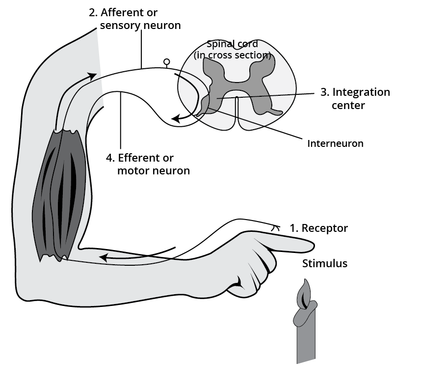
Reflex Action and Reflex Arc
Reflex action is an immediate involuntary stereotyped response. The neural elements involved in reflex action follow an arc called the reflex arc. Reflex arc or reflex pathway includes:
Receptor (sensory cell receiving stimulus)
↓
Afferent or sensory neuron
↓
The dorsal root of the spinal cord
↓
Connecting neuron in CNS
↓
Efferent or motor neuron
↓
The ventral root of the spinal cord
↓
Effector organ
Human Eye
The human eye is spherical, with the front one-fifth of it visible and the rear four-fifths hidden inside the orbit of the eye. There are three layers to it:(i) the outermost sclera (dense connective tissue),
(ii) middle choroid (supplied with blood vessels),
(iii) innermost retina.
The choroid lines the sclera's posterior portion. The choroid enlarges anteriorly to generate the ciliary body. The choroid and cornea split apart in front of the ciliary body, generating the iris, a visible, colored part of the eye. Seen just behind the pupil, a solid, transparent, crystalline biconvex lens is retained by ligaments that are connected to the ciliary body.
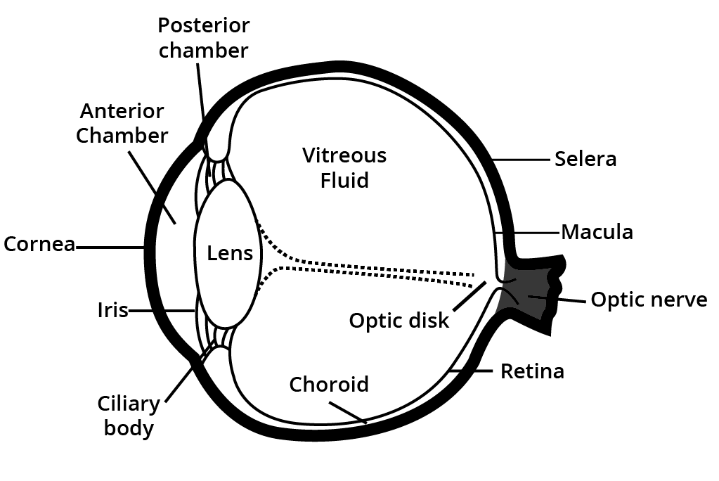
The optic nerve is the one that runs from the eye to the brain. This area is referred regarded as a blind spot because it lacks photoreceptors. The lateral to the blind area, where only cones are tightly packed, is where the macula lutea is located.
The area in the cornea between the lens and a fluid known as aqueous humor is termed the aqueous chamber. The fluid is filled with this fluid. As a result, the fluid that fills the vitreous chamber—which is located between the lens and the retina—is referred to as vitreous humor.
Rods and cones are the two different types of photoreceptor cells. These proteins are light-sensitive. Rods are more sensitive to low light, but cones are more sensitive to very bright light and color vision. Rhodopsin is the name of the protein found in rods. Specifically, cones react to the colors red, green, and blue.
CBSE Class 11 Biology Notes Chapter 21 PDF
The neural control and coordination class 11 notes, are available in PDF format via the link below. When studying this chapter, students can refer to these notes as a helpful reference. It covers crucial subjects on the Neural Control and Coordination.
Do you need help with your homework or preparing for exams?
Study without using the internet
Benefits of CBSE Class 11 Biology Notes Chapter 21
Comprehensive Understanding – The notes simplify complex topics like the nervous system, neurons, synaptic transmission, and reflex actions, making them easier to grasp.
Exam-Oriented Preparation – Aligned with the CBSE syllabus and exam pattern, these notes cover essential topics frequently asked in board exams and competitive exams like NEET.
Quick Revision – Well-structured summaries, key points, and diagrams help in quick last-minute revisions before exams.
Concept Clarity – The notes explain neural coordination in detail, strengthening conceptual understanding for both theoretical and application-based questions.
PYQ Practice – Important previous year questions (PYQs) and sample questions help students get familiar with the types of questions asked in the exams, improving their problem-solving skills.
CBSE Class 11 Biology Notes Chapter 21 FAQs
What is neural control and coordination?
What brain function is coordination?
What is the function of neural control?