CBSE Class 11 Biology Notes Chapter 17 Breathing and Exchange of Gases

CBSE Class 11 Biology Notes Chapter 17: In Class 11 Biology, Chapter 17 talks about "Breathing and Exchange of Gases." It's all about how living things get oxygen from the air and release carbon dioxide. We'll learn how this happens in different animals, from small ones to big ones like us.
We'll also look at how our lungs and other organs help us breathe. Understanding this helps us see how important it is to get oxygen into our bodies and get rid of carbon dioxide. It's like learning how our bodies work to keep us alive and healthy!CBSE Class 11 Biology Notes Chapter 17 PDF
The breathing and exchange of gases class 11 notes are accessible as PDF files that you can download and view at your convenience on any device. Once downloaded, the file can be read offline. When needed, these notes can aid in the student’s comprehension and memory of the material. This CBSE Class 11 Biology Notes Chapter 17 PDF is a great resource to help you do well on your tests.CBSE Class 11 Biology Notes Chapter 17 Breathing and Exchange of Gases PDF
CBSE Class 11 Biology Notes Chapter 17 Breathing and Exchange of Gases
Respiratory Organs
Understanding the diversity of respiratory mechanisms across different organisms is crucial in comprehending their adaptations to varied habitats. Below is a table summarizing some organisms and their respiratory organs:| Organism | Respiratory Organ(s) |
|---|---|
| Terrestrial Mammals | Lungs |
| Fish | Gills |
| Earthworms | Moist skin |
| Insects | Tracheal system (spiracles and tracheae) |
| Amphibians | Lungs, skin, and buccopharyngeal cavity |
| Birds | Lungs and air sacs |
| Plants | Stomata and lenticels (for gas exchange) |
Human Respiratory System
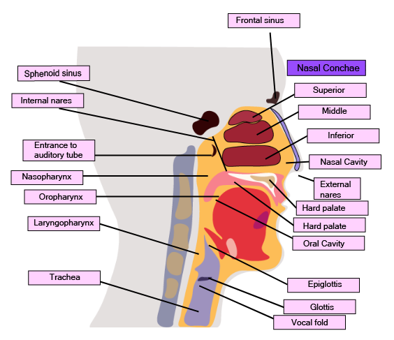
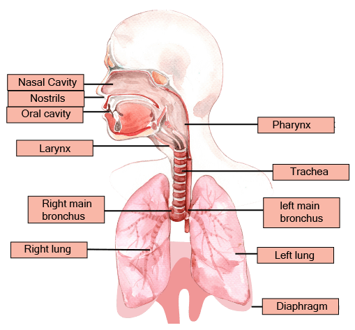 The human respiratory system is a complex network of organs and structures that facilitate the exchange of gases between the body and the environment. It all starts with the external nostrils, which lead into the nasal cavity. Inside the nasal cavity, specialized cells secrete mucous, a slimy fluid that helps capture dust and other particles from the inhaled air, keeping the nasal passages moist and clean.
The nasal cavity extends into the pharynx, a junction where the respiratory and digestive systems meet. The pharynx is divided into three sections: the oropharynx, nasopharynx, and laryngopharynx. Each section serves a specific purpose, such as allowing food to pass through (oropharynx) or facilitating airflow (nasopharynx).
The glottis, located at the bottom of the nasopharynx, is protected by the epiglottis, a cartilaginous flap that prevents food from entering the windpipe or trachea during swallowing. The trachea is a thin-walled tube that runs in front of the esophagus and carries air to and from the lungs.
The human respiratory system is a complex network of organs and structures that facilitate the exchange of gases between the body and the environment. It all starts with the external nostrils, which lead into the nasal cavity. Inside the nasal cavity, specialized cells secrete mucous, a slimy fluid that helps capture dust and other particles from the inhaled air, keeping the nasal passages moist and clean.
The nasal cavity extends into the pharynx, a junction where the respiratory and digestive systems meet. The pharynx is divided into three sections: the oropharynx, nasopharynx, and laryngopharynx. Each section serves a specific purpose, such as allowing food to pass through (oropharynx) or facilitating airflow (nasopharynx).
The glottis, located at the bottom of the nasopharynx, is protected by the epiglottis, a cartilaginous flap that prevents food from entering the windpipe or trachea during swallowing. The trachea is a thin-walled tube that runs in front of the esophagus and carries air to and from the lungs.
Labelled Diagram of Nasal and Throat Cavity
 The larynx, often referred to as the voice box, is situated at the upper end of the trachea and is primarily responsible for producing sound. It is supported by cartilage rings, including the thyroid cartilage and cricoid cartilage.
In males, the thyroid cartilage is more prominent and can be seen as the Adam's apple protruding from the neck. Within the larynx, there are two mucous membrane folds known as vocal cords. These cords vibrate when air passes through, creating sounds for speech and other vocalizations.
Meanwhile, the trachea, or windpipe, is a tube-like structure that connects the larynx to the lungs. Its walls are reinforced by C-shaped rings made of cartilage, which provide support and prevent collapse of the trachea, ensuring the passage of air remains open even when there is less air pressure within.
The larynx, often referred to as the voice box, is situated at the upper end of the trachea and is primarily responsible for producing sound. It is supported by cartilage rings, including the thyroid cartilage and cricoid cartilage.
In males, the thyroid cartilage is more prominent and can be seen as the Adam's apple protruding from the neck. Within the larynx, there are two mucous membrane folds known as vocal cords. These cords vibrate when air passes through, creating sounds for speech and other vocalizations.
Meanwhile, the trachea, or windpipe, is a tube-like structure that connects the larynx to the lungs. Its walls are reinforced by C-shaped rings made of cartilage, which provide support and prevent collapse of the trachea, ensuring the passage of air remains open even when there is less air pressure within.
Labelled Diagram of Trachea
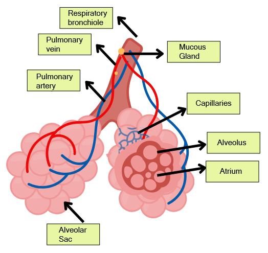
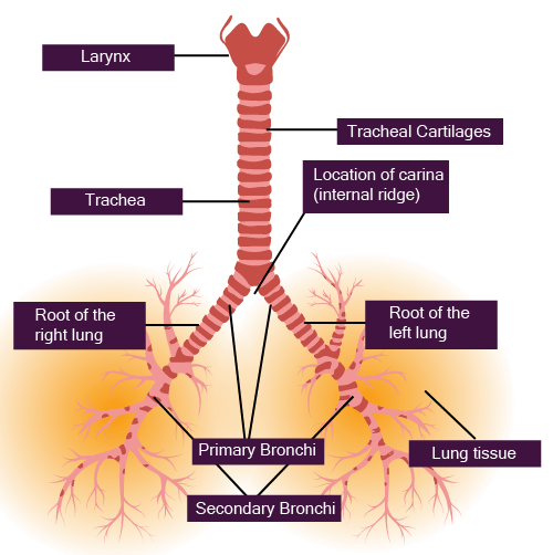 After the trachea, the air passages divide into two bronchi, with each bronchus leading to one lung. Within the lungs, these bronchi further branch out into smaller tubes known as bronchioles, which extend throughout the lung tissue. The bronchioles eventually divide into numerous tiny ducts called alveolar ducts. At the end of these ducts are clusters of thin-walled air sacs called alveoli. These alveoli resemble bunches of grapes, with each cluster of alveoli known as an infundibulum.
Labelled Diagram of Alveoli
 The respiratory system plays a crucial role in supplying oxygen to the body and removing carbon dioxide, which is a waste product of cellular metabolism. It consists of several components, including the airways (such as the bronchi and bronchioles) and the alveoli, where gas exchange takes place. The lungs are protected by a double-layered membrane called the pleura, which contains pleural fluid to reduce friction during breathing.
The respiratory system can be divided into two main parts: the conduction part and the exchange part. The conduction part includes all the respiratory structures from the nostrils to the alveoli and is responsible for conducting atmospheric air to the lungs. It also helps in filtering, moisturizing, and warming the air before it reaches the lungs. On the other hand, the exchange part is where oxygen diffuses into the bloodstream and carbon dioxide diffuses out of the bloodstream into the lungs.
Located in the chest cavity, the lungs are surrounded by the spine, sternum, ribs, and diaphragm. Changes in the volume of the chest cavity, caused by the movement of the diaphragm and rib muscles, result in changes in lung volume, which are essential for breathing.
The process of breathing, or lung ventilation, involves the inhalation of atmospheric air rich in oxygen and the exhalation of alveolar air rich in carbon dioxide. Gas exchange occurs in the alveoli, where oxygen and carbon dioxide diffuse across the alveolar membrane into and out of the bloodstream, respectively. These gases are then transported through the blood to various organs and tissues, where oxygen is used for cellular metabolism and carbon dioxide is produced as a byproduct. This exchange of gases between the blood and body tissues is essential for cellular respiration, which provides the energy needed for various metabolic processes in the body.
The respiratory system plays a crucial role in supplying oxygen to the body and removing carbon dioxide, which is a waste product of cellular metabolism. It consists of several components, including the airways (such as the bronchi and bronchioles) and the alveoli, where gas exchange takes place. The lungs are protected by a double-layered membrane called the pleura, which contains pleural fluid to reduce friction during breathing.
The respiratory system can be divided into two main parts: the conduction part and the exchange part. The conduction part includes all the respiratory structures from the nostrils to the alveoli and is responsible for conducting atmospheric air to the lungs. It also helps in filtering, moisturizing, and warming the air before it reaches the lungs. On the other hand, the exchange part is where oxygen diffuses into the bloodstream and carbon dioxide diffuses out of the bloodstream into the lungs.
Located in the chest cavity, the lungs are surrounded by the spine, sternum, ribs, and diaphragm. Changes in the volume of the chest cavity, caused by the movement of the diaphragm and rib muscles, result in changes in lung volume, which are essential for breathing.
The process of breathing, or lung ventilation, involves the inhalation of atmospheric air rich in oxygen and the exhalation of alveolar air rich in carbon dioxide. Gas exchange occurs in the alveoli, where oxygen and carbon dioxide diffuse across the alveolar membrane into and out of the bloodstream, respectively. These gases are then transported through the blood to various organs and tissues, where oxygen is used for cellular metabolism and carbon dioxide is produced as a byproduct. This exchange of gases between the blood and body tissues is essential for cellular respiration, which provides the energy needed for various metabolic processes in the body.
Mechanism of Respiration
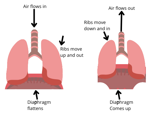
Breathing, the exchange of atmospheric air between the lungs and the outside environment, is essential for supplying oxygen to the body and removing carbon dioxide. This process involves two main phases: inspiration (inhalation) and expiration (exhalation), which are facilitated by the expansion and contraction of the lungs. During inspiration, the diaphragm and intercostal muscles contract, causing the chest cavity to expand. This increases the volume of the lungs, leading to a decrease in pressure within the lungs compared to the atmosphere. As a result, air flows into the lungs through the nose and mouth. Conversely, during expiration, the diaphragm and intercostal muscles relax, allowing the chest cavity to return to its resting position. This reduces the volume of the lungs, increasing the pressure within them relative to the atmosphere. Air is then expelled from the lungs through the nose and mouth. Extra-abdominal muscles can assist in enhancing the strength of both inhalation and exhalation. Typically, a healthy individual breathes around 12-16 times per minute, which equates to approximately 10 liters of air per minute. Respiratory rate can be measured using a device called a spirometer or respirometer.Diagram Showing Mechanism of Respiration
 Respiratory volumes and capacities play a important role in understanding lung function and respiratory health. Here are some key definitions and measurements related to respiratory volumes and capacities:
Tidal volume (TV): This refers to the amount of air that enters or exits the lungs during a single breath under normal conditions. For adults, the average tidal volume is around 500 milliliters (ml).
Inspiratory reserve volume (IRV): IRV is the additional volume of air that can be inhaled beyond the tidal volume during a forced inhalation. It typically ranges from 2000 to 3000 ml in healthy individuals.
Expiratory reserve volume (ERV): ERV represents the extra volume of air that can be exhaled after a normal exhalation. In healthy adults, ERV is approximately 1100 ml.
Residual volume (RV): RV is the volume of air that remains in the lungs even after forceful exhalation. It ensures continuous gas exchange in the lungs and usually amounts to about 1100 to 1200 ml.
Pulmonary capacity (PC): Pulmonary capacity is a collective term for various lung capacities.
- Inspiratory capacity (IC): IC denotes the maximum amount of air a person can inhale after a normal exhalation. It is calculated by adding tidal volume and inspiratory reserve volume (TV + IRV).
- Expiratory capacity (EC): EC represents the maximum amount of air that can be exhaled after a normal inhalation (TV + ERV).
- Functional residual volume (FRC): FRC refers to the volume of air remaining in the lungs after a normal exhalation. It comprises expiratory reserve volume and residual volume (ERV + RV).
- Vital capacity (VC): VC indicates the maximum amount of air that can be exhaled forcefully after the deepest inhalation. It includes inspiratory reserve volume, expiratory reserve volume, and tidal volume (IRV + ERV + TV).
- Total lung capacity (TLC): TLC represents the maximum volume of air that the lungs can hold. It includes all lung volumes: inspiratory reserve volume, expiratory reserve volume, tidal volume, and residual volume (IRV + ERV + TV + RV).
Gas Exchange
Gas exchange in the lungs primarily occurs in the alveoli, where oxygen from inhaled air diffuses into the bloodstream, while carbon dioxide from the blood diffuses into the alveoli to be exhaled. This exchange relies on simple diffusion driven by pressure and concentration gradients. The efficiency of gas exchange is influenced by factors such as the solubility of gases and the thickness of the membrane. Oxygen and carbon dioxide have different partial pressures (pO2 and pCO2, respectively) in various parts of the respiratory system. In atmospheric air, the partial pressure of oxygen is around 160 mm Hg, while the partial pressure of carbon dioxide is much lower, around 0.3 mm Hg. As blood moves through the lungs, it encounters areas with higher oxygen partial pressure and lower carbon dioxide partial pressure compared to the blood. This gradient facilitates the diffusion of oxygen into the bloodstream and the release of carbon dioxide from the blood into the alveoli. Understanding these partial pressures and their variations in different parts of the respiratory system is crucial for comprehending the mechanisms of gas exchange and respiratory physiology.Diagrammatic Representation of Gas Exchange Between Alveoli and Other Body Parts
The process of gas exchange, particularly the diffusion of oxygen and carbon dioxide across the alveolar membrane, is facilitated by several factors. Firstly, the structure of the diffusion membrane plays a critical role. It consists of the squamous epithelial cells lining the alveoli, the endothelial cells of the capillaries surrounding the alveoli, and the basement membrane between these layers. This membrane is incredibly thin, measuring less than a millimeter in total thickness, which enables efficient gas exchange. Moreover, the partial pressure gradient of gases drives the diffusion process. Despite the lower partial pressure of oxygen compared to carbon dioxide, the efficiency of gas exchange is high due to the large surface area of the alveoli and the rich blood supply in the pulmonary capillaries. Physiological variables such as concentration gradients, blood flow, and ventilation-perfusion matching also contribute to efficient gas exchange. These variables ensure that oxygen is delivered to tissues in proportion to their metabolic needs and that carbon dioxide is efficiently removed from the bloodstream. Overall, the coordinated interplay of these factors optimizes the exchange of gases across the alveolar membrane, ensuring that oxygen is delivered to tissues for cellular respiration while carbon dioxide is effectively eliminated from the body.Gas Transportation
Oxygen and carbon dioxide, vital gases for cellular function, are transported through the bloodstream in different ways.Oxygen Transportation: In the bloodstream, oxygen is primarily carried by red blood cells (RBCs), with about 97% binding to the iron-containing pigment hemoglobin, forming oxyhemoglobin. The remaining 3% of oxygen is transported dissolved in plasma. Hemoglobin has four iron-containing parts, allowing each molecule to bind with four oxygen molecules. The binding of oxygen to hemoglobin is influenced by factors such as carbon dioxide partial pressure, hydrogen ion concentration, and temperature.
The relationship between hemoglobin saturation and oxygen partial pressure is depicted by the oxygen dissociation curve, which shows an S-shaped curve. In the alveoli, where oxygen partial pressure is high and carbon dioxide partial pressure is low, oxyhemoglobin forms. Conversely, in tissues with low oxygen partial pressure, high carbon dioxide partial pressure, high hydrogen ion concentration, and higher temperature, oxygen dissociates from hemoglobin, allowing oxygen to be released to the tissues. Under normal conditions, 100 mL of oxygenated blood carries about 5 mL of oxygen.Carbon Dioxide Transportation: Carbon dioxide is transported in various forms in the blood. Approximately 20-25% is carried by hemoglobin as carbamino-hemoglobin, proportional to carbon dioxide partial pressure. Carbon dioxide binding to hemoglobin is influenced by oxygen partial pressure, with more binding occurring in tissues where carbon dioxide partial pressure is high and oxygen partial pressure is low. When carbon dioxide partial pressure is low and oxygen partial pressure is high, as in the alveoli, carbon dioxide dissociates from carbamino-hemoglobin.
The majority of carbon dioxide (about 70%) is converted into bicarbonate ions by the enzyme carbonic anhydrase abundant in red blood cells. This enzyme is responsible for the conversion of carbon dioxide into bicarbonate ions, which can easily move from tissues to the alveoli for elimination. Additionally, about 7% of carbon dioxide is transported dissolved in plasma, while the remaining 5-7% is transported bound to hemoglobin.Regulation of Respiration: The respiratory rhythm and rate are regulated by the nervous system to meet the body's oxygen demand. The respiratory center, located in the medulla oblongata of the brain, coordinates respiration. It consists of several neurons and includes the respiratory rhythm center, pneumotaxic center, and chemosensitive area. These centers respond to various stimuli, including oxygen and carbon dioxide levels in the blood, to adjust the breathing rate and depth accordingly.
Respiratory Rhythm Center
The respiratory system's regulation involves specialized centers in the brain that coordinate breathing.Respiratory Rhythm Center: Located in the dorsal region of the medulla oblongata, this center generates the basic respiratory rhythm. Neurons in this center send signals to the diaphragm, the primary muscle responsible for inspiration, causing it to contract and initiate inhalation.
Pneumotaxic Center: Found dorsally in the upper pons of the brain, the pneumotaxic center modulates the functions of the respiratory rhythm center. It regulates the number and depth of breaths by sending signals that can increase the breathing rate. Strong signals from this center shorten both inspiration and expiration, thereby altering the respiratory rate.
Chemosensitive Center: Situated near the respiratory rhythm center, the chemosensitive center is highly sensitive to changes in carbon dioxide (CO2) and hydrogen ion concentrations. Elevated CO2 and hydrogen ion levels trigger increased inspiratory and expiratory signals. Although oxygen levels do not directly affect respiratory signals, the chemosensitive center responds to increased CO2 and hydrogen ions.
Receptors in the aortic arch and carotid artery detect changes in CO2 and hydrogen ion concentrations and transmit signals to the respiratory rhythm center, prompting adjustments in the breathing process to eliminate these substances.Disorders of the Respiratory System: Various respiratory diseases and disorders affect respiratory function.
Asthma: Characterized by difficulty breathing and wheezing due to inflammation of the bronchi and bronchioles, asthma is a common respiratory condition.
Emphysema: A chronic obstructive lung disease, emphysema involves abnormal distension or inflation of the alveolar walls, leading to loss of elasticity. Alveolar walls degenerate, and alveoli merge, causing increased lung size and difficulty exhaling. Cigarette smoking is a major cause of emphysema.
Occupational Respiratory Disorders: Pulmonary diseases resulting from exposure to harmful substances in the workplace are known as occupational respiratory disorders. Examples include silicosis, caused by chronic exposure to silica dust in industries like mining, and asbestosis, resulting from prolonged exposure to asbestos dust in asbestos factories.
Benefits of Breathing and Exchange of Gases Class 11 Notes
- These notes provide a clear explanation of how our body breathes and exchanges gases, making it easier for students to understand the complex processes involved.
- With simplified explanations and diagrams, these notes help in clarifying concepts related to respiratory organs, mechanisms of breathing, gas exchange, and more.
- Studying these notes can help in exam preparation by covering all the essential topics outlined in the CBSE syllabus for Class 11 Biology.
- The notes are well-organized, making them ideal for quick revision before exams or tests. Many notes include practice questions and answers, allowing students to test their understanding and practice for exams.
- Diagrams and illustrations included in the notes help in visualizing the respiratory system and processes, aiding in better retention of information.
CBSE Class 11 Biology Notes Chapter 17 FAQs
What is the respiratory system?
What are the main organs of the respiratory system?
How does gas exchange occur in the lungs?
How is oxygen transported in the blood?
What factors affect the binding of oxygen to hemoglobin?