
Meiosis Of Cell Cycle
Cell cycle of Class 11
Meiosis is characterized by two successive divisions of the cytoplasm and nuclei (Meiosis I and Meiosis II), accompanied by only one replication of the chromosomes.
A double division which occurs in a diploid cell (nucleus) and gives rise to four haploid cells (nuclei), each having half the number of chromosomes as compared to the parent cell.
Interphase Similar to that of mitosis except that S-phase is prolonged and a distinct G2 phase is either short or absent.
DNA replicates during S-phase and each chromosome comes to have two chromatids.
Karyokinesis
The nucleus undergoes two divisions -
Meiosis-I/Reduction/Heterotypic division – No. of chromosomes becomes half and genetically different due to crossing over in daughter nuclei.
Meiosis-II/Equational/Homotypic division – No. of chromosomes remain same and similar as produced after the end of first division.
Meiosis-I: Comprises of four substages
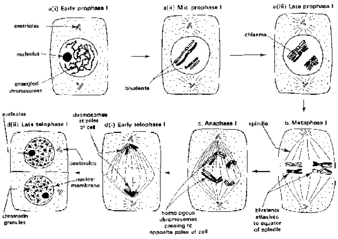
Fig. Stages of the first meiotic division
Prophase - I
More complicated and prolonged.
Divided into five subphases - leptotene, zygotene, pachytene, diplotene and diakinesis.
1. Leptotene/Leptonema
Nucleus enlarges - Chromatin fibres of interphase shorten and elongated chromosomes become clear, which possess a string of swollen areas called chromomeres.
Chromosomes are replicated but the chromatids are not distinguishable due to the presence of a nucleoprotein core between them.
In many animal cells the chromosomes show a peculiar arrangement called bouqet stage - The ends of chromosomes converge towards the side having replicated centrosomes.
Both centriole pairs develop astral rays and one of them begins to move to the opposite side.
In cells undergoing meiosis there are two sets of chromosomes i.e., chromosome number is diploid. There are two similar chromosomes of each type known as homologous chromosomes.
The two homologous chromosomes contributed by different parents (paternal and maternal) resemble each other in the presence of their-centromeres, chromomeres, shape and size.
2. Zygotene/Zygonema
The two homologous chromosomes get attached to each other laterally due to the development of nucleoprotein core between them.
Pairing is such that the genes of same character present on chromosomes come to lie opposite exactly.
The process of attachment of the homologous chromosomes is known as Synapsis or Syndesis forming Synaptinemal complex. Depending on the place of origin of pairing or synapsis is :
(i) Procentric (from centromeres towards ends)
(ii) Proterminal (from ends towards centromeres)
(iii) Intermediate (at various places between centromeres and ends)
On account of synapsis chromosomes form pairs or Bivalents which are half the no. of chromosomes.
Spindle appratus develops in late prophase and is completed in metaphase. It disappears in late anaphase but mostly in early telophase. It is astral/amphiastral (i.e, with two asters) and arises from centrosome
(centrioles) in animal cells and some lower motile plants. In most of plants, it is mostly anastral (i..e., poles without aster) and arises from cytoplasmic tubulin proteins by gelation. The spindle is made up of
spindle fibres and astral rays which are derived from microtubules. Each spindle fibre has 4-20 microtubules. Chemically these fibres, are made up of (i) Sulphur rich tubulin protein (90-95%), (ii) RNA (3-5%), (iii) ATPase, (iv) traces of lipids, actin and myosin. The spindle helps in the migration of chromatids towards poles.
3. Pachytene/Pachynema
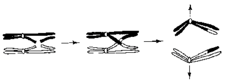
Fig. Crossing over of genes
Bivalents shorten. Each bivalent has 4 chromatids - 2 of each chromosome. Chromatids belonging to same chromosome are called as Sister chromatids while chromatids belonging to two different chromosomes are non-sister chromatids.
In the presence of enzyme endonuclease breaks develop in individual chromatids and process is called nicking. Nicks get healed in most of the cases, but in one out of 1000 - gaps develop in the region of nicks by enzyme exonuclease.
Separation of chromatid segments occurs in between two gaps by U-Protein or enzyme Unwindase.
The separated segments of non-sister chromatids may exchange position and phenomenon is known as crossing over, a process of exchange of genetic material or chromatid segments between two homologous chromosomes. Crossing over occurs in Pachytene.
Separated chromatid segments soon get reunited with the help of an enzyme R-protein and process is called reannealing.
4. Diplotene/Diplonema
Nucleoprotein fusion complex of synapsed chromosomes dissolves and the homologous chromosomes separate except in the region of crossing over.
Chromatids become distinguishable (tetrad stage).
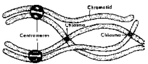
Fig. Diagram interpreting the photomicrograph
The points of attachment after partial dissolution of nucleoprotein complex are known as Chiasmata which may be terminal or interstitial. Crossing over is observed in diplotene.
Homologous Chromosomes may appear cross like, ringlike or chain like depending on position of chiasmata.
In fish, amphibian, reptile and avian oocytes, there is uncoiling of chromosomes which becomes so marked that the greatly enlarged nucleus assumes an interphase appearance and bivalent chromosomes may attain lampbrush configuration.
A long lasting period e.g., in the fifth month of prenatal life, human oocytes have reached this stage and remain in it till ovulation occurs after many years.
5. Diakinesis
No. of chiasmata diminishes during this period.
By the end, homologues are held together only at their ends and process is known as terminalisation.
Nucleolus degenerates and simultaneously nuclear envelope disintegrates.
Metaphase - I
Colourless bipolar spindle apparatus appears in the region of degenerated nucleus.
Spindle microtubules become attached to the kinetochores.
Each homologue is attached to one of the poles by the homologous kinetochore.
Bivalents arrange themselves on equator. Limbs of the chromosomes are usually short and lie horizontally on equator.
Since there are two centromeres in each bivalent the centromeres of all bivalents produce a double metaphasic plate.
Distribution of bivalents is at random so that the individual paternal and maternal chromosomes can face either of two poles of the spindle.
Each chromosome gets attached to the spindle pole of its side by chromosomal fibre/tactile fibril which arises in the region of centromere / kinetochore.
Fibres of homologous chromosomes are always in opposite direction.
Anaphase - I
Homologous chromosomes break their connections and separate out and process is known as disjunction.
Separated chromosomes or univalents are also called dyads because each of them consists of two chromatids which lie at an angle to each other.
Chromosomes move towards the spindle poles along the path of their tactile fibrils.
At the end two groups of chromosomes are produced, with each group having half the number of chromosomes present in the parent nucleus.
Telophase - I
Polar groups of chromosomes arrange themselves in haploid or dyad nuclei
Chromosomes elongate, nucleolus is formed followed by appearance of nucleoplasm and nuclear envelope.
Elongated chromosomes usually remain straight and do not enter interphase.
In some cases telophase is completely omitted (e.g. trillium) when anaphase chromosomes directly enter the metaphase of homotypic division.
Cytokinesis may or may not follow Meiosis I.
