
Internal Structure Of Root
Share
Internal Structure Of Root
Anatomy Of Flowering Plants of Class 11
Root is part of the plant axis which normally grows below the soil surface and functions as an anchoring and absorbing organ. It has a comparatively simple internal organization than that of a stem due to the absence of nodes and internodes and lateral appendages.
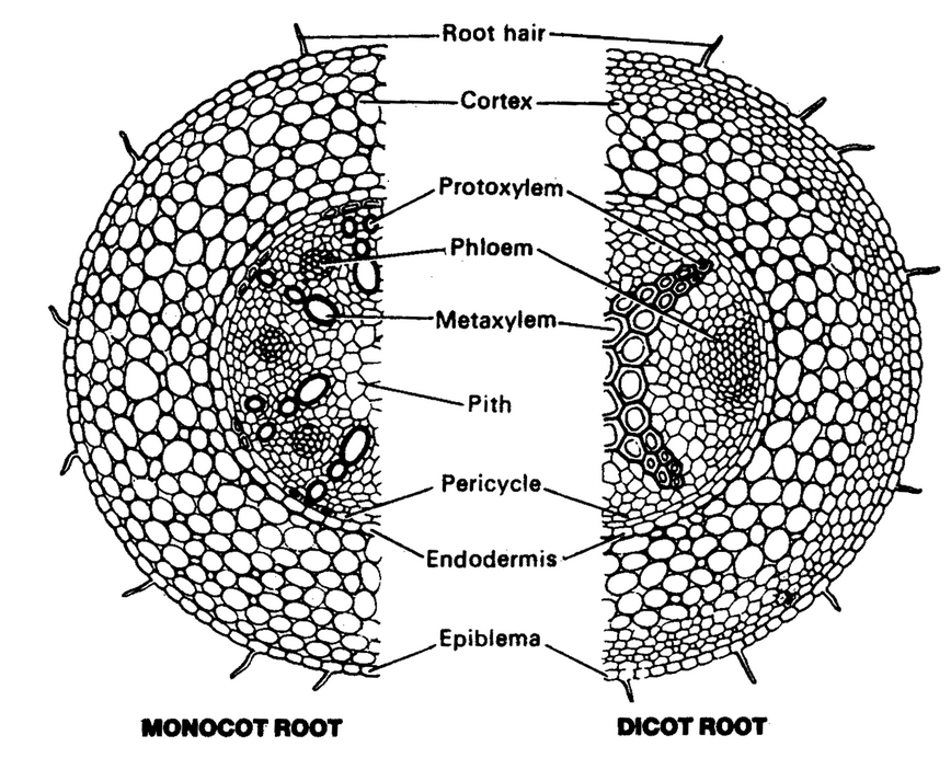
The arrangement of tissues from the periphery to the centre of a primary root is as follows:
Epiblema
This is the outermost layer of tightly placed thin walled and non cutinized cells. The outer wall of the cells of epiblema gives rise to unicellular root hair. These hairs help in absorption of water from the soil.
Epiblema is usually uniseriate, but in aerial roots of some orchids and epiphytic aroids multiseriate epidermis, called velamen, is present.
Cortex
The root has a histologically simple cortex usually composed of thin walled isodiametric parenchymatous cells with many intercellular spaces. The cortical cells are usually devoid of chloroplasts but they store starch.
Some roots (e.g., Iris) develop a specialized layer beneath the epiblema, called exodermis. It arises from subepidermal layers of the cortex. The cells of exodermis have suberized cell walls.
Endodermis
Endodermis delimits cortex from the stele. It is composed of closely packed living cells: which are characterized by the presence of casparian strips in their radial walls.
Some cells of endodermis, usually the ones which lie opposite to protoxylem, remain thin walled. These cells, called passage cells or transfusion tissue allow radial diffusion of water.
Anatomical differences between dicot root and monocot root
|
Character |
Dicot root |
Monocot root |
|
Pericycle |
Gives rise to lateral roots, phellogen and a part of vascular cambium. |
Gives rise to lateral roots only. |
|
Vascular bundles |
Usually 2-6. (Exception Ficus bengalensis having 14 v.bs.). Vessels appear angular or polygonal in transverse section. |
Usually 6-20. (Exception Allium cepa having 4 v.bs.) Vessels appear oval or rounded in transverse section. |
|
Conjunctive tissue |
Parenchymatous; forming the most part of vascular cambium. |
Mostly sclerenchymatous but not forming vascular cambium. |
|
Cambium |
Develops later on. |
Does not develop. |
|
Pith |
Absent or poorly developed. |
Large, well developed and parenchymatous. |
|
Secondary growth |
Occurs |
Does not occur. |
Pericycle
Pericycle occurs beneath the endodermis and it forms the outer boundary of the primary vascular cylinder of the root. It is usually a single layer of cells.
Vascular system
The vascular bundles of roots are radial with alternating strands of xylem and phloem separated by parenchymatous conjunctive tissue. The xylem is exarch.
In dicot roots, the number of xylem bundles usually varies from 2 to 6 and in monocot roots, it usually ranges from 6 to 20. Depending on the number of xylem bundles, roots are designated as monarch (with one xylem bundle), diarch (two bundles), triarch (three bundles), tetrarch (four bundles), pentarch (five bundles) or polyarch (many bundles).
Pith
Pith occupies a very small area in the centre of the root. It consists of thin walled parenchymatous cells with intercellular spaces.
In many dicot roots, due to the development of metaxylem vessels in the centre, pith is obliterated.
secondary growth in dicot root
