
Human Eye
Share
Human Eye
Sensory System of Class 11
A fluid-filled ball with about 21 mm (17.5 mm in just born) antero-posterior diameter (optical or visual axis) lodged in the bony orbits of sphenoid bone with a cushion of fat.
About 1/6th of eye ball (1/3rd in frog and 1/5 in rabbit) projects outside while the rest part remains within the socket. This anterior part is covered with transparent cornea. Each eye is protected by upper and lower-eye lids with eye lashes.
Eye Lids (Palpebrae)
The extended folds of skin at the dorsal (upper) and ventral (lower) edges of the orbit are beset with eyelashes. Its under surface is soft epidermal layer like a mucous membrane that slides over the cornea.
The upper or 1st eyelid is larger and movable (in frog lower eyelid is larger).
In fishes the eyelid is absent while in snake it is a fixed transparent cover upon the eye ball.
A thin epidermal layer called conjunctiva is extended over the cornea between base lines of the eyelids, when infected causes conjunctivitis (or red eye).
Nictitating membrane (3rd eyelid) is a transparent, semilunar, secondarily folded upon the cornea. It provides additional protection to the eye in water, bushes or burrows. It is vestigeal in man as a small muscular elevation in the inner corner of eye called as plica semilunaris.
Eye lashes are the hairs present on the margins of both the eyelids to keep the dirt away.
Eye (or Ocular) Muscle
Three pairs extraocular muscles attached to the external surface, (the fascia) of the eye ball move it in different directions. These are superior and inferior rectus; anterior and posterior rectus; superior and inferior oblique muscles.
If any pair has one muscle of unequal length it causes the defect called squint eye (or strabismus).
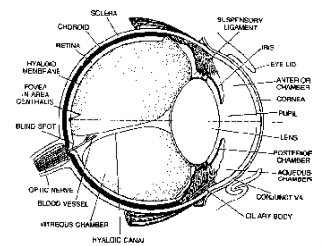

Fig. L.S. of Human Eye (Diagrammatic) Fig. Extraocular muscles of human eye
Associated Glands
Lacrimal gland (tear gland): Consists of chain of three small glands located in the outer angle of each eye. Tear flows into the lacrimal duct through small pore, lacrimal punctum to pass into the nasal chamber of its side.
Tear is a salty fluid, that contains amino acids, glucose and lysozyme.
A continuous layer of tear is always present upon the cornea to keep it moist, and away from the dust and other foreign particles. It also nourishes cornea and acts as a medium for refraction of light; lysozyme in it is antibacterial. In human tear secretion begins after four months of birth.
Tearing is stimulated by injury, irritation, emotions etc.
- Meibomian gland (tarsal gland) is a modified sebacious gland located on the margin of eyelids. Its oily secretion makes a film over the tear to prevent it’s fall from the surface of cornea.
- Zeis gland is also a modified sebacious gland in the hair follicle of eye-lashes to keep them smooth and water proof by its oily secretion.
- Glands of Moll are the modified sweat gland on the inner surface of eyelids found only in human, of uncertain function.
