
Structure of Human Eye
Sensory System of Class 11
Structure of Eye
The eye ball is made of three layers: (i) Sclerotic coat or Sclera (ii) Choroid layer (iii) Retina
Sclerotic coat (fibrous tunic):
The outermost fibrous layer, also called as ‘white of eye’, the frontal exposed part is transparent non-vascular cornea which receives nutrition from aqueous humour.
It provides shape and support to the eyeball and also surface (fascia) for the insertion of eye muscles.
The circular line of joint between sclera and cornea is called limbus or sclero-corneal junction.
In frog the sclera is cartilaginous while in some birds it is bony.
Choroid layer (Vascular tunic or Uvea)
The middle, pigmented and vascularised layer of the eyeball
makes the nutritive and screen layer preventing the entry of light
.png)
Anteriorly thin distinguishes as ciliary body and iris.
At the circular margin near cornea the ciliary body projects into the interior of eyeball as thick bulging part with blood vessels and ciliary
muscle helps in suspending lens through suspensory ligament (zonula of Zinn).
Ciliary muscles control the curvature of lens to focus objects at different distances.
From blood capillaries in ciliary body the substances from blood pass into aqueous humour which is collected back through canal of
Schlem into the venules.
Blockage of the canal of Schlem causes rise in the pressure of aqueous humour which is a disease called glaucoma.
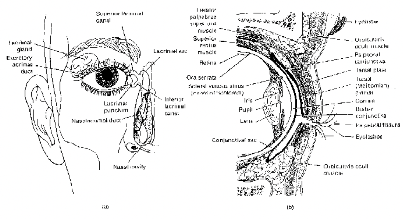
Fig. Detailed view of anterior cavity
Iris (arrows show the flow of aqueous humor)
Front part of choroid layer beyond ciliary body below cornea is a circular diaphragm called iris with an aperture in the middle called pupil through which light enters the eye.
Two sets of antagonistic muscles in the iris, circular muscle and radial muscle decrease and increase the pupil diameter respectively according to the amount (intensity) of light.
Tapetum
In many vertebrates the innermost layer of choroid, close to retina, is specially modified to reflect the light back upon retina, it gives glare (or shine) in the eye.
In deep sea elasmobranchs and many mammals it contains light reflecting crystals of guanin hence called tapetum lucidum.
In carnivores and lower primates the tapetum layer has crystals of other organic substance and called tapetum cellulosum.
The tapetum helps in vision in darkness and also shows a means of aggression.
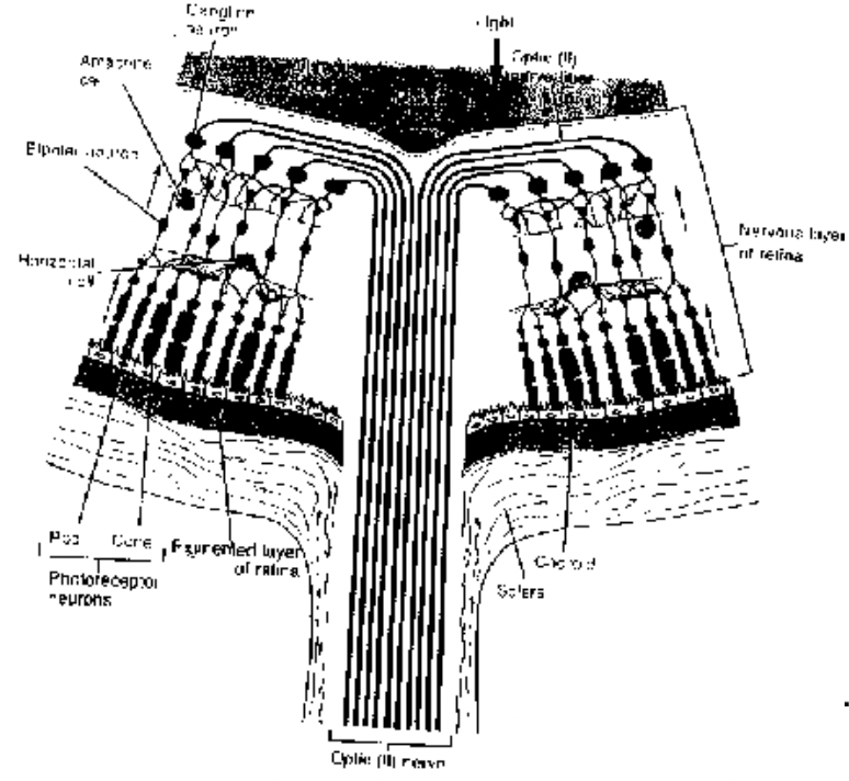
Retina (Neurosensory layer)
The innermost epithelial layer has 3 parts, pars optica, pars ciliaris and pars iridica. The margin of pars optica which extends over ciliary body is serrated and called ora serrata. Organised in 10 layers the outermost cell layer is of pigmented cell followed by sensory receptor (rod and cone) cell layers and four types of neurons including bipolar neuron and ganglionic neuron whose axons bunch form optic nerve. Other neurons are horizontal cells and amacrine cells.
The rods and cones synapse with bipolar cells, which on other end synapse with ganglionic neurons. These junctions form the outer plexiform layer and inner plexiform layer respectively.
Horizontal cells (always inhibitory) connect one receptor cell to other in the outer plexiform layer, hence generate ‘lateral inhibition’.
Amacrine cells connect one ganglionic neuron to another in the inner plexiform layer. More than 30 types have been identified so far.
Muller cells (glial cells) are present in between these cells and provide them with support, nutrition etc.,
Ganglionic neurons are of 3 types : W (40%), X (55%) and Y.
The site on retina where axons converge to form optic nerve which pierces out of eyeball is called blind spot since no image formation takes place here for no sensory cells in this part.
There is a yellow spot on retina which matches the line of optical axis of lens is called macula lutea having only cone cells, in the middle of it there is a shallow depression called fovea centralis, the spot for sharp vision.
Rod cells are rod-shaped, filled with visual purple (or rhodopsin) associated with dim light vision. Cone cells are small, cuboidal filled with visual violet (or iodopsin) associated with bright light and color vision containing three types of color pigments these are of 3 types : erythrolabe, chlorolabe and cyanolabe with red, green and blue colours respectively.
In color-blind persons the red or green cones may be either missing or very less in number. When red cones are absent (= protanope) the red object appears green and in absence of green cones (= deuteranope) the green color appears red.
In nocturnal animals rod cells are more numerous while in diurnal animals it is opposite, e.g. in owl and bat cone cells are absent while in crow, sparrows, squirrel only cone cells are present.
In human retina rod cells (11-12 crore) are much larger in number than the cone cells (65-68 lakh). The number of cone cells in fovea centralis is 15 lakh/mm
.png)

Lens
A biconvex ectodermal transparent structure enclosed in a capsule remains behind the pupil attached with the ciliary body by suspensory ligament.
Receives nutrition from aqueous humor.
It divides the eye ball into two chambers:
- Anterior aqueous chamber, containing aqueous humor (watery fluid) with glucose, amino acids and salts constantly replaced, provides nutrition to lens, cornea and others and also acts as refractive medium.
- Posterior vitreous chamber with jelly-like vitreous humor containing collagen, vitrein protein and hyaluronic acid. A narrow hyloid canal runs obliquely through it upto blind spot, provides support to the eye ball from its interior. Once formed during embryonic development it remains through out. Cataract Either due to disease or aging the denaturation of protein of lens makes it opaque to obstruct the entry of light through it, corrected by replacing defective lens by artificial lens.
