
Development of Mammal
Share
Development of Mammal
Embryonic Development of Class 12
Development of Mammal
Follows same basic plan and sequences of events as that of frog.
Due to different type of egg and viviparous development there are differences.
Being amniote fetal membranes, amnion, chorion, yolk sac and allantois develop.
Cleavage
Holoblastic-equal type and the pattern is biradial, indeterminate type.
Takes place during passage of zygote through fallopian tube to uterus.
During 2nd cleavage division one of the two blastomeres divide little later resulting in 3-cell stage for a short while. But, soon typical 4-cell stage appears and subsequently morula is formed.
In many mammals zona pellucida remains intact housing the morula.
Developing embryo acquires nutrition from the uterine fluid/blood of mother from the very beginning.
Blastula formation
Rearrangement of blastomeres form blastula (blastodermic vesicle) with blastocoel.
At this stage the zona pellucida disintegrates and rapid growth of embryo begins.
The blastocoel signifies :
- The site where ancestral embryo used to lodge nutrients as yolk.
- Space used to acquire more and more fluid with increasing dimension of the embryo. This results in the expansion of outer layer of blastodermic vesicle (also called blastocyst) as thicker layer.
After one week of fertilization, blastocyst get implanted in the uterine wall.
Peripheral cells of morula form trophoblast or trophectoderm which draws food for the embryo from uterine wall. This has only trophic and protective function and forms foetal membranes and part of placenta, has no role in the formation of embryo.
Inner cell mass forms body of the embryo, initially distinct as outer epiblast and inner hypoblast. The trophoblastic cells above the epiblast is called cells of Rauber.
Now, the blastula develops trophoblastic villi and gets attached to the endometrium of uterus.
The uterine wall at this time undergoes profound changes to form decidua, distinct as :
- Decidua basalis – between embryo and uterine serosa
- Decidua capsularis – between embryo and lumen of uterus
- Decidua parietalis – remaining part of decidua.
Gastrulation
The sequence of forming respective germinal layers is different from frog, the order is as follows:
Formation of Endoderm:
With increasing size of the inner cell mass and blastodermic vesicle a group of cells detach (delamination) from here and push into the blastocoel, get arranged along the inner wall of blastocoel, divide rapidly and forms a complete inner layer of endoderm.
Now the erstwhile blastocoel becomes the lumen of archenteron. This differentiates into two parts – one within the body of embryo, forming gut and the other projects out as distal yolk sac.
After shifting of endodermal cells the rest part of inner cell mass forms embryonic disc.
Formation of Mesoderm:
At the caudal margin of embryonic disc the cells divide rapidly to become thick mass.
Subsequently, these cells start migrating in the area behind endoderm to form mesoderm.
Formation of Ectoderm:
After the formation of mesoderm the remaining cells of the embryonic disc proliferate and get arranged in a layer outside the mesoderm to form ectoderm.
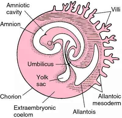
Fig.Early stages of embryonic development in a mammal
Organogenesis
Different parts/organs of the body are formed by further continuation of differentiation (specialization). Localized groups of cells are sorted out in an orderly manner with unique precision. After gastrulation the first phase is neurulation, during which neural plate is formed by the involution of presumptive neural ectoderm cells.
By the interaction of these ingressing cells the foundation of organogenesis is laid. This process is called induction. Such cells which induce the differentiation of organ forming cells are called organizer or inducer. The organizer concept was introduced by Spemann (1869-1941) and got Nobel Prize in 1935.
Structures and organs of the adult body gradually take their characteristic shapes and forms.
