
Placenta
Share
Placenta
Embryonic Development of Class 12
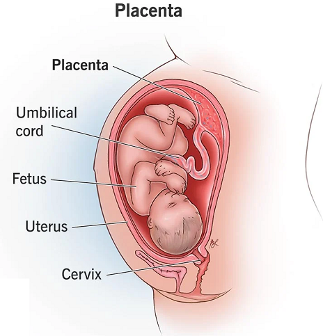
Placenta
Placenta is an organ which establishes physiological connection between mother and foetus through which only selective exchange of substances from blood takes place. It doesn’t allow the direct passage of maternal blood into foetus body because :
Mother’s blood contains variety of substances (e.g. nitrogenous wastes, protein and other chemical) that may harm or kill the embryo.
The blood of mother at high pressure may destroy the vital organs of embryo in the beginning.
The female hormone in mothers blood will not allow the male baby to develop.
This is formed as an important apparatus in viviparous development in mammals during pregnancy. It acts as ultrafilter ; soluble organic and inorganic substances, nutrients, some hormones, antibodies against diphtheria, smallpox, scarlet fever can pass through it. Respiratory gases and nitrogenous waste also pass through it.
As endocrine gland it synthesises large quantities of protein and becomes the major source of progesterone itself.
Secretes hCG (human chorionic gonadotropin), chorionic thyrotropin, corticotropin, somatotropin, mammotropin, estrogen.
hCG level in urine is indicator for pregnancy test, it also stimulates corpus luteum to continue to secrete progesterone until the end of pregnancy.
In the last stage of pregnancy it secretes relaxin that facilitates childbirth (parturition).
The umbilical cord connects the foetus to the placenta.
After implantation various stages and types of placenta are formed in eutherians as follows :
Trophoblastic villi
This is formed as temporary connection with uterine wall in blastocyst stage for some time later replaced by yolk sac placenta.
Yolk sac placenta (or choriovitelline placenta) : The yolk-sac is more developed and fused with chorion, only found in Metatherians.
Allanto-chorionic (or allantoic) Placenta :
The true placenta, formed by the fusion of allantois with chorion ; forms corresponding villi with uterine wall, thus both maternal and foetal parts contribute, however, there is no fusion of foetal and maternal blood.
In different mammals the structure varies according to the level of contacts between maternal and foetal parts.
The types of placenta on this basis and the pattern of distribution of villi on the body surface of embryo, are depicted in the following chart.
Types of Placenta
Classification of placenta according to the nature of the foetal membranes involved
- Yolk sac placenta (chorion-vitelline). Placenta is formed from yolk sac and chorion, e.g., Kangaroo and opossum (both are metatherians or marsupials).
- Chorioallantoic placenta. Placenta is derived from allantois and chorion; e.g., most eutherian mammals.
- Chorionic placenta. Placenta is formed from chorion, e.g., human beings.
Classification of placenta according to the histology. Six tissue barriers in placenta are :
- Endothelium of foetal blood vessels
- Foetal connective tissue
- Trophoblast
- Uterine epithelium
- Uterine connective tissue and
- Endothelium of maternal blood vessels.
Five histological types of placenta are present:
- Epitheliochorial placenta. Simplest type. All six tissue barriers (layers) of the placenta are present, e.g., horse, ass and pig.
- Syndesmochorial placenta. Uterine epithelium is absent; with five placental barriers, e.g., cow, sheep, goat, buffalo, camel and giraffe.
- Endotheliochorial placenta. Uterine epithelium and uterine connective tissue are absent; with four placental barriers, e.g., carnivores (dog, cat, lion, tiger, fox, bear and mongoose).
- Haemochorial placenta. All the three uterine tissue barriers (uterine epithelium, uterine connective tissue and endothelium of maternal blood vessels) are absent; with three placental barriers, e.g., lemur, apes, monkeys and men.
- Haemoendothelial placenta. All the three uterine tissue barriers and two foetal tissue barriers (foetal connective tissue and trophoblast) are absent; with only one placental barrier, e.g., rabbit, rat and guinea pig.
Classification of placenta according to the fate of uterine placenta
- Non-deciduate placenta. No part of uterine placenta is shed in the after birth, e.g., horse, ass and zebra.
- Deciduate placenta. Some part of uterine tissue is passed out as decidua in the after birth, e.g., humans.
- Contra-deciduate placenta. Placenta is non-deciduate and even the foetal placenta is absorbed, .e.g., Talpa (mole) shrew and Perameles (bandicoot).
Classification of placenta according to the distribution of villi on chorion
- Diffuse placenta. The villi remain scattered all over the surface of the chorion, pig. horse and lemur.
- Cotyledonary placenta. The villi are arranged in separate tufts or patches called cotyledons, e.g., cow, goat, sheep and deer.
- Intermediate placenta. The villi are arranged in cotyledons as well as scattered, e.g., camel and giraffe.
- Zonary placenta. The villi form an in- complete (example racoon) or complete girdle encircling the blastocyst, e.g., cat, dog, seal and elephant.
- Discoidal placenta. Villi occur on a small disc-shaped area of the blastocyst, e.g., rat, rabbit, bear and bat.
- Metadiscoidal placenta. Villi first occur all over but later become restricted to one or two discs, (a) Mono Discoidal placenta. Villi are restricted to one circular disc, e.g., rabbit and man. (b) Discoidal placenta. Villi are restricted to two discs, e.g., monkey and apes.
Pregnancy
In first trimester (3 months) basic structure of the body of embryo is formed with all systems.
Viral infection (e.g. by Rubella) causes German measles in the foetus.
Exposure of certain chemicals (teratogens) may cause abnormalities in the embryo. e.g., phocomelia.
Childbirth Occurs in Three Stages
Illustrates the three stages of childbirth, dilation, expulsion, and placental.
Stage 1: the dilation stage, gets its name from the dilation of the cervix. This phase begins when uterine contractions start and generally lasts 6–12 hours; but it can last much longer.
Stage 2: the expulsion stage, begins after the cervix is dilated to 10 centimeters and the baby is engaged. At this time, uterine contractions usually occur every 2 or 3 minutes and last 1–1.5 minutes each.
Stage 3 : The final stage of delivery is the placental stage. It results in the expulsion of the placenta. The placenta remains attached to the uterine wall for a short while. It is then expelled by uterine contractions, usually within 15 minutes of childbirth.
Changes in baby
Expansion of lung and start of breathing.
Closure of ductus aorticus and foramen ovale.
Adult pattern of blood flow through heart, aorta and pulmonary arteries.
In some babies these switch over may not be completed due to inadequate synthesis of nitric oxide.
Secretion of Milk
3-4 days after delivery breasts begin to secrete milk in sufficient amount, stimulated by prolactin.
Its release from the teats is stimulated by oxytocin or due to suckling stimulus by baby.
Milk contains an inhibitory peptide. If breasts are not fully emptied peptides accumulate and inhibit further production of milk.
The first milk, colostrum, secreted just after the child birth contains lots of proteins and antibodies that provide passive immunity to the newborn.
