
Electrical Instruments
Technology for Medical Applications of Class 12
Both nerve and muscle cells show electrical property of excitability and conductivity. They transmit information in the form of action potentials,
which in turn produce measurable electric signals that can be detected at the surface of the body by sensitive electronic circuits called biopotential amplifiers.There are various instruments to record such potentials or follows :
Electrocardiograph
It is used to record ECG which is the combination of electrical and mechanical signals a result of stimulation and contraction of the heart muscle during cardiac cycle.
Einthoven (1906), first used string galvanometer to record the human heart activity.
The simplest type of ECG monitor is a cardioscope. Its graphical record is called electrocardiogram. Each part of the cardiac cycle produces its own characteristic peak denoted by P, Q, R, S and T.
P-wave denotes atrial activity i.e. depolarisation at SA-node followed by atrial systole. QRS complex denotes ventricular activation by depolarisation at AV-node followed by ventricular systole R is the tallest wave.
T-curve gives ventricular recovery or repolarisation, and also considered as pause. PR-interval of 0.16 second and QT-interval of 0.32 second show the normal state Used to detect the rate, rhythm and condition of heart beat, and can help diagnose any deviation from the normal pattern like cardiac arrhythmia, heart block, asystole hypertrophy of cardiac muscle, ischaemia, myo-cardial infarction. Echocardiography is used for obtaining an image of the structure of heart using ultrasound. Doppler echocardiogrpahy allows indirect measurement of the flow (velocity) of blood as it passes through the heart.
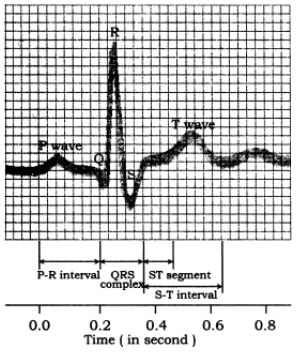
Fig. Electrocardiogram
Electromyograph (EMG)
Recording of electrical activity in muscle is used to detect disorders such as muscular dystrophy or defect of innervation such as neuropathy or reticulopathy.Actual site of nerve damage can be located in case of nerve injury.
Electroencephalograph (EEG)
Discovered by Hans Berger (1929); used for recording electrical signals from brain. Small electrodes connected to amplifier are stuck to the patients scalp which receive the signals and feed into EEG machines, recording takes about 45 minutes. Waves of different types are generated as a result of synchronized activity of millions of neurons
of the cerebral cortex.
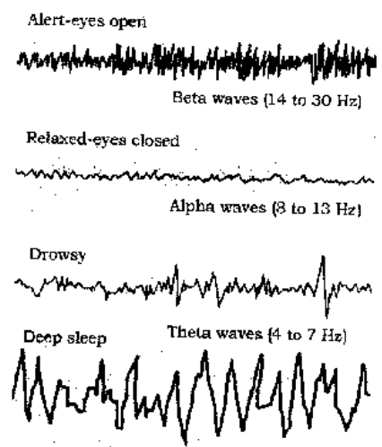
(alpha) waves : Low frequency (8-13 Hz) wave appear during relaxed state of mind or closed eyes.
(beta) waves : High frequency (14-30 Hz) waves occur in normal alert or awaken state, associated with mental activity in frontal area of brain. Still higher frequency (30-80 Hz) waves show highly alert brain or focussed on something.
(theta) waves : Low frequency (4-7 Hz) waves shows drowsiness or early phase of sleep or appears during emotional stress, also found in children between 2-5 years age.
(delta) waves : Lowest frequency (0.5 to 3 Hz) occur during deep sleep or in the infants. Used for diagnosing conditions like epilepsy, encephalitis, dementia, brain tumor, depth of anaesthesia; also detect brain death, effects of drug on brain and metabolic disorders, sleep disorders like insomnia, somnambulism, narcolepsy etc. During REM sleep -waves appear.
During epilepsy or pathological brain condition a short duration high frequency (30 Hz) waves of high voltage appears followed by abnormally low frequency and low voltage waves.
SQUID (Super conducting quantum interference device) is also known as magneto-encephalography (MET) helps for studying even weaker magnetic fields of brain.
Polygraphy
Polygraph, a simple, portable multichannel monitors records qualitative changes in many physiological parameters at a time, like, cardiac variables (ECG), heart pulse rate (HR), relative blood pressure (BP), rate and depth of breathing, and the resistance of skin to the conduction of electricity, activity of eye (electroculograph) Used as lie detector, monitoring stages of sleep, and electrophysiological behaviour of the brain and its dysfunction.
Autoanalyser (automatic analysis by continuous flow)
Computerised instrument, used for biochemical analysis of samples of blood, urine and other body fluids like cerebro-spinal fluid, saliva, sweat etc. to estimate glucose, urea, cholesterol, enzymes and other proteins.
Biochemical tests are performed through electrical sensors (electrodes) to assess the function of particular organ like liver or kidneys. Has full control of reaction temperature, sample volume, reagents, and transfer of fluids. Timing of these functions are supervised by micro-processor.
Endoscopy (end : within; skopein : view) Endoscope consists of long flexible tube attached to handset. Tube is inserted through an opening in the body, and its tip is ‘steered’ to its destination.
Bundles of optical fibres transmits light to the tip where an image is formed on an array of light sensitive cells, called charge coupled device (CCD). Photoelectrical signals through camera are sent to the videomonitor where a magnified image is seen. One channel carries water and air to wash and dry the surgical area. Miniature surgical instruments like forceps and microscissors are carried to the site of surgery through a parallel channel, controlled by a cable.
Gastroscope : used to examine stomach for an ulcer.
Laparoscope : detect cysts or infections of the uterus, fallopian tube and ovaries.
X-ray Radiography
Discovered by Roentgen (1895); also known as Roentgenography. Use of X-ray to take photograph called radiography provides two dimensional image of the internal body parts.
Tissues of different densities shows different images on the radiography. A more dense tissue absorbs or obstructs more X-rays causing opacity. Less dense tissues are radioluscent.
Bones, tooth being radio-opaque are most suitable for study by this technique. Helps to diagnose pulmonary tuberculosis, pneumonia, stomach ulcers, intestinal obstruction, stone in gall bladder, urinary bladder, ureter and kidney etc. Hollow structures like gut, blood vessels can be viewed by filling them with certain chemicals like salts of barium, iodine etc.
Drawbacks
Image of different organs get superimposed on to the flat sheet of film. Hazard of causing mutation and cell injury leading to cell death.
Angiography (angeion-vessel; graphein – to record) X-ray is, coupled with image intensifiers to provide real image, which can be viewed by CRT (cathode ray tube) during video monitor surgery.
Provides view of blood flow in vessels and indicates the presence of blockages. A modern version of it is digital subtraction angiography (DSA) in which angiograph of heart or blood vessels is taken and stored in computer. Then 2nd angiograph is taken after injecting a contrast (radio-opaque) agent like iodine into blood. First image is digitally subtracted from the second, which provides a clear outline of the blood flow to heart, brain or kidneys.
CT-Scan (Computerised tomographic scanning) or CAT-Scan (Computerised axial tomography)
Sir Godfrey N. Hounsfield and Allen M. Cormack received Nobel Prize (1979) for their discovery of CT-Scan while, theoretical basis was
given by G. N. Ramachandran Modified X-ray imaging in which computer reconstructs the image of organ or parts seen. It gives 3-D view of cross-section of an organ in a series.
Advantage of CT-Scan over X-rays
Tissues having minor differences in their densities can be recognised as separate entities.
Less hazardous; no superimposition of images. Helps in diagnosis of diseases of highly sophisticated parts i.e. brain, spinal cord and localizing the exact position depth, size etc.
Helps in tumour identification, determining the feasibility of operative treatment.
PET Scan (Positron Emission Tomographic Scanning)
Developed by Louis Sokoloff (1985). Diagnostic technique based on detection by positrons (positive electron) emitted from radio-isotopes like C11, N13, or O15, generated by the cyclotron.
These isotopes are incorporated into biological molecules like glucose, amino acids, CO2 and NH3 through blood-drip. These substances are found in higher concentration in the parts/tissues that are metabolically more active.
Magnetic Resonance Imaging (MRI)/ Nuclear Magnetic Resonance (NMR)
Developed by Felix Bloch and Edward M. Purcell (1946) independently Provides the best pictorial information without exposing the patient to hazardous effect of radiations.
Magnetic resonance is generated by subjecting the nuclei of H-atom to strong external magnetic field. H-atoms of the water molecules form the most abundant source of protons in the body, hence detects, water content of the tissues. As biological systems have abundant H-atoms, NMR signals produced are picked up by the receiver coils and converted to digital signals fed into the computer for image construction. The patient is laid in the centre of the magnetic rings where a very strong magnetic field, (about 70,000 times stronger than that of the earth) is generated by using large water cooled resistive magnets or super-conductive magnets with liquid helium. Teeth and bones, which contain little water do not appear in MRI. Used to detect tiny lesions of multiple sclerosis of brain and spinal tissue; to examine joint injuries and slip-disc in vertebral column; can also visualise cancerous tumors. Lauferbur Mansfield received Nobel Prize (2002) for use of MRI in medical diagnosis.
NMR (MRI) superior to CT-Scan because
Forms images from various angles while CT-Scan give mainly cross-sectional images.
Does not expose patients to hazards of ionizing radiations. Using spectroscopy, tissue metabolism can be studied by NMR.
Provides better differentiation among soft tissues. Nuclei of C, Na and P can also be used instead of H nuclei. Provides 3-dimensional images that reflect the metabolic and chemical activity of tissues being studied.
Used for measuring regional cerebral blood volume, blood flow, metabolic rates, levels of glucose and O2. detecting tumours, locating the origin of epileptic activity within brain and for examining brain function in various mental illnesses, locating colour processing centres in visual cortex of brain.
Disadvantages : This technique is very expensive, not widely available, does not shows the image of bone and calcium.
Ultrasound Scanning or Sonography
High frequency sound wave (1-15 million Hz) is generated by piezo-electric effect when electric potential is applied to the crystals of lead zirconate titanate. Ultrasound waves when applied over the surface of any organ/tissue it gives echoimage of that organ/tissue. Echo-picture of solid organs is always better than air filled cavities like pleura, lungs. Used for diagnosis of stones in gall bladder, biliary tract, kidney, ureter etc. lesions, cyst, tumour (solidification) or cirrhosis of liver. Detection of pregnancy and disease of uterus like endometriosis, fibromyoma, carcinoma of ovary
