
Some representatives of Nematoda
Classification of Animals Non Chordates of Class 11
Rhabditis
Many species of Rhabditis are free-living & semi-parasitic forms. Rhabditis maupasi lives in soil.
The males are smaller than females.
The male has a single tubular testis, however the female has two ovaries.
Life cycle is simple & direct.
Enterobius (Oxyuris)
Enterobius vermicularis is commonly known as pinworm.
It is most common parasitic nematode of man throughout the world. Its incidence, however, is greater in women & children.
Adult worms live in the caecum, appendix and colon.
Sexes are separate. Males are short & they have curved end with a single spicule. Females are long & they have straight pointed end.
The mail is monorchic & the female didelphic. Life cycle direct.
The anal itching takes place in Enterobius infection because female worm lay eggs in perianal region.
Pinworm causes entrobiasis involving loss of appetite, insomnia, restlessness, abdominal pain & appendicitis.
Ancylostoma (Hookworm)
Most dangerous parasitic round worms of man. They differ from Ascaris in the absence of lips but they have three chitinous plates that bear sharp cutting teeth.
Hookworms flourish under primitive conditions where people go bare foot, modern sanitary conditions do not exist & human faeces are deposited in the ground. Larva enters the human body from moist soil by boring through the skin of the feet.
The adult worms live parasitically in the intestine of man where they suck blood, lymph, bits of mucous membrane & tissue fluids from the lining of the intestinal wall.
It causes acute anaemia due to extensive injury to the host intestinal wall. It also retards growth of the body in the children. It causes mental & physical deficiency. The disease is called ancylostomiasis.
Wuchereria (Filarial worm)
Wuchereria bancrofti lives as an endoparasite in the lymphatic vessels & lymph nodes of human beings.
It is a digenetic parasite. Human being is a primary host whereas Culex or Aedes mosquito is the intermediate host.
Wuchereria feed on blood and lymph.
The female gives rise to juveniles called microfilariae.
When mosquito bites the infected person these juveniles reach the mid gut of the mosquito along with blood. After penetrating the stomach wall they reach the haemocoel and finally migrate to the proboscis of
the mosquito. When the infected mosquito penetrates its proboscis into the skin of the human being, the juveniles leave the proboscis, and then reach the lymph glands through the lymphatic and blood vascular system.
Sexes are separate. The female is larger than male. Tail of male is coiled like tendril and has a pair of unequal copulatory spicules. Female has an anus while male bears cloacal aperture.
In the lymph glands, the juveniles develop into adults. It blocks the lymphatic system resulting in inflammation of lymph glands and other pathological conditions.
It causes a disease known as elephantiasis in the legs and arms.
Dracunculus medinensis
Commonly known as guinea worm. Occurs in subcutaneous tissue of man. The toxic secretions of the parasite causes a blister on the skin of the host. It is a digenetic parasite. The primary host is human being & Cyclops (water flea) is the intermediate host. Sexes are separate. The parasite causes itching, diarrhoea, nausea, vomiting & eosinophilia.
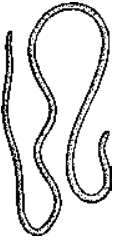
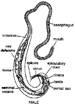
Fig. Dracunculus medinesis Fig. Trichuris trichiura (Male)
Trichuris (Whip worm)
The parasite resides in the caecum, appendix & colon of man, especially children, in warm moist climates. The infection spreads through contaminated food & water containing embryonated eggs. Its infection causes abdominal pain, diarrhoea & anaemia.
Ascaris (Round worm)
It is a monogenetic parasite, occurs in the human small intestine. It is more common in children. The food of the worm consists of semi-digested food of the host, the blood & the fluid of the alimentary canal of the host.
External Morphology : The body is elongated & cylindrical, tapering at both ends. Running along the entire length of the body are four longitudinal lines-one mid-dorsal, one midventral & two lateral. These
lines are impressions of syncytial epidermis which is much thickened all along the length.
The mouth is triradiate aperture, guarded by three broad lips, one mid-dorsal & two sub-ventral. The lips are denticulate & bear sensory papillae. The drosal lip has two double sensory papillae & each ventro-
lateral lip has one double sensory papilla. Each ventrolateral lip bears towards its lateral side, a single papilla, near which lies a reduced papilla known as amphid. The amphids are chemoreceptors whereas
other sensory papillae are tangoreceptors.
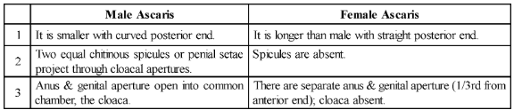
The excretory pore is situated mid-ventrally, a little behind the mouth. Sexes are separate. The differences between male & female Ascaris are:
Body wall. The body wall is made up of 3 layers, an outer cuticle, a middle epidermis and an inner layer of longitudinal muscles.
Circular muscles are altogether absent.
Digestive system.
It is complete. It extends from mouth to anus. It comprises of a short pharynx or oesophagus representing the fore-gut, a long intestine or
midgut, & a short rectum or hind-gut. The fully or partially digested food is sucked by muscular pharynx. Digestion is completely extracellular. Excess of food is stored mainly as glycogen & a little fat in the syncytial epidermis. The intestinal cells may digest food intracellularly. Food of Ascaris is chyme. Ascaris secretes amylase, protease and lipase enzyme to digest the food material.
Respiration : Usually anaerobic.
Excretory system. They excrete through H-shaped longitudinal excretory canals (also known as renette cells). The worm is ammonotelic & ureotelic.
Nervous system :
The nervous system comprises of:
(a) a richly ganglionated nerve ring or circumenteric ring encircling the pharynx, and
(b) nerves extending anteriorly as well as posterioly from the nerve ring. The ganglia located on the nerve ring are :
(i) A single dorsal ganglion.
(ii) A pair of sub-dorsal ganglia, one on each side of the dorsal ganglion.
(iii) A pair of lateral ganglia, each subdivided to form six lateral ganglia.
(iv) A pair of large ventral ganglia.
Eight anterior & eight posterior nerves extend from the ring.
Reproductive system.
Sexes are separate. The differences between male & female Ascaris has been discussed earlier.
Male Ascaris is monorchic (single testis). Testis is telogonic (gametogonia are budded off from proximal end of the gonad & undergo gametogenesis). The spermatozoa are amoeboid.
Female Ascaris is didelphic (two ovaries). Ovaries are telogonic. The ova are amoeboid in shape.
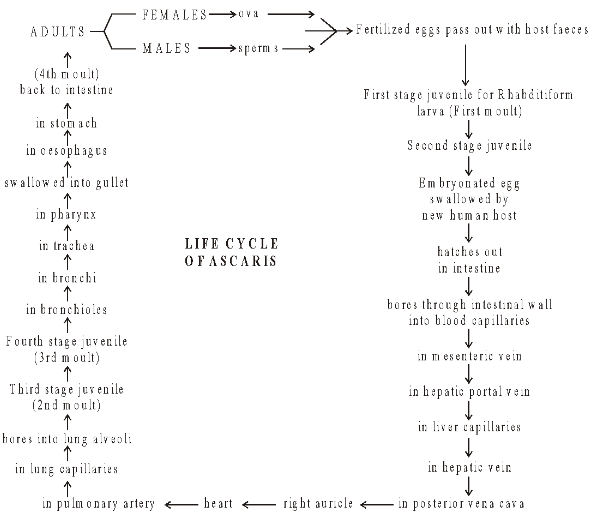
Life History
Copulation takes place in the small intestine of the host. After fertilization zygotes reach the uterus. Zygote are oval & brown coloured enclosed within chitinous sheath. Outermost albuminous coat is secreted by
uterus, which is warty. It is called mammilated egg. It has lipids & glycogen globules as reserve food material. Mammilated egg is covered by three layers. The innermost layer is made up of lipid; middle layer
is made up of chitin whereas outermost layer is made up of albumen. Only outermost layer is secreted by wall of uterus, remaining layers are secreted by fertilized egg. Cleavage takes place outside the body.
Cleavage is holoblastic, spiral & determinate.
Parasitic Adaptations :
(a) Protective cuticle.
(b) Anaerobic respiration.
(c) Simple digestive system.
(d) Simplest receptors.
(e) Profuse breeding capacity.
Parthogenesis : Ascaris causes ascariasis. Symptoms are nausea, vomiting, diarrhoea, loss of appetite, insomnia. In acute infections it may affect nervous system causing epilepsy, unconsciousness, may also cause appendicitis.
Therapy : Chenopodium oil is commonly used to get rid of Ascaris. Other anti-helmintic drugs are alcopar, antipar, etc.
