
Biochemical Aspects
Share
Biochemical Aspects
Movement and Locomotion of Class 11
During contraction actin and myosin combine to form actomyosin, this was shown in vitro by Szent Gyorgi (1946), won Nobel Prize for this work.
There are three basic pathways for providing energy to be utilised in muscle contraction.
- The hydrolysis of ATP provides free energy to be converted to mechanical energy by interaction of actin and myosin. Here myosin plays the role of ATPase.
- The ATP molecule present in muscle cell actually lasts for only few seconds of muscle action. Hence muscle mainly depends upon other phosphate molecule creatine phosphate (CP) as source of energy stored in it. This acts as reacharger of ADP.
- After the CP of reserve is exhausted muscle resorts to glycolysis of glycogen. Whose end product is lactic acid that causes fatigue.
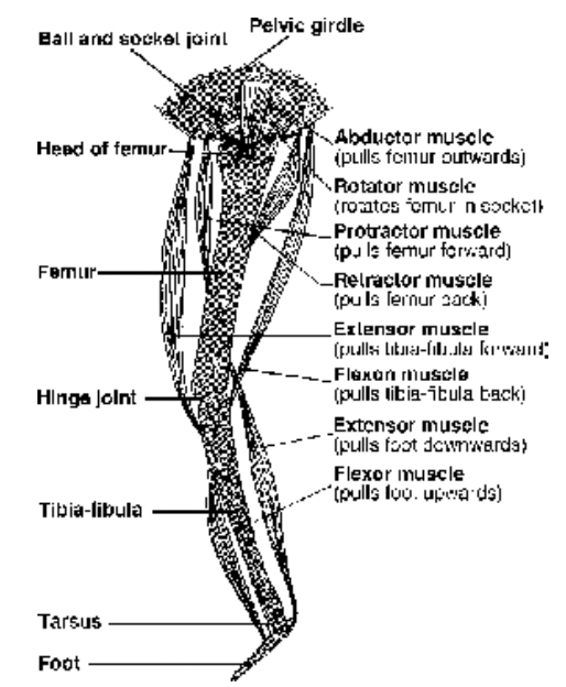
Fig. A small selection of the more importantmuscles found in the hind leg
Oxygen debt : Till the entire amount of creatine phosphate is recovered through regeneration of ATP by oxidative phosphorylation body remains in oxygen debt and to repay it the person keeps on panting after a strenuous exercise.
Cori cycle : The accumulated lactic acid diffuses out of muscle through blood and reaches liver where it is reconverted to glycogen. i.e., cycle operating in liver and muscle.
Troponin and Tropomyosin are two regulatory proteins present on thin filament and are essential for the actin–myosin interaction i.e., muscle contraction.
Ca++ and Mg++ ions are also essential for this.
ATP was discovered in muscle by Fiske and Subba Row (1929).
The category of skeletal muscle based upon the types of movement brought about by them in the body is as given below. These are generally present as antagonistic pair.
The antagonist muscle are operated in opposite way i.e., when one contracts the other relax, e.g. biceps of the arm at elbow joint is flexor while triceps is its antagonist hence acts as extensor.
- Flexor & Extensor : Flexor brings the articulating bone closer to body e.g. folding of arm at elbow joint; the extensor causes stretching or extension of the limb
- Pronator & Supinator : Pronator rotates the fore arm to turn the palm downward or backward; the Supinator moves it opposite.
- Abductor & Adductor : Abductor pulls the bone away from the main axis of the body, e.g., deltoideus; adductor brings it closer, e.g., latissimus dorsi.
- Elevator & Depressor : Elevator raises the parts up e.g., masseter lifts the lower jaw, the depressor pulls the parts down e.g., depressor mandibulae.
- Dilator & Sphincter : Dilator enlarges the opening size while sphincter (circular muscle) closes it.
