
Skeletal System in Man
Movement and Locomotion of Class 11
Human skeleton is made of 206 bones which have formed from some 300 bones through fusion. The bones are arranged into axial and appendicular skeleton. Number of bones in child = 350
Axial Skeleton
It is part of endoskeleton which occurs along the middle longitudinal axis of the body. Axial skeleton has 80 bones forming four structures — skull, vertebral column, sternum and ribs.
Skull (29 bones)
It constitutes skeleton of the head region. Skull consists of four parts — cranium, facial bones, hyoid and ear bones.
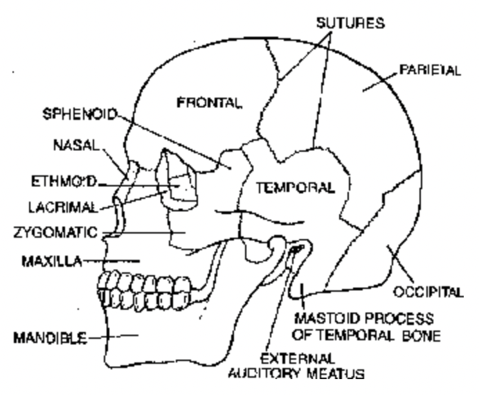
Fig. Skull of Human (lateral view)
Cranium (Brain Box, 8 bones). It is the bony structure which encloses the brain and protects it. It is hollow and nearly rounded. Bones constituting the cranium are called cranial bones. They are eight in number.
(i) Frontal-1 (ii) Parietal-2 (iii) Occipital-1 (iv) Temporal-2 (v) Sphenoid-1 (vi) Ethmoid- 1.
Cranial bones are flattened bones. They are jointed with one another by fixed interdigited joints called sutures. At the base of skull occipital has a very large opening called foramen of magnum (largest foramen). Through foramen of magnum brain communicates with spinal cord. There are two lateral protuberances or occipital condyles, one on each side of foramen magnum. Because of the presence of two occipital condyles, human skull is called dicondylic. The two occiptial condyles are articulated with atlas vertebra by hinge joints for performing nodding movements. Each temporal bones bear opening of external auditory meatus. Skull bones also possess fontanelles. Fontanelles are the soft areas present in foetal cranium. An infant may have many fontanelles at birth but generally six are recognised. Anterior fontanell or Frontal fontanell is largest and it closes by 18 to 24 months of age. Neurocranium is part of skull having brain and sensory capsules. Temporal bone has styloid process, zygomatic process and mastoid process. It also posses and external auditory meatus.
- Ear Ossicles (6 bones). Each side or temporal region contains an auditory capsule or middle ear. It has three bones. These bones are small in size and named as ear ossicles or ear bones. They are outer malleus (hammer shaped), middle incus (anvil shaped) and innermost stapes (stirrup shaped). These bones are helpful in the amplification of sound by 20-22 times. Malleus is derived from articular bone.
- Facial Bones (14 bones). Facial bones constitute front part of skull alongwith skeleton of lower jaw, hard palate and nose. They are fourteen in number. Incus from quadrate bone and
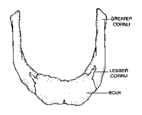
Fig. Human hyoid bone
(i) Nasals-2 (ii) Maxillae-2 (iii) Zygomatic (cheek bones)-2 (iv) Lacrimals-2 (v) Palatines-2 (vi) Inferior nasal conchae-2 (vii) Vomer-1 (viii) Mandible-1.
Frontal is common bone to both cranium and face. Maxillae form the upper jaw whereas mandible forms the lower jaw. Upper jaw is fused with cranium. Lower jaw or mandible is
horse-shoe shaped bone which bears two processes on each side, a pointed one named coronoid and a rounded one called condyloid. Mandible is strongest and largest bone of face.
Movable mandible helps in the mastication of food as well as speech. Jaws have sockets for the fixation of teeth.
- Hyoid Bone (Tongue Bone; 1 bone). It lies at the base of tongue and above the larynx. It is not joined to the rest of skeleton except to styloid process of temporal bone by muscles with the help of certain muscles. It is horse shoe-shaped slender bone. Upper portion of hyoid bone has a swelling known as the greater cornu. The lower portion has small projection known as lesser cornu. Hyoid bone and the tongue together form the hyoid apparatus. Hyoid bone is fractured during strangulation.
Vertebral column : It is our backbone which extends in the mid axis of the back (posterior) part of our trunk from head to the lower (inferior) extremity of trunk. Together with the sternum and rib, it forms the supporting frame work of our trunk. It support and rotate the head, suspends the viscera, protect vital organs, provides attachment to limb girdles, facilitates some movement of the trunk and houses the spinal cord.
Curvatures of vertebral column : In spite of the erect posture of body, the vertebral column of man is not perfectly straight. It displays four curves to enhance balancing power and firmness for upright posture of body. The curvatures are cervical, thoracic, lumbar and pelvic (=sacral). At birth, the whole column curves with the convexity towards the back. By the time, the infant stands, this convexity persists only in thoracic and sacral regions. These are called primary curvatures. A cervical curvature results from the effort on an infant to hold erect (3 to 4 months old infant). A lumbar curvature results when the infant learns to walk during the age of about 10 to 18 months. Cervical and lumbar curvatures are concave towards the back.
Vertebral column of man
- Made up of pieces of bones known as vertebrae.
- Vertebrae of man are acoelus i.e. Centrum is flat and without cavity.
- Vertebral column also known as spinal column or backbone.
- Number of vertebrae are thirty three (33) in vertebral column.
- Cervical vertebrae are seven (7).
- Thoracic vertebrae are twelve (12).
- Lumbar vertebrae are five (5).
- Sacral vertebrae are five (5) in child but fuse to form sacrum, caudal vertebrae are four (4) but form coccyx in adult.
In the adult stage of man number of vertebrae are as follows –
|
Cervical (in neck) |
7 |
|
Thoracic (in chest or thorax) |
12 |
|
Lumbar ( in loins) |
5 |
|
Sacral (in upper part of pelvis) |
1 (formed of five fused vertebrae) |
|
Coccygeal or Coccyx (in lower part of pelvis) |
1 (formed of four fused vertebrae) |
Atlas vertebra
- First cervical vertebra.
- Body is formed by centrum and vertebral arch.
- It supports the globe of the head like the earth by the atlas (super man).
- Centrum is absent.
- Neural spine reduced.
- Transverse process are long.
Axis vertebra
- Second cervical vertebra.
- Centrum acoelus.
- Odontoid process present.
- It is pivot for rotation of atlas and head around odontoid process.
- Transverse process small.
Cervical vertebra
- Long neural spine.
- Centrum acoelus.
- Transverse process are large.
- Vertebrarteal canals present.
Thoracic vertebra
- Centrum acoelus.
- Neural canal is formed by union of two neural arches.
- Neural spine is a flat & long directed backward.
- Club shaped transverse process.
- Neural arch with superior articular process.
- Two demifacets for articulation of head of a rib are present.
Lumber vertebra
- Centrum acoelus.
- Neural spine well developed.
- Transverse process are thin and long.
- Small accessory process present near the root of each transverse process.
- It is the largest vertebrae.
Sacrum
- It is fusion of five vertebrae.
- Ventral curtualine increases pelvic capacity.
- Transverse process is much modified into a broad sloping mass project laterally from the body.
- Sacral canal is formed by sacral vertebral foramina.
- In female sacrum is shorter and wider.
- In birds some of the vertebrae are fuse to form synsacrum. [Last thorasic+ Lumber+ Sacral+ One or two caudal]
Coccyx
- It is formed by fusion of four caudal vertebrae.
- It is last section of backbone.
- It is small triangular bone.
- Two coccygeal cornua project up to articulate with sacral cornua.
- Rudimentary transverse process.
Ribs
There are twelve pairs of ribs which join the thoracic vertebrae of the vertebral column on the dorsal side and the sternum on the ventral side to form the thoracic basket. Each rib is
composed of two parts — sternal and vertebral part. Sternal part of rib is attached to the sternum in front. The vertebral part of rib is attached to the vertebral column at the back. Each
rib. has a head, a tubercle (both for the attachment to thoracic vertebrae), neck, a shaft (body) and costal cartilage (for the attachment with sternum).
Ribs are of three sub types—true, false and floating.
- True Ribs. First seven pairs of ribs are known as true ribs as they are directly attached to sternum as well as vertebral column. Their size increases gradually from first to seven. The rib is longest.
- False Ribs. 8th, 9th and 10th pair of ribs are called false ribs as they are attached to one another as well as to the costal side of 7th pair of ribs. False ribs are also called vertebrochondrial ribs.
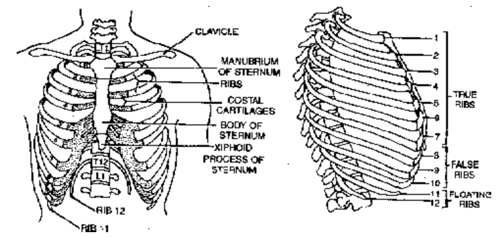
Fig. Thoracic cage (Anterior & Lateral views)
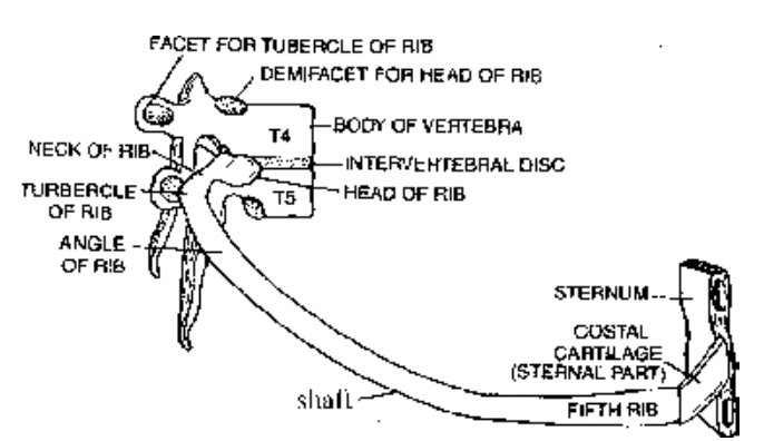
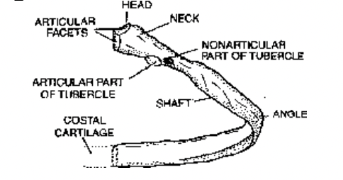
Fig. Fifth right rib as it articulates with vertebral column Fig. Fifth right rib as seen from
posteriorly and with sternum anteriorly posterior aspect
- Floating Ribs. 1lth and 12th pairs of ribs are known as floating ribs because their one end is joined to vertebral column whereas the second end is free. Floating ribs play a very important role to protect the kidneys.
Functions :
- Ribs make the human thorax wider from side to side than from the front to back. It is an adaptation for the erect posture and helps to maintain equilibrium.
- They protect two delicate organs, heart and lungs.
- Ribs provide attachment to intercostal muscles that take part in breathing movements.
- Floating ribs protect the kidneys.
Sternum (Breast Bone)
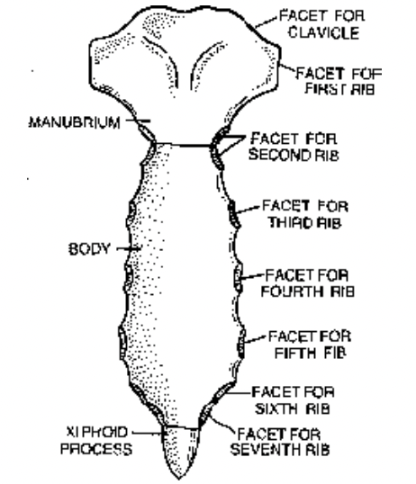
- Manubrium. It is wide flat plate which forms the upper part of sternum. It has facets for attachment to clavicles (collar bones) and first pair of ribs, 2nd pair of rib also.
- Body (Mesosternum). It is elongated middle part of sternum. Body of sternum is often equated with blade of dagger while manubrium is equated with handle of dagger. Body has facets for attachment of 3-7 rib pairs.
- Xiphoid (Xiphisterum). It is lower part of sternum which is cartilaginous in the young but becomes ossified in adults. Normally, it is small and pointed but can be bifid, curved or deflected. Xiphoid is often equated with point of the dagger.
Functions
- (Sternum is component of rib cage.
- It has facets for attachment of clavicles and ribs.
- It protects diaphragm.
- Sternum has red bone marrow and is important area of haemopoiesis.
