
Disorder of Skeletal System
Share
Disorder of Skeletal System
Movement and Locomotion of Class 11
Arthritis.
It is painful stiffness and inflammation of joints caused by infection, allergy, deficiency or hormonal imbalance. Arthritis is of three types—rheumatoid arthritis, osteoarthritis and gouty arthritis.
- Rheumatoid Arthritis. It is caused by inflammation of synovial membrane caused by bacterial infection, allergy or hormonal imbalance. Diagnosis of the disease is carried out by finding rheumatoid factor which resembles immunoglobulin IgM. Inflammation of synovial membrane leads to its thickening and excessive secretion of synovial fluid. It puts a pressure on the joint and the joint becomes painful. Later the synovial membrane starts secreting an exudate which forms abnormal granules over the membrane. The exudate is called pannus. The granules cause erosion of cartilage surface. The fibrous tissue attached to bones becomes ossified. Movements become extremely painful, ultimately resulting in immobilisation of the joint. Heat treatment and physiotherapy are helpful in the early stages. A completely damaged joint is replaced surgically. Staphylococcus bacteria which causes this also effects heart valves (Rheumatic heart disease).
- Osteoarthritis. It is a form of arthritis in which one or many joints undergo degenerative changes like loss of articular cartilage and proliferation of bone and cartilage in the joint. It is very common in old age, where degeneration of hyaline cap by the regular use and deposition of calcium makes the surface hard. This causes one bone to move over the other bone and leads to pain in joints. Osteoarthiritis is common in the joints of hands, knees and spine.
- Gouty Arthrititis or Gout. It is a disease associated with an inborn error of uric acid metabolism that increases its production or interferes with uric acid excretion. Thus, uric acid concentration increases in blood. Excess uric acid is converted into monosodium urate crystals that precipitate from the blood and become deposited in joints and other tissues. Men are more often affected than women.
Osteoporosis.
It is commonly an age dependent systematic disorder in which bone mass is reduced and the bones undergo microarchitectural deterioration. The bones become fragile and prone to fracture. The various reasons are (i) Post-menopausal hormonal imbalance in women. (ii) Decrease in organic matter of bones in old age. (iii) Pregnancy, (iv) Hormonal imbalance like thyrocalcitonin, parathyroid and sex hormones. (v) Prolonged cortisone treatment, (vi) Dietary deficiency of Ca2+ and vitamin D. Osteoporosis leads to severe pain commonly in the lower back. Bones become easily fractured.
Bursitis.
It is the inflammation of bursa, the connective tissue structure surrounding a joint, i.e., synovial joint. It is due to infection, injury or excessive or traumatic exercise. The chief symptom is severe pain of the affected joint, particularly on movement.
Dislocation :
The displacement of any articular surface of joint from its normal position, like coming out of ball-like head from the socket of the other bone. Ligaments are also damaged. There is a severe pain and inflammation. Movement is restricted. Resetting of the joint is required.
Strain.
Twisting of tendons between bone and muscle. It is less severe.
Sprain.
It is twisting, stretching or tearing of ligament. Ligament has poor power of regeneration. It causes severe pain. It is a minor disorder, which can however, become chronic.
Prolapsed Disc (Slipped Disc).
Displacement of intervertebral disc from its normal position. It is due to mechanical injury or wrong sitting posture. It causes severe pain due to pressure on adjacent nerves.
Paget’s disease
Irregular thickening and softening of bones with increased vascularity, especially in bones of skull, pelvis and extremities. In this osteoclastic resorption is massive and new bone formation by osteoblasts is extensive.
Fracture.
It is a break in a compact bone due to mechanical injury. It is very rare in children because their bones have more organic matter and are quite flexible. Fracture is common in old age because of less organic matter and more inorganic matter, whereby their bones become hard and brittle.
Fracture is of following types :
- Simple. Bone is completely broken into two parts.
- Compound. Bone completely breaks into two parts and the fractured pieces pierce into the skin.
- Comminuted. When a bone breaks down into more than two pieces, it is also called multiple or comminuted fracture.
- Evulsive. A chip breaks out but remains attached to ligament.
- Green Stick Fracture. When a bone undergoes simple crack but the bone remains intact. It is very common in children, having high recovering power.
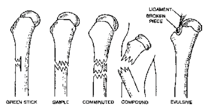
Fig. Types of fractures
Miscellaneous Points
Achondroplasia Imperfect ossification within cartilage of long bones during fetal life; also called fetal rickets. It causes a form of dwarfism.
Craniology. Study of skulls.
Arthrology. Study of joints.
Largest Foramen in Humans. Foramen magnum
Sharpey’s Fibres. Collagenous fibres holding bones and teeth.
Craniotomy any surgery that requires cutting through the bones surrounding the brain.
Necrosis Death of tissues or organs; in the case of bone, results from deprivation of blood supply resulting from fracture, extensive removal of periosteum in surgery, exposure to radioactive substances, or other
causes.
Osteitis Inflammation or infection of bone.
Osteoarthritis The degeneration of cartilage, allowing the bony ends to touch and, form the friction of bone against bone, creating a bony reaction that worsens the friction and worsens the condition; usually
associated with the elderly.
Osteoblastoma A benign tumor or bone and cartilage.
Osteoma A benign bone tumor.
Osteosarcoma A malignant tumor composed of osseous tissue.
Pott’s (POTS) disease Inflammation of the backbone, caused by the microorganism that produces tuberculosis.
Elbow joint is an example of hinge as well as pivot joint.
Osteology. Study of skeleton.
Femur. Longest bone in humans. Stapes. Smallest bone in humans.
Tibia. Strongest bone in humans. Tibio-fibula. Longest bone in case of Frog.
Skull in Animalia
- (i) Monocondylic. With one occipital condyle, e.g., Reptiles and Birds.
- (iii) Dicondylic. With two occipital condyles, e.g., Amphibia and Mammalia.
Wish Bone. It is V-shaped bone formed by the fusion of clavicle and interclavicle in birds. It is also named as merry thought bone or Furcula.
Vertebrae in Vertebrates
- Acoelous or Amphiplatyan. Flat centrum at each end, e.g., Rabbit.
- Procoelous. Concave centrum at front side, e.g., Frog and Lizard.
- Opisthocoelous. Concave centrum at hind side, e.g., Salamander.
- Amphicoelous. Concave centrum at both ends of vertebra, e.g., 8th vertebra of Frog.
- Heterocoelous. Saddle shaped or concavo-convex centra e.g., Birds.
Neurocranium. A part of skull having brain and sensory capsules.
Largest Foramen. Foramen of Magnum at the base of cranium from where brain communicates with spinal cord.
Cervical Vertebrae in Mammals : Majority of mammals have seven cervical vertebrae. Some exceptions are :
(i) Two-Toed Sloth—6 (ii) Three-Toed Sloth—9 (iii) Manatee—6 (iv) Ant Bear—8.
Sella Turcica. It is depression on the upper surface of sphenoid bone of skull, which is meant for lodging the pituitary gland.
Weberian Ossicles. Small bones developed from 1st four vertebrae of bony fishes like carp and cat fish. These connect air bladder to internal ear and act as a barometer.
Differences between Male and Female Skeleton of Humans
| S.NO. | Male Skeleton | Female Skeleton |
| 1. | It is heavy and long. | It is light and short. |
| 2. | Pelvic cavity is narrow. | It is wide and deeper. |
| 3. | Greater pelvis is deep. | Greater pelvis is shorter. |
| 4. | Heavy-and thick pelvis. | It is light and thin. |
| 5. | Coccyx is less movable. | It is more movable. |
| 6. | Pubic arch is less than 90°, i.e., acute. | Pubic arch is more than 90°, that is, obtuse. |
| 7. | Joint surfaces are longer. | Joint surfaces are smaller. |
| 8. | Long, narrow and concave sacrum. |
Sacrum is short, flat, broad and having forward curvature in lower parts. |
| 9. | Ischial tuberosity is turned inward. | Ischial tuberosity is turned outward. |
| 10. | Obturator foramen in pelvic girdle is circular. | It is oval in shape. |
| 11. | Sciatic notch is narrower. | It is wider. |
| 12. | Pelvic inlet and outlet are smaller and narrow. | These are larger and wider. |
| 13. | Anterior superior iliac spines lie close to each other. | They are wide apart. |
Erect Posture of Humans
- Head. Head lies vertically at the tip of vertebral column, which is semimobile. Base of head is having a wide passage called foramen of Magnum, by which it communicates with rest of the body.
- Neck. Semi-long reck with cervical vertebrae, provides mobility in all directions.
- Height. Height is due to presence of longer skeletal system and provides a wider range of vision.
- Fore Limbs. Powerful metacarpals and phalanges have gripping power. Palm of hand is wide providing a large area for balance, gripping etc. Opposable thumb providing better tooling power and multifarious use of hand.
- Backbone. Two convex and two concave curvatures in the backbone make the centre of gravity between the heels, in order to maintain balance and make walking over the legs much easier.
- Thorax. It helps to provide equilibrium as the sides are wider than region from front to back.
- Pelvis. It is a bowel-like structure, provides good support to the lower abdominal visceral organs.
- Hind Limbs. Leg bones are much stronger than the arm bones as they have to lift the entire weight of the body. Broad and flat feet provide stability and arches in feet provide springness.
