
Connective Tissue
Animal Tissues of Class 11
Origin : Formed by mesoderm of the embryo.
Most abundant tissue in the body, forming about 30% of body mass.
Consists of few cells, an extensive matrix and a rich blood supply.
Provides structural framework and support to different tissues.
Two general types of connective tissue are found - embryonic connective tissue and adult connective tissue.
Embryonic connective tissue is found in embryo, consists of mesenchyme and mucous connective tissue. Wharton’s jelly present in the umbilical cord of foetus is a mucous connective tissue.
The adult mucous connective tissue occurs in comb of cock and vitreous body of eyeballs.
Connective tissue plays a vital role in body defence, tissue repair, fat storage and transport of materials etc. Three components of connective tissue are - intercellular medium, connective tissue cells and fibres.
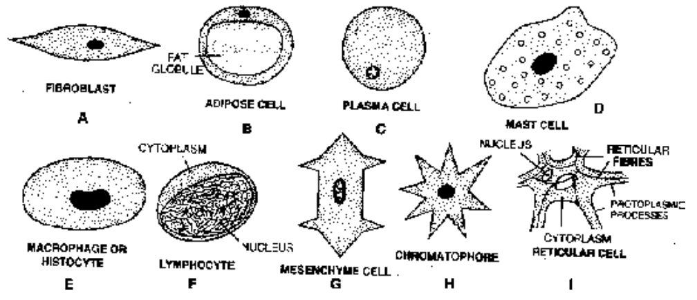
Fig. Different types of cells in the connective tissues
Types
(i) Intercellular Medium (ground substance) mainly a mixture of carbohydrates and proteins with various forms of mucopolysaccharides. The most common mucopolysaccharide ground substance is hyaluronic acid.
(ii) Connective tissue cells The cells are of different types
(a) Fibroblasts produce various types of fibres and matrix.
(b) Adipose cells (Adipocytes or Lipocytes) store fat.
(c) Plasma cells (Plasmatocytes) synthesize antibodies. Plasma cells are also called ‘Cart Wheel Cells’ because thin chromatin in the nucleus forms four or five clumps giving the nucleus a resemblance to a cart wheel.
(d) Mast cells (Mastocytes) produce histamine, heparin and serotonin. Mast cells are related to basophils of the blood. Histamine dilates the walls of blood vessels in inflammatory and allergic reactions while
heparin checks clotting of blood inside the blood vessels. Serotonin acts as a vasoconstrictor to arrest bleeding and to increase blood pressure.
(e) Macrophages (Histiocytes) ingest cell debris, bacteria and foreign matter. Macrophages are derived from monocytes.
(f) Lymphocytes ingest cell debris, bacteria and foreign matter.
(g) Mesenchyme cells give rise to various type of conective tissue cells.
(h) Chromatophores (Pigment cells) are found in the dermis of the skin where they impart colour to the animal.
(i) Reticular cells. They form reticular tissue and are phagocytic in nature.
(iii) Connective Tissue Fibres : These are of three types : (a) Collagenous or collagen fibres (white fibres) are made up of collagen protein. When boiled in water collagen changes into gelatin. These fibres
occur in bundles and are unbranched and inelastic. (b) Elastic fibres (yellow fibres) are formed of a protein called elastin. These fibres are branched and elastic. (c) Reticular fibres. These fibres are delicate,
branched and inelastic. They are made up of reticulin protein. They always form a net work.
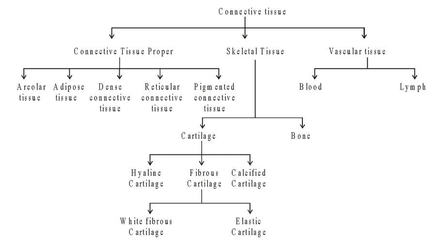
Connective Tissue Proper
- Binds different organs and their parts together.
- Extracellular material consists of insoluble protein fibres lying in a jelly-like transparent matrix.
Areolar tissue (Loose Connective tissue)
Most widely distributed tissue in body
Structure
Consists of ground substance, the matrix, white yellow and reticular fibres and cells like fibroblasts, mast-cells, macrophages, lymphocytes, plasma cells, mesenchyme cells,
chromatophores.
Location
Present as subcutaneous tissue around muscles, nerves and blood vessels, in submucosa of GIT and respiratory tract etc.
Also forms the internal framework (=stroma) and packing material of many organs.
Functions
Acts as a binding material.
Provides strength, elasticity and support to various tissues.
Provides rapid diffusion of materials and migration of wandering cells towards areas of infection and repair.
Adipose Tissue
Specialized type of loose connective tissue where fibroblasts are modified for fat storage.
Synthesises, stores and metabolizes fat.
Poor conductor of heat, it reduces heat loss through the skin.
In frog, adipose tissue forms the fat bodies.
The adipose tissue is found in the subcutaneous tissue, around the heart, kidneys, eyeballs, mesenteries and omenta.
Prominent adipose tissue sites are: subcutaneous fat (paniculus adiposes), blubber of whales, hump of camel.
Adipose tissue has 2 types of fat: white (yellow) fat and Brown fat.
White fat contains cells with a single large fat globule (monolocular).
Brown fat contains fat cells with several small fat globules (multilocular). Brown color is because of the presence of iron-containing cytochrome pigments.
- Brown fat is found in hibernating animals and new born babies. On oxidation, it yields about 20 times more energy than ordinary white fat.
Excessive accumulation of fat is called adiposis.
Dense Connective Tissue
It is of two types - white fibrous and yellow elastic connective tissue.
White fibrous connective tissue
Mainly consists of white collagenous fibres in bundles.
Fibroblasts are present in rows between the bundles.
Found at the joint between skull bones and makes them immovable.
Principal component of tendons and ligaments.
Tendon is a very dense, strong fibrous connective tissue made of collagen fibres. Tendon connect bones to a skeletal muscle.
Ligaments connect bones together.
Sprain is caused by excessive pulling of ligaments.
Elastic connective tissue
- Mainly made of freely branching elastic fibres which give the tissue a yellowish colour.
- Fibroblasts are present only in spaces between the fibres.
- Component of the cartilage of larynx, the elastic walls of arteries, trachea.
- Yellow elastic ligaments form the ligamenta flava of the vertebral column and true vocal cords.
Reticular Connective Tissue
- Consists of stellate reticular cells, having protoplasmic processes which join to form a cellular network.
- Forms stroma of many organs including the liver, spleen and lymph nodes. It is also found in bone marrow and lamina propria of the gut wall.
- Provides strength and support to many organs.
- Helps to bind together the cells of smooth muscles.
- The reticular cells are phagocytic and form defence mechanism of the body.
Pigmented Connective Tissue
- Pigment cells (chromatophores or melanophores) are irregular in shape containing yellowish brown, black or blue melanin granules.
- Melanin is produced by cells called melanocytes.
- Chromatophores simply phagocytose, engulf the melanin from melanocytes like macrophages.
- Present in the choroid, ciliary body, iris of the eye and dermis of the human skin.
- Pigmented connective tissue gives colour to the structures.
