
Nervous Tissue
Animal Tissues of Class 11
Nervous tissue originate from embryonic ectoderm.
Nervous tissue contain two types of cells - neurons and neuroglia.
The specialized property of nervous tissue are excitability and conductivity. They show response to any given stimuli.
Neurons form structural and functional unit of the nervous system. Human nervous system has about 100 billion neurons.
Form the longest cells in human body (largest is oocyte).
There is no division of neurons after birth : possess least power of regeneration.
A neuron consists of three distinct portions
(i) Cell body; (ii) dendrites; (iii) axon
The cell body of a neuron is called cyton, perikaryon or soma. It contains abundant granular cytoplasm and a large nucleus.
Cytons of most neurons occur in brain.
The structures present in neuroplasm characteristic of neurons are Nissl’s bodies or trigoid granules.
Nissl granules are rich in RNA and are concerned with protein synthesis.
Cyton is concerned with metabolic maintenance and growth.
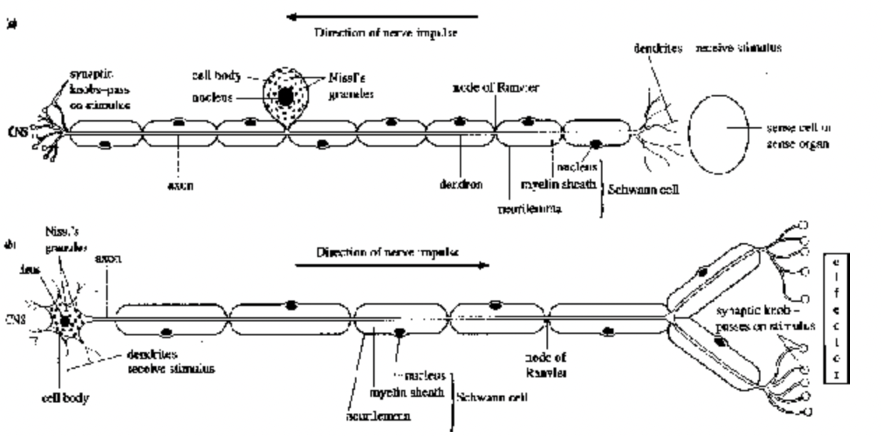
Fig. (a) Sensory neurone, (b) Motor neurone
Dendrites are highly branched extensions of the cytoplasm of the cell body
.
Function of dendrites is to conduct an impulse towards the cell body.
Dendrites are afferent processes because they bring nerve impulse to the cyton.
Axon is a longer process and branches distally into many fine filaments called telodendria.
The knobbed ends of telodendria are called endbulbs, axon terminal or buttons.
An axon usually originates from the cell body as a small conical elevation called the axon hillock.
The cytoplasm of an axon is called axoplasm and surrounded by a plasma membrane called axolemma.
Nissl bodies are absent in the axon and axon hillock. Axon is an efferent process because it conducts impulse away from cell body to another neuron or tissue.
The extended axon or dendrite of a neuron is called a nerve fibre.
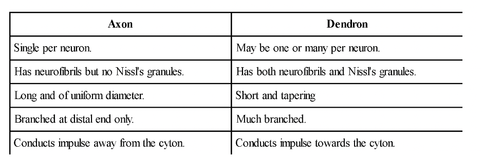
Table : Differences between Axon and Dendron
Two types of axons are myelinated and non-myelinated.
The myelinated or medullated or white fibre is surrounded by a phospholipid covering called myelin sheath.
The myelin sheath is secreted by Schwann cells.
Myelin sheath is a layer covering of vertebrate nerve fibre.
At certain portions myelin sheath is absent and form constrictions called nodes of Ranvier.
Myelin serves as an insulating material. It causes saltatory conduction of impulses.
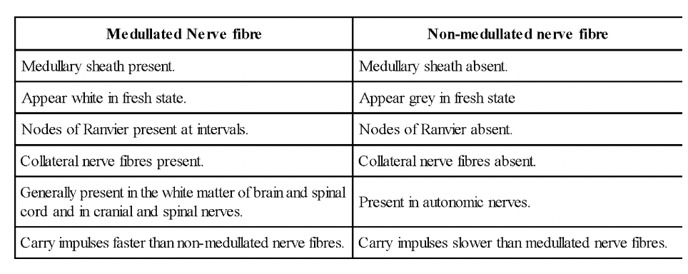
Table : Differences between Medullated and Non-medullated fibres
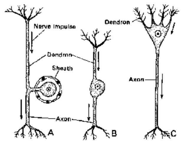
Neurons are classified on the basis of nature and number of their processes. Four types of neurons are (i) non-polar (ii) unipolar (iii) bipolar and (iv) multipolar neurons.
Unipolar non-polar neurons are found in Hydra. It has only one process extending from the cell body as found in embryos and dorsal root ganglia of adult vertebrates.
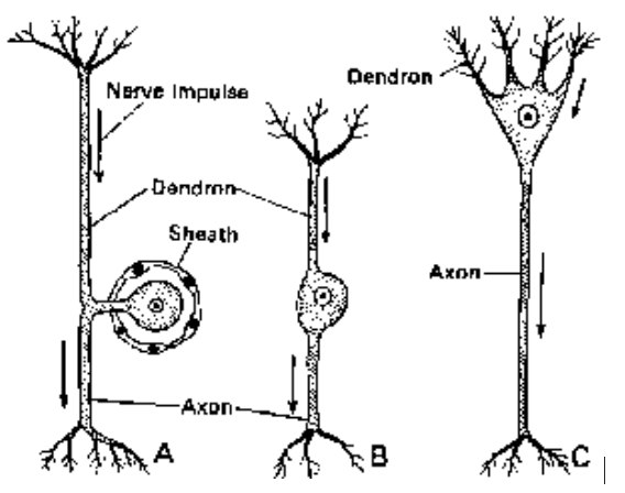
Fig. Different types of Neurons
Bipolar neurons have a single dendrite and axon at opposite poles of the cyton. These are found in the retina of eyes, olfactory epithelium and inner ear.
Multipolar neurons have several dendrites and one axon. Most neurons in the brain and spinal cord are multipolar.
On basis of function, neurons are of 3 types: Sensory neurons, Motor neurons and Interneurons.
Interneurons (Adjustor Neurons) : Present in the CNS and occur between sensory and motor neurons for distant transmission of impulses. They are neither sensory nor motor.A nerve is a bundle of nerve fibres outside the CNS.Fine fibrous connective tissue surrounding individual nerve fibre is called endoneurium.The parallel bundles of nerve fibres are called fasciculi surrounded by perineurium.The entire nerve is surrounded by a vascular connective tissue called epineurium.The nerves are of 3 types according to the nature of the fibres they are composed of:
(a) Sensory (Afferent Nerve) : They contain sensory fibres olfactory, optic and auditory cranial nerves.
(b) Motor (Efferent Nerves) : They contain motor nerve fibres occulomotor, pathetic, abducens, spinal accessory and hypoglossal cranial nerves.
(c) Mixed nerves : They contain both sensory and motor nerve fibres. Trigeminal, facial, glossopharyngeal, and vagus cranial nerves. Most spinal nerves are mixed nerves
Neuroglia forms the connective tissue of nervous system which performs support and projections.
Three different types of neuroglia cells in CNS are astrocytes, oligodendrocytes and microglia.
Astrocytes are star-shaped cells with numerous processes. They are of 2 types - fibrous and protoplasmic astrocytes.
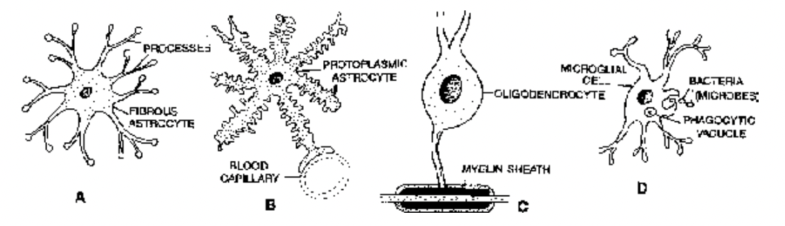
Fig. Different kinds of neuroglial cells. A. fibrous astrocyte, B. Protoplasmic astrocyte,
C. Oligodendrocyte, D. Microglial cell
Fibrous astrocytes are mainly found in white matter of CNS and their processes are thin and asymmetrical.
Protoplasmic astrocytes are mainly found in grey matter of CNS and their processes are thicker and symmetrical.
Astrocytes communicate with one another through calcium channels.
Play a role in maintenance of the blood brain barrier. They also repair damaged areas of nervous tissue.
Oligodendrocytes resemble astrocytes but processes are fewer and smaller. They occur near the medullated nerve fibres and near the surfaces of soma of neurons. They form myelin sheaths around the axons that lie with the CNS.
Microglia are small cells with few processes. They are phagocytic in nature and are derived from mesoderm of the embryo. They are more numerous in grey matter than in white matter and act as scavangers.
They may migrate to injured areas of nervous tissue and function as small macrophages.
Neuroglia are of clinical interest because they are a common source of tumors of the nervous system.
Table : Differences between neurons and neuroglia
Ependymal cells
These cells are arranged as an epithelial layer. One cell thick, which lines the ventricles (cavities of the brain) and the central canal of the spinal cord. The cells vary from squamous to columnar according to their
location. Their free surface bears numerous microvilli and cilia. The microvilli help in the absorption of cerebrospinal fluid. The movements of the cilia contribute the flow of the cerebrospinal fluid. The
ependymal cells possess one or more long processes towards opposite side which penetrate the nervous tissue.
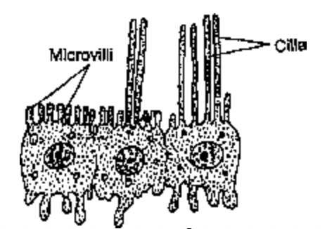
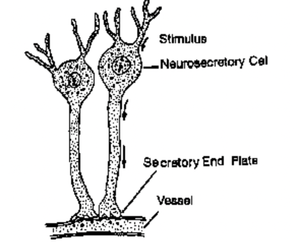
Fig. Ependymal Cells Fig. Neurosecretory Cells
Neurosecretory Cells
These specialized nerve cells function as endocrine organs. They release chemicals from their axons into the blood instead of synaptic cleft. Neurosecretory cells of the hypothalamus of the vertebrate brain
secrete neurohormones (=releasing factors). These neurohormones are carried from the hypothalamus to the anterior lobe of the pituitary gland where they regulate the secretion of pituitary hormones such as
ACTH, TSH, GH, LH, FSH and prolactin.
The terms like Myo and Sarco are used in context with muscles.
Myoblasts - give rise to muscle fibres.
Myocytes (sarcocytes) - are muscle cells.
Myofibrils - fine fibrils present in the muscle fibre, these are made up of myofilaments.
Sarcoplasm - Cytoplasm present in muscle cells.
Sarcolemma - Plasma membrane of muscle fibres.
Contractility and electrical excitability are the most specialized properties of muscular tissue.
Functions of Muscular tissue
Responsible for the movement of the body parts and locomotion of the individual.
Muscles bring about peristalsis in intestine, heart beat, production of sound etc.
Facial expression depends on muscles.
Supports the bones and other structures.
Makes up about 40% of the body weight in mammals, classified into three types:
(a) Skeletal
(b) Visceral
(c) Cardiac
Skeletal muscle is attached to bones (except tongue muscles and muscles of upper part of oesophagus). It is also known as striated, somatic, phasic or voluntary muscle.
Visceral muscle is located in viscera. It is also known as non-striated, smooth, or involuntary muscle.
Cardiac muscle is found in wall of the heart. It is striated and involuntary.
Striated Muscles
- Muscle fibre has stripped appearance when viewed under microscope.
- Muscle fibres are formed by myoblast. The nucleus undergoes repeated divisions.
- A stripped muscle fibre is multinucleated and it is a syncytium (coenocyte).
- Fine fibrous connective tissue surrounding individual muscle fibre is called endomycium.
- The parallel bundles of muscle fibres are called fasciculi, surrounded by perimycium.
- The entire muscle is usually wrapped with a fibrous connective tissue called epimycium.
- The smooth endoplasmic reticulum of a muscle cell is called sarcoplasmic reticulum. It stores Ca2+.
- Major component of striated muscle is water (75%).
- The proteins present in muscles are actin, myosin, troponin, myoalbumin, myoglobulin and myogen.
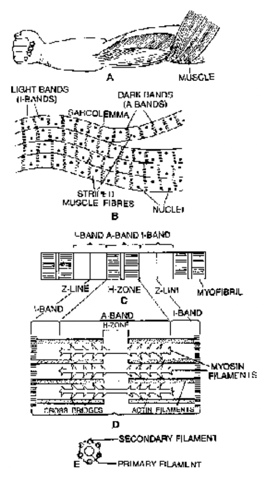
Fig. Structure of striated muscle at different levels of magnification
- The non-protein components of muscle are ATP, phosphocreatine, creatine and urea.
- Potassium is abundantly present in muscles. The minerals present in traces are sodium, calcium, phosphorus and magnesium.
- Muscles are the site of storage of glycogen.
- An iron-containing pigment called myoglobin or muscle haemoglobin transports oxygen in muscles.
Structure of striated muscle
- Skeletal muscle fibre possess transverse striations in the form of regular alternate dark A (anisotropic) and light I (isotropic) bands.
- At the centre of I band is a fine, dense Z-line or Krause’s membrane, dobber’s dot or dobber’s disc.
- Z-line divides the myofibrils into functional units called sarcomeres, it is the area between two Z-lines.
- H-zone is found at the centre of A-band, M-line or Hensen’s line is in the middle of H-zone.
- Two types of protein filaments in muscle are myosin and actin.
- Myosin filaments are thick and located inside A-bands. Actin filaments are thin and more numerous, present mainly inside I-band.
- Junction between muscle and motor nerve is known as neuromuscular or myoneural junction.
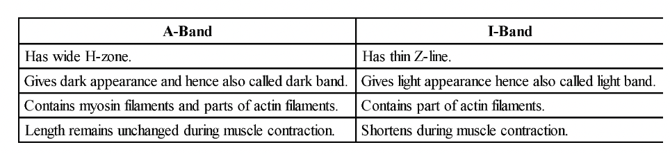
Difference between A and I-bands
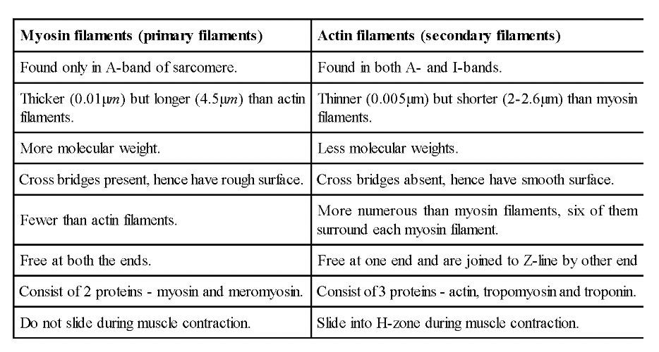
Differences between Myosin and Actin filaments
Contraction of Striated Muscle
1. Sliding filament theory of muscle contraction was proposed by Andrew Huxley and Ralph Niedergerk.
2. The contraction of muscle is brought about by a sliding movement of actin filaments over myosin filaments (Hugh Huxley and Ian Hanson).
3. During muscle contraction, both I band and H zone progressively shorten and eventually disappear.
4. Release of calcium ions from sarcoplasmic reticulum trigger the muscle contraction process.
5. Calcium is an essential element for the contraction of muscles.
6. In the presence of Ca2+ and energy from ATP, actin and myosin interact forming actomyosin. Contraction of a muscle is caused by actomyosin.- The energy for contraction of muscle is obtained from ATP. In a resting muscle, ATP combines anaerobically with creatine to form creatine phosphate. When muscle contracts creatine phosphate breaks down to produce ATP. During muscle contraction, conversion of pyruvic acid to lactic acid proceeds anaerobically.
- Muscles become rigid after death, the condition is known as rigor mortis. Rigor mortis is due to permanent irreversible contraction, established by permanent link between actin and myosin and fall in the concentration of ATP molecules.
- Twitch is a rapid, jerky response to stimulus.
- Tetanus is a sustained contraction of muscle by a wave of stimuli.
- When length of a muscle remains constant but the tension increases sharply, contraction is known as isometric.
- When tension of a muscle remains constant but muscle shortens, the contraction is known as isotonic.
- Muscles of eye lid contract for shortest duration.
- When there is a drop in the force of contraction or muscles fail to contract after prolonged stimulation, it is known as fatigue. The muscle fatigue occurs after strenous exercise due to the accumulation of lactic acid.
- Cardiac muscles do not fatigue.
- Lactic acid of muscles is converted to glycogen in liver by Cori’s cycle.
Smooth muscles
- Also known as visceral, non-striated, non-skeletal or involuntary muscles.
- Most widely distributed muscles in the body, eg-gastro-intestinal tract, uterus, urinary bladder, iris, ciliary body, blood vessels etc.
- Smooth muscle fibre is spindle-shaped, thick in the middle and thin at either ends, uninucleated and has no sarcolemma. Contraction, under the control of ANS.
- Functionally smooth muscles are of 2 types - single-unit smooth muscle and multi-unit smooth muscle.
- Single-unit muscle fibres are composed of muscle fibres closely joined together, contract as a single unit. eg - urinary bladder and GIT.
- Multi-unit smooth muscles are composed of more-independent muscle fibres, contract as separate units eg-hair root muscles and muscles in the wall of blood vessels.
- Smooth muscles never connect with skeleton.
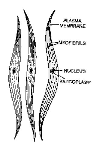
Fig. Nonstriated muscle fibres
Cardiac Muscles
- Found in the wall of the heart and in the wall of large veins (eg-pulmonary veins and superior vena cava) at the point of entry in the heart.
- Cardiac muscles have long, cylindrical, branched and uninucleated cells divided at places by intercalated discs. Nucleus is located near the centre. They lack definite sarcolemma.
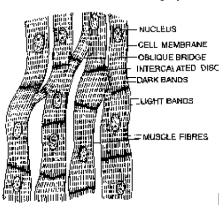
Fig. Cardiac Muscle Fibres
- The fibres have some lateral branches, known as oblique bridges to form contractile network.
- Intercalated discs, function as boosters of contraction wave and permit the wave of muscle contraction to be transmitted from one cardiac fibre to another.
- Striated, involuntary, contract quickly and do not get fatigued.
- Supplied with both central and autonomic nervous system.
- Richly supplied with blood capillaries.
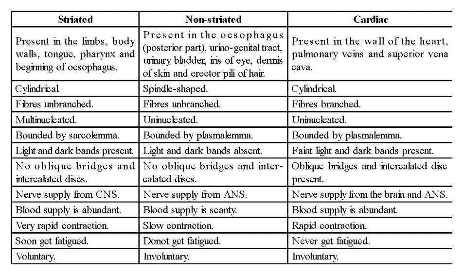
Differences between striated, Non-striated and Cardiac Muscles
Other Contractile Cells
Cells
1. Myoepithelial cells
Present in salivary glands, mammary glands and sweat glands.
Form a sort of epithelium called myoepithelium.
Cells contract to expel saliva, milk and sweat.
Innervated by autonomic nerves.
Resemble smooth muscle cells and are involuntary.
Arise from ectoderm.
2. Myofibroblasts
Cells resemble fibroblasts but contain actin and myosin arranged as in smooth muscles and are contractile. They are specialized contractile fibroblasts.
The contraction of wounds is caused by the shortening of myofibroblasts.
3. Pericytes
Found around capillaries and venules.
Contain actin and myosin.
Pericytes give rise to myofibroblasts and to mesenchymal tissue which can differentiate into fibroblasts and can form new blood vessels.
