
Glandular Epithelium
Share
Glandular Epithelium
Animal Tissues of Class 11
- Glands in a vertebrate body may originate from ectoderm, mesoderm or endoderm.
- Cells of the gland are secretory in nature.
- Zymogen granules appear in the cytoplasm of the secretory cells.
- A gland can be classified on the basis of presence and absence of ducts, number of cells, shape of secretory unit, mode of secretion and nature of secretion.
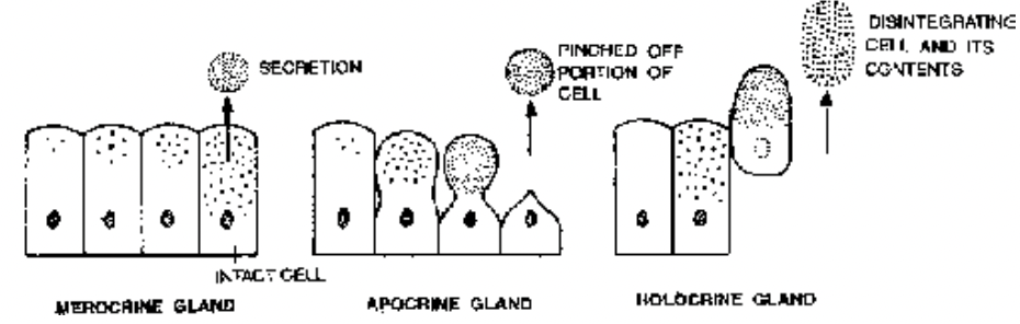
Fig. Types of glands according to their modes of secretion
(a) Nature of Secretion
1. Mucous Glands : They secrete slimy mucin-rich liquid called mucus, e.g., goblet cells, sublingual salivary gland. Mucus secreting cells are called mucocytes. Mucocytes have frothy cytoplasm. Nucleus is basal and flattened.
2. Serous Glands : The glands produce clear, watery, albuminoid secretion having glycoproteins, enzymes and lysozyme, e.g., parotid salivary gland, intestinal glands, sweat glands. The secretory cells are called serocytes. Serocytes possess granules.
3. Mixed Glands : The glands have more than one component. Some components produce mucus while other form albuminoid secretion, i.e., both mucocytes and serocytes are present, e.g., submandibular salivary glands, gastric glands.
(b) Mode of Secretion
1. Merocrine Glands : Glands in which the secreting cells remain intact during the formation and discharge of secretion, e.g., goblet cells, most sweat glands, salivary glands. The terms epicrine (Gk. epi-upon, krinein-to separate) and eccrine (Gk. ekkrenein– to expel) are also sometimes used for merocrine glands. The secretion is poured out through exocytosis and other modes of active transport. cAMP is also involved.
2. Apocrine Glands : Secretion first accumulates below the free surface. The distal part of cytoplasm containing the accumulated secretory product is then pinched out, e.g., sweat glands of axilla and genito-anal regions.
3. Holocrine Glands : The glandular cells first get filled up with secretory products. They then burst or disintegrate to liberate the secretory products, e.g., sebaceous glands.
4. Mixed Glands : They operate more than one mode of secretion simultaneously. Mammary gland cells secrete lipid by apocrine method and protein casein by merocrine method. Sweat glands of axilla have both apocrine and eccrine cells (Ito, 1988).
(c) Site of Secretion
1. Exocrine Glands : Glands which drain out their secretion to the body surfaces, i.e., exterior and surfaces continuous with it, e.g., integumentary surface, alimentary tract, respiratory tract, urinary and genital tracts. The secretion is passed out either by ducts or poured directly over the substrate, e.g., gastric glands, mucous glands, intestinal glands, salivary glands, sweat glands, sebaccous glands, mammary glands.
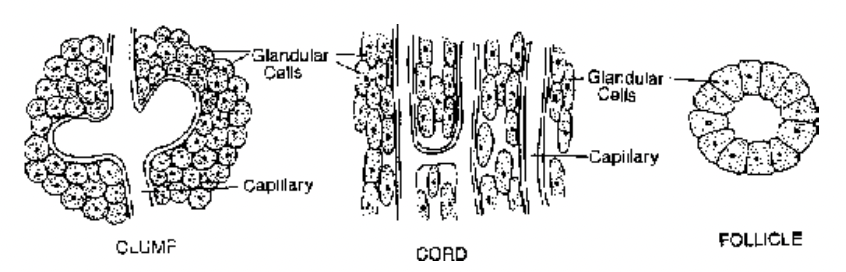
Fig. Types of groupings of endocrine cells
2. Endocrine Glands : Glands which pour their secretion internally into blood-vascular system directly. Commonly called ductless glands as they lack draining ducts, e.g., thyroid, adrenal, parathyroid, thymus,
pituitary, pineal. Endocrine cells can also occur individually at places. Most of them are paracrine as found in digestive tract.
3. Paracrine Glands : These glands produce local hormones, tissue hormones or parahormones. The hormones do not pass into blood but get bound to receptors of nearby cells of different types to influence their functioning, e.g., acetylcholine, kinins, prostaglandins. Many hormone producing glands of gastrointestinal tract are paracrine in nature. Some of them have both endocrine and paracrine activity, e.g.,
gastrin.
4. Heterocrine Glands : The glands have both exocrine and endocrine secretions, e.g., pancreas.
(d) Cell Number
1. Unicellular Glands : Isolated single cells function as glands. They are of two types:
Mucocytes secrete mucus. At the time of liberation of mucus, the apical region of the cells develop a sort of small cup for exocytosis. Hence, mucocytes are also called goblet, chalice or caliciform cells.
Serocytes produce clear albuminoid secretion generally having enzymes and lysozyme.
2. Multicellular Glands : The glands are formed from a patch or epithelium of secretory cells. They are of two types:
(i) Laminar glands : the secretory cells occur in a patch or layer present on the surface without any invagination. Most endocrine glands are laminar in structure.
(ii) Diverticulate glands : develop an invagination. Secretory cells lie in the invagination. They open on the surface with or without a duct. Diverticulate glands are mostly exocrine. Commonly, an exocrine gland possesses two parts, a duct and secretory part. Both are epithelial in origin.
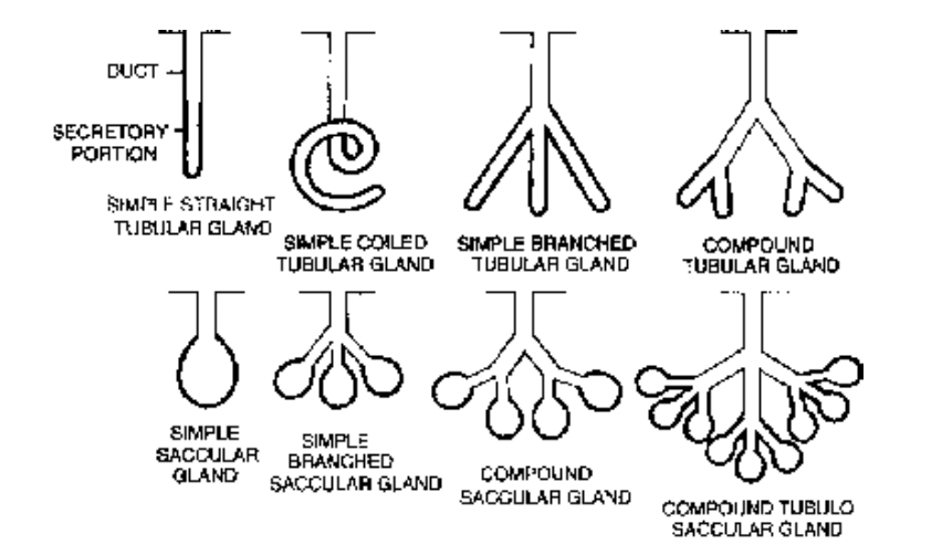
Fig. Structural types of Exocrine Glands
(e) Shape of secretory unit
1. Simple tubular glands : The secretory portion is straight and tubular e.g., crypts of Lieberkuhn.
2. Simple branched tubular glands : The secretory portion is branched and tubular eg gastric and uterine glands.
3. Simple coiled tubular glands : The secretory portion is coiled eg sweat glands.
4. Simple alveolar/saccular glands : The secretory portion is flask-like eg seminal vesicles of mammals and cutaneous glands of frog.
5. Simple branched alveolar/saccular glands : The secretory portion is branched and flask-like eg sebaceous glands.
6. Compound tubular glands : The secretory portion is tubular eg bulbourethral glands and liver.
7. Compound alveolar/sacular glands : The secretory portion is flask-like. eg salivary glands (sublingual-and submandibular).
8. Compound tubulo-alveolar glands : The secretory portion is both tubular and flask-like eg salivary gland (parotid) and pancreas.
Modified Epithelia
- The epithelium contains specialised columnar cells which can pick up external stimuli with the help of sensory hair or cilia present on their free ends.
- The stimulus is transferred to nerve fibres present on the inner ends of sensory or neuro-epithelial cells.
- Sensory cells are surrounded by a number of non-receptive supporting cells.
- Found in internal ear, nasal mucosa and taste buds.
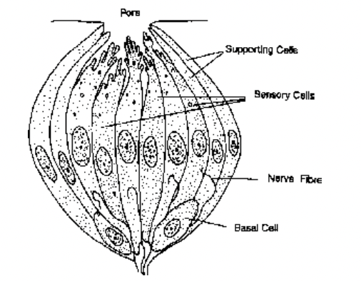
Fig. Neuroepithelium. V.S. Taste Bud
(ii) Germinal Epithelium
- Occurs in testis and ovary.
- Germinal epithelium of human testis is represented by single layered wall of seminiferous tubules. In ovary it occurs on the outside.
- Specialised to produce sex cells or gametes.
(iii) Pigmented Epithelium
- Lies at the back of retina in contact with choroid.
- Made of cuboidal cells, rich in melanin granules.
(iv) Myo-epithelium
- The cells possess the ability to contract.
- The cells are called myoepitheliocytes or basket cells. They are fusiform or stellate in outline. Lines secretory and duct portions of salivary, sweat, lacrimal and mammary glands.
- A basement membrane is present below the epithelium. The cells possess actin and myosin fibrils like those of smooth muscle cells. They also contain cytokeratin-like epithelial cells.
- The cells are commonly ectodermal in origin.
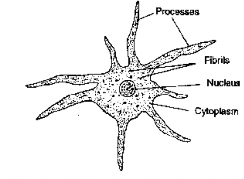
Fig. A myoepitheliocyte
- Contraction of myoepithelial cells help in flow of glandular secretion into larger channels.
- Myo-epithelial cells also produce components of basement membrane and growth factors for glandular epithelium.
