
Covering and Lining Epithelium
Animal Tissues of Class 11
Classified on the basis of arrangement of layers, cell shapes and functions.
Simple/Unilaminar Epithelium
- Made up of single layer of compactly arranged cells with underlying non-cellular basement membrane.
- Occurs over moist surfaces which are not subjected to wear and tear.
- Concerned with absorption, secretion, diffusion and movement of materials.
(a) Simple squamous epithelium
- Consists of flat, tile-like, polygonal cells with a centrally located flattened nuclei. It is also known as pavement epithelium.
- A tissue similar to simple squamous is endothelium. It lines the heart, blood vessels and lymph vessels and forms the wall of capillaries.
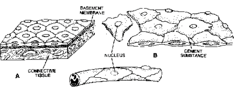
Fig. A. Simple squamous epithelium, B. Endothelium of blood capillary
Location :
- Present in terminal bronchioles and alveoli of lungs, wall of the Bowman’s capsule, descending limbs of loop of Henle and membranous labyrinth.
- Mesothelium is a type of squamous epithelium that lines the thoracic and abdomino-pelvic cavities and covers the viscera.
- The cells of endothelium and mesothelium may become wavy, hence these epithelia are called tessellated.
Functions :
Protection, excretion, gas exchange and secretion of coelomic fluid.
(b) Simple Cuboidal Epithelium
Consists of a single layer of cuboidal cells with equal height and width and spherical nucleus.
The cells of cuboidal epithelium often form microvilli on their free surface. This gives a brush-like appearance to their free border called brush-bordered cuboidal epithelium.
In ovaries and seminiferous tubules of the testes, it is called germinal epithelium because it produces gametes (ova and sperms respectively).
Location : The cuboidal epithelium is present in the small salivary and pancreatic ducts, thyroid vesicles, parts of membranous labyrinth, proximal and distal convoluted tubules of the nephrons of kidneys, ovaries, seminiferous tubules of testes, and ciliary bodies, choroid and iris of eyes. Other sites of cuboidal epithelium are the inner surface of the lens and the pigment cell layer of the retina of the eye.
Functions : Protection, secretion, absorption, excretion and gamete formation
.png)
Fig. A. SImple cuboidal epithelium; B. Simple columnar epithelium of intestine
(c) Simple Columnar Epithelium
- Consists of tall and columnar cells, height exceeding width. Nuclei are elongated and situated in the basal part.
- Simple columnar epithelium lines the stomach, small and large intestine, digestive glands and gall bladder.
- Columnar epithelium is found in small intestine and has striated border (brush-border) which increases the absorptive capacity of the intestine.
- Certain cells of this epithelium contain mucus (a slimy substance) and are called goblet (or mucous) cells as they look like goblet. The epithelium containing mucus secreting cells, alongwith the under lying supporting connective tissue is called mucosa or mucous membrane. The latter is present in the stomach and intestine.
- The intestinal mucosa (= mucous membrane) has microvilli to increase the absorptive surface area and is called brush-bordered columnar epithelium which is highly absorptive.
Functions :
(i) Secretion of mucus by goblet cells.
(ii) Absorption by cells lining small intestine and gall bladder.
(iii) Components of most glandular epithelia.
(iv) Protection
(d) Simple Ciliated Epithelium
- The cells bear numerous delicate hair-like outgrowths, the cilia, arising from basal granules.
- Mucus secreting goblet cells also occur in the ciliated epithelium.
- The cilia remain in rhythmic motion and create a current to transport the materials which come in contact with them.
The ciliated epithelium is of two types:
(i) Ciliated Columnar Epithelium :
Comprises columnar cells which have cilia on the free surface.
Lines most of the respiratory tract and fallopian tubes (oviducts).
Also lines the ventricles of the brain and the central canal of the spinal cord. It is also present in tympanic cavity and auditory tube (eustachian tube).
(ii) Ciliated Cuboidal Epithelium
Consists of cubical cells which have cilia on the free surface. It occurs in certain parts of nephrons of the kidneys.
Functions : Maintain a flow of mucus or liquid or suspended particles in one direction. In the respiratory tract the cilia help to push mucus towards the pharynx (throat). In the oviducts the cilia help to move an
egg towards the uterus. The cilia of the ventricles (cavities) of the brain and central canal of the spinal cord help to maintain the circulation of cerebrospinal fluid present there. In the nephrons of the kidneys, cilia keep the urine moving.
(e) Pseudo-stratified Epithelium
The cells are columnar, but unequal in size. The long cells extend up to free surface. The short cells do not reach the outer free surface.
The long cells have oval nuclei, however, short cells have rounded nuclei.
Mucus secreting goblet cells also occur in this epithelium.
Although the epithelium is one cell thick, yet it appears to be multi-layered which is due to the fact that the nuclei lie at different levels in different cells. Hence, it is called pseudostratified epithelium.
Occurs in the large ducts of certain glands such as parotid salivary glands and the urethra of the human male. Also present in the olfactory mucosa.
The long cells have cilia at their free surface, however, the short cells are without cilia. Occurs in the trachea and large bronchi. The movements of the cilia propel the mucus and foreign
particles towards the larynx.
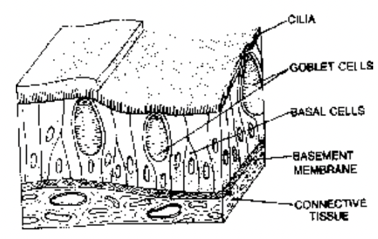
Fig. Ciliated pseudostratified epithelium
Functions : Protection, secretion, movement of secretions from glands, urine and semen in the male urethra and mucus loaded with dust particles and bacteria from the trachea towards the larynx.
Stratified Epithelia
Has many layers of epithelial cells, however, the deepest layer is made up of columnar or cuboidal cells. Classified on the basis of the shape of the cells present in the superficial layers. It is of four types.
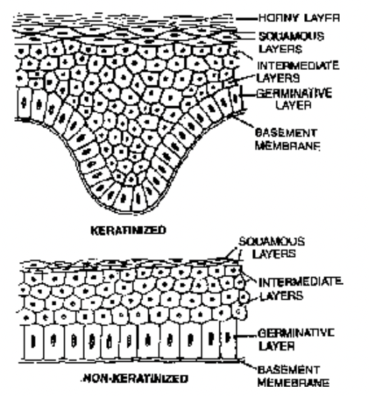
Fig. Keratinised and non-keratinised stratified epithelium
(a) Stratified Squamous Epithelium
With many layers of squamous cells; Two types are non-keratinized and keratinized.
Non-keratinizing epithelium is found on wet surfaces that are subjected to considerable wear and tear and do not perform the function of absorption.
Keratinized epithelium contains keratin, a highly insoluble fibrous protein with water-proofing qualities.
This epithelium is also resistant to friction and bacterial invasion.
The cells in the deepest (basal) layer are columnar or cuboidal with oval nuclei. It is called stratum germinativum (malpighian layer). The cells of this layer divide by mitosis to form new cells. The new cells gradually shift outward.
In the middle layers, the cells become polyhedral with rounded nuclei. These are called intermediate layers.
The superficial layers are flat with transversely elongated nuclei. These layers are called squamous layers. It is of two types:
(i) Keratinized Stratified Squamous Epithelium
In the outer few layers, the cells replace their cytoplasm with a hard, waterproof protein, the keratin. The process is called keratinization. These layers of dead cells are called stratum corneum or horny layer.
The deeper layers have living polygonal cells.
The horny layer is shed at intervals due to friction.
Occurs in the epidermis of the skin of land vertebrates.
(ii) Non-keratinized Stratified Squamous Epithelium
It does not have keratin. It is unable to check water loss and provides only moderate protection against abrasion (scraping).
Occurs in the oral cavity (buccal cavity), tongue, pharynx, oesophagus, anal canal, lower parts of urethra, vocal cords, vagina, cervix (lower part of uterus), conjunctiva, inner surface of eye lids and cornea of eye.
(b) Stratified Cuboidal Epithelium
Outer layer of cuboidal cells and basal layer of columnar cells.
Forms the epidermis of fishes and many urodels (tailed amphibians such as salamanders.)
Also lines the sweat gland ducts and larger salivary and pancreatic ducts.
(c) Stratified Columnar Epithelium
Columnar cells in both superficial and basal layers.
Covers the epiglottis and lines mammary gland ducts and parts of urethra.
(d) Stratified Ciliated Columnar Epithelium
Outer layer consists of ciliated columnar cells and basal layer of columnar cells.
Lines the larynx and upper part of the soft palate.
Transitional Epithelium (=Urothelium)
Consists of 4 to 6 layers of cells. The cells of deepest (= basal) layer are columnar or cuboidal.
The cells of middle layer are polyhedral or pear-shaped. The cells of the surface layer are large and globular or umbrella-shaped.
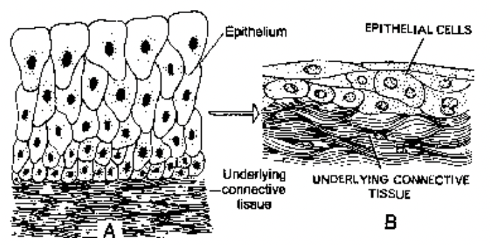
Fig. A & B. Transitional epithelium in wall of nondistended and distended urinary bladder
There is no germinative layer or basement membrane but shows mitosis.
The cells of inner most (=basal) layer rest on underlying connective tissue.
The cells of transitional epithelium and underlying connective tissue are very stretchable. When this eipithelium is stretched all the cells become flattened.
Location : Found in the renal calyces, renal pelvis, ureters, urinary bladder and part of the urethra. Because of its distribution, it is also called urothelium (epithelium present in the urinary system).
Functions :
Permits distention. The transitional epithelium of the urinary bladder can be stretched considerably without being damaged.
When stretched, it appears to be thinner and the cells become flattened or rounded.
Also protective in function.
Functional Classification of Epithelial Tissues
Sensory Epithelium : Found in the retina of eye, internal ear, nasal chamber and tongue. It perceives stimuli and conducts impulses.
Germinal Epithelium : Present in the ovaries and testes. Its cells produce gametes (ova and sperms).
Pigmented Epithelium : Found in the retina and posterior part of the iris of eye. They have pigment which gives colour.
Glandular Epithelium : Present in glands such as gastric glands, intestinal glands, etc. and secretes fluid (secretion).
Absorptive Epithelium : Found in the stomach, intestine, and nephrons of kidneys. It helps in the absorption of food in stomach and intestine and liquid materials in the nephrons.
