
Cell Membrane
Structure of Cell of Class 11
Its presence was first recognised by Nageli & Cramer (1855) who gave the term ‘plasma membrane’
It was experimentally confirmed by E. Overton (1899) suggesting that it is made of lipid.
J.O. Plowe (1931) called it plasmalemma.
Gorter and Grendel (1925) postulated that it is made of “bimolecular lipid layer”.
Every cell is bounded by membranous covering which separates, the contents of cells from their external environment, controlling exchange of materials such as nutrients and waste products between the two.
They also enable separate compartments to be formed inside cells in which specialised metabolic processes such as photosynthesis and aerobic respiration can take place.
Membranes also act as receptor sites for recognising hormones, neurotransmitters and other chemicals, either from the external environment or from other parts of the organism.
A thin, delicate membrane of about 70 Å to 100 Å thickness, made almost entirely of proteins and lipids. The most common lipids are phospholipids.
Each phospholipid molecule consists of a polar head containing phosphate, and two non-polar hydrocarbon tails from the fatty acids used to make the molecule. The head is hydrophilic (water-loving) and the
tails are hydrophobic (water-hating). In presence of water they form a bilayer.
Membrane proteins have been classified as integral (intrinsic) or peripheral (extrinsic) according to the degree of their association with the membrane and the methods by which they can be solubilized.
Peripheral proteins are separated by mild treatment, are soluble in aqueous solutions, and are usually free of lipids e.g. spectrin of erythrocytes, cytochrome c of mitochondria.
Integral proteins represents more than 70% of the membrane proteins and require drastic procedures for isolation. e.g. most membrane-bound enzymes, drug and hormone receptors, histocompatibility antigens
(glycophorins).
Various models about its structure are based upon its biochemical and biophysical properties:
1. Lamellar or Sandwitch Model : by Danielli and Davson According to this two layers of phospholipids were covered with proteins on the two sides.Phospholipid molecules are at right angles to the plane of
the membrane. Their charged hydrophilic heads are associated with the ionized groups of the protein, present in the form of thin sheets. The sheets of protein and hydrophobic properties of the membrane make it selectively permeable.
2. Unit Membrane Concept : Robertson (1959) found in his EM studies, membrane as a three layers (trilaminar) structure consisting of two electron-dense zones of 2 nm thickness, separated by a middle light zone of 3.5 nm width. He also supported the model of Danielli and Davson with little modification.
The dark zone consists of protein and head part of lipid and middle light zone consists of the tail part of lipid of both the layers together. Also called as “rail-track” model.
Davson-Danielli suggested proteins to be in the globular (alpha) configuration, but in Robertson’s model it is in the extended (beta) configuration.
The three-layered-protein membrane is a fundamental structural unit in all the cells, termed as ‘unit membrane’ to denote its cellular universality.
The cell membrane consists of a single unit membrane while the double membraned coverings of the mitochondrion, chloroplast, endoplasmic reticulum, nucleus and Golgi apparatus consists of two unit
membranes in paired arrangement and in close apposition. The grana of the chloroplast is interpreted as multilayered structure made up of unit membranes closely packed and stacked on top of another.
3. Fluid mosaic model – Singer and Nicolson (1972) : The small protein molecules float about in a fluid phospholipid bilayer. The scattered protein molecules resemble a mosaic but, since the phospholipid bilayer is fluid, the proteins form a fluid mosaic pattern.
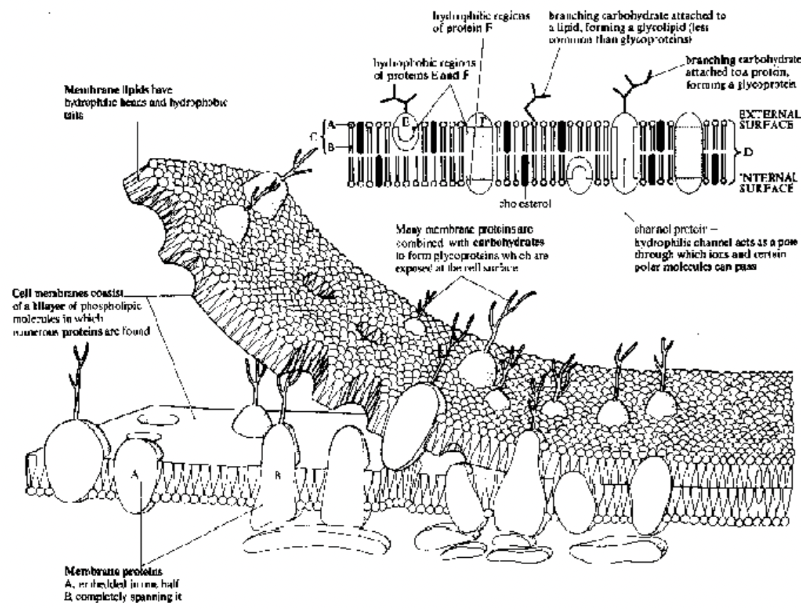
Fig. Fluid mosaic model of membrane structure
Expressed as ‘Protein icebergs in a sea of phospholipids”.
Modifications
Microvilli : Finger-like evaginations of plasma membrane which increase surface area of cells for absorption and perform movement with the help of actin filament.
Caveolae : Cave-like invaginations of the cell surface to increase surface area.
Lomasomes/Border bodies : Discovered by Moore and Mc Aear. Mesosome-like structures located in between the cell wall and cell membrane.
Occur in fungi and concerned with cell wall synthesis.
Cell Surface Junctions
Desmosomes : Thickened circular areas on plasma membrane of adjacent cells with radiating fibrils (tonofibrils) in cytoplasm. Generally occur in epithelial cells and provide cell adhesion.
Tight junction or Zona Occludence : Outer most layer of two plasma membranes at the site of attachment fuse completely. Fused membrane reveals a 5-layered structure. Permeability of ions is more at tight junction. Tight junction occur in nerve tissues at synapses.
Gap Junction or Nexus : Fine protein channel (connexon) join two plasma membrane of adjacent cells. Exchange of ions between cells may take place by protein channels. Desmosomes, Tight junction and Gap junction occurs in animals cell.
Plasmodesmata : Strasburger (1901) Cytoplasmic connection between two adjacent cells through pores in cell wall and cell membrane. Plasmodesmata are characteristic of multicellular plants and they
maintain continuity of cytoplasm between adjacent cells.
Functions:
The phospholipid bilayer provides the basic structure of membranes. It also restricts entry and exit of polar molecules and ions. The other molecules have a variety of functions like:
Channel proteins and carrier proteins are involved in the selective transport of polar molecules and ions across the membrane.
Enzymes on epithelial cells, lining some parts of the gut contain digestive enzymes in their cell surface membranes.
Receptor molecules for chemical signalling between cells. e.g. hormones are chemical messengers which circulate in the blood but only bind to specific target cells which have the correct receptor sites.
Neurotransmitters, the chemicals which enable nerve impulses to pass from one nerve cell to the next, also fit into specific receptor proteins in nerve cells.
Antigens act as cell identity markers or ‘name tags’. They are glycoproteins, i.e. proteins with branching carbohydrate side chains like ‘antennae’. There is an enormous number of possible shapes to these side
chains, so each type of cell can have its own specific markers. This enables cells to recognise other cells, and to behave in an organised way, during development of tissues and organs in multicellular organisms.
Foreign antigens can also be recognised and attacked by the immune system.
Proteins in photosynthesis and respiration take part in the energy transfer systems that exist in the membranes of chloroplasts and mitochondria respectively.
Cholesterol acts like a plug, reducing the escape or entry of polar molecules through the membrane.
Transport Across the Cell Surface Membrane
Cell membranes are only about 7 nm wide but they present barriers to the movement of ions and molecules, particularly polar (water-soluble) molecules such as glucose and amino acids that are repelled by the
non-polar, hydrophobic lipids of membranes. This prevents the aqueous contents of the cell from escaping.
Transport across membranes must still occur to
- to obtain nutrients.
- to excrete waste substances.
- to secrete useful substances.
to generate the ionic gradients essential for nervous and muscular activity.
to maintain a suitable pH and ionic concentration within the cell for enzyme activity.
There are four basic mechanisms, namely diffusion, osmosis, active transport and bulk transport.
Diffusion
Movement of molecules or ions from a region of their higher concentration to a region of their lower concentration down a diffusion gradient. The process is passive, it does not require energy and happens spontaneously.
Occurs by the random motion of molecules which is due to their kinetic energy (energy of movement). Each type of molecule moves down its own diffusion gradient independently of other molecules. e.g.
oxygen diffuses from the lungs into the blood while at the same time carbon dioxide diffuses in the opposite direction.
Three factors in particular affect the rate of diffusion :
1. The steepness of the diffusion gradient, the steeper the gradient, the faster the rate of diffusion.
2. The greater the surface area of a membrane through which diffusion is taking place, the greater the rate of diffusion.
3. Rate of diffusion decreases rapidly with distance. Diffusion is therefore only effective over very short distances. It is essential that membranes are thin so that molecules or ions can cross them rapidly.
The respiratory gases, oxygen and carbon dioxide, diffuse rapidly through membranes. Water molecules, although very polar, are small enough to pass between the hydrophobic phospholipid molecules without
interference. However, ions and larger polar molecules such as amino acids, sugars, fatty acids and glycerol are repelled by the hydrophobic region of the membrane and diffuse across extremely slowly.
Facilitated Diffusion
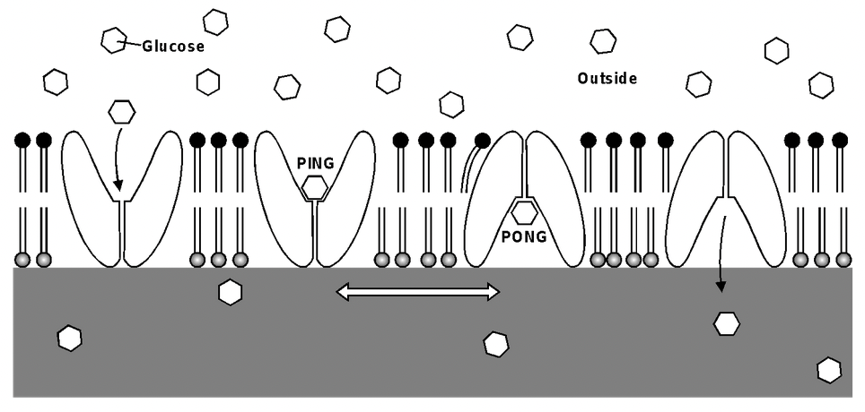
Fig. Facilitated diffusion through a carrier protein
Some ions and polar molecules can diffuse through special transport proteins called channel proteins and carrier proteins.
Channel proteins contain water-filled hydrophilic channels or pores whose shape is specific for a particular ion or molecule. Alternately several proteins combine, forming a channel between them.
Channel proteins have a fixed shape. Diffusion can occur through the channel in either direction.
Disease cystic fibrosis is caused by a fault in a protein which acts as a chloride ion channel.
Carrier proteins undergo rapid changes in shape. They exist in two forms, known as the ‘ping’ and ‘pong’ states. The binding site faces outwards in one state and into the cell in the other state.
Osmosis
The passage of water molecules from a region of their higher concentration to a region of their lower concentration through a partially permeable membrane. A form of diffusion in which only water molecules move.
The tendency of water molecules to move from one place to another is measured as the water potential, represented by the symbol (Greek letter psi). Water always moves from a region of higher water potential to one of lower water potential. Solute molecules reduce . The extent by which they lower is known as the solute potential.
Active Transport
Active transport is the energy-consuming transport of molecules or ions across a membrane against a concentration gradient.
Energy is required because the substance must be moved against its natural tendency to diffuse in the opposite direction.
Movement is usually in one direction only, unlike diffusion which is reversible.
Active transport is achieved by carrier proteins situated in the cell surface membrane.
The carrier proteins involved in active transport need a supply of energy to keep changing shape which is provided by ATP from respiration.
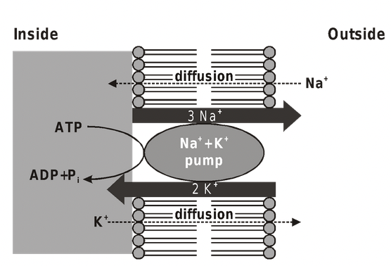
Fig. Sodium-Potassium Pump
Sodium Potassium Pump (Na+ – K+ pump)
The pump is a carrier protein which spans the membrane from one side to the other.
On the inside it accepts sodium and ATP, while on the outside it accepts potassium. For every 2K+ taken into the cell, 3Na+ are removed.
The transfer of sodium and potassium across the membrane is brought about by changes in the shape of the protein.
A potential difference is built up across the membrane, with the inside of the cell being negative.
Potential difference tends to restrict the entry of negatively charged ions (anions) such as chloride which explains why the chloride concentration inside red blood cells is less than outside despite the fact that
chloride can diffuse in and out by facilitated diffusion.
Positively charged ions (cations) tend to be attracted into cells.
Both concentration and charge are important in deciding the direction in which ions cross membranes.
Essential in controlling the osmotic balance of animal cells (osmoregulation).
If the pump is inhibited, the cell swells and bursts because a build-up of sodium ions results in excess water entering the cells by osmosis.
Important in maintaining electrical activity in nerve and muscle cells and in driving active transport of some other substances such as sugars and amino acids.
Also, high concentrations of potassium are needed inside cells for protein synthesis, glycolysis, photosynthesis and other vital processes.
Active transport is associated with epithelial cells, as in the gut lining and kidney tubules (nephrons), because these are active in secretion and absorption.
Bulk Transport
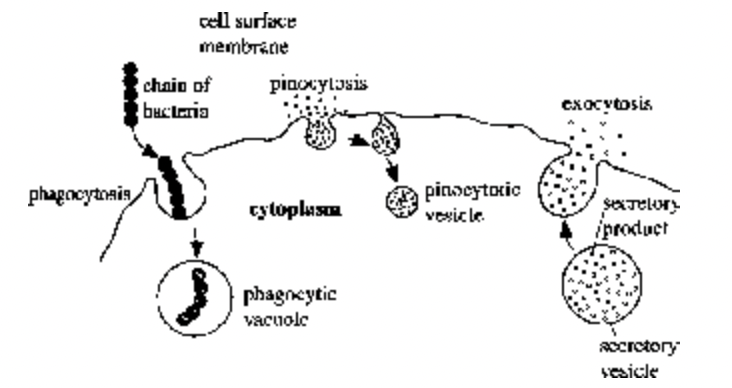
Fig. Endocytosis & Exocytosis
Endocytosis and exocytosis are active processes involving the bulk transport of materials through membranes, either into cells (endocytosis) or out of cells (exocytosis).
Endocytosis occurs by an infolding or extension of the cell surface membrane to form a vesicle or vacuole. It is of two types:
Phagocytosis (‘cell eating’) Material taken up is in solid form. Cells specialising in the process are called phagocytes and are said to be phagocytic. e.g., some white blood cells take up bacteria by phagocytosis.
The sac formed during uptake is called a phagocytic vacuole.
Pinocytosis (‘cell drinking’) Material taken up is in liquid form. Vesicles formed are often extremely small. Pinocytosis is used by the human egg cell to take up nutrients from the surrounding follicle cells.
Exocytosis is the reverse process of endocytosis. Waste materials may be removed from cells, such as solid, undigested remains from phagocytic vacuoles, or useful materials may be secreted. Secretion of enzymes from the pancreas is achieved in this way. Plant cells use exocytosis to export the materials needed to form cell walls.
