
Mitochondria
Structure of Cell of Class 11
Mitochondria (Gr. mitos, thread; chondrion, granule) are organelles found in the cytoplasm of all eukaryotic cells.
Mesosome of bacteria is analogous to mitochondrion in eukaryotes.
First seen by Kolliker (1850) in muscles and he called them ‘sarcosomes’.
Flemming (1882) described these organelles as ‘filia’.
Altmann (1890) observed these structures and named them ‘bioplasts’.
Benda (1898) stained these organelles with crystal violet and renamed them ‘mitochondria’.
Michaelis (1900) used Janus green B as a vital stain to observe mitochondria in living cells.
A single mitochondrion is found in Microsterias, a unicellular green algae.
Provide an energy-transducing system by which the chemical energy contained in foodstuffs is converted, by oxidative phosphorylation, into high-energy phosphate bonds (ATP).
Rod-shaped, with a diameter of about 0.5-1 µm and a variable length that may range up to 7 µm.
There are 1000 to 1600 mitochondria in a liver cell and 300,000 in some oocytes. Green plants contain fewer mitochondria than animal cells.
Distribution of mitochondria may be related to their function as suppliers of energy.
Their orientation in the cell may be influenced by the organization of the cytoplasmic matrix and vacuolar system.
Contains several ribosomes, soluble proteins, enzymes of Kreb’s cycle and nucleic acids.
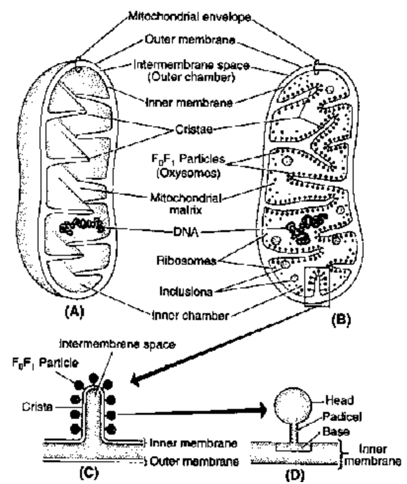
Fig. A. Mitochondrion cut open to show its membranes and cristae; B. Internal structure of mitochondrion;
C. An enlarged crista; D. An enlarged F0F1-particle
Structure :
An outer limiting membrane, about 6 nm thick, surrounds the mitochondrion.
An inner membrane is separated from it by a space of about 6 to 8 nm, that projects into the mitochondrial cavity complex as infoldings called mitochondrial crests.
Inner membrane divides the mitochondrion into two chambers:
(i) The outer chamber contained between the two membranes and in the core of the crests.
(ii) The inner chamber filled with a relatively dense proteinaceous material usually called the mitochondrial matrix.
The mitochondrial crests are, in general, incomplete septa or ridges that do not interrupt the continuity of the inner chamber; thus, the matrix is continuous within the mitochondrion.
Within the mitochondrial matrix are small ribosomes, soluble proteins, and circular DNA, enzymes of Kreb’s cycle.
Small tennis racket like bodies consisting of F0 or base piece (40 × 110 × 115 Å), a stalk (50 Å long) and a round headpiece or F1 (80–100 Å in diameter) are attached upon the cristae with a gap of 100 Å. These
are also called elementary particles or oxysomes or Moran Fernandez particles. The F0 subunit is the channel protein through which proton (H+) passes back to the matrix from peri-mitochondrial space. The
F1 subunit contains the enzyme ATP synthetase which forms ATP from ADP by utilizing the potential energy of proton. This process is called oxidative phosphorylation.
Four complexes, together with mobile components (NADPH, CoQ and cytochrome c), constitute complete respiratory chain in this inner membrane, hence called as energy transducing membrane.
Presence of DNA and ribosomes in the mitochondria makes these organelles independent for production of some of their own proteins.
Multiplication of the mitochondria is by binary fission - a prokaryotic, bacterial character.
Mitochondria have character of changing structure. This is dependent upon the physiological activity of the cell as well as the activity occurring in the organelle.
Inactive or orthodox state, when ATP concentration is low or the respiratory chain is inhibited, the matrix of the mitochondria occupies a larger area.
Active or condensed state when mitochondria are actively engaged in photo-phosphorylation and electron transport, the cristae are more randomly distributed and the inter-membrane space remains highly enlarged.
Distribution :
With the help of cytoplasmic streaming mitochondria move in cytoplasm found in middle piece of sperm between contractile element of muscle cells, dividing cells etc.
Numbers :
a. 1-mitochondria in Chlorella Microsterias, Trypanosoma
b. 20–24 mitochondria in sperm c. 300–400 mitochondria in kidney
d. 500–1000 mitochondria in liver e. 50,000 mitochondria in giant Amoeba (Choas Choas)
f. 1.4 to 1.5 lac mitochondria in egg of sea urchin
g. 3 lac mitochondria in oocytes of some amptobians and
h. 5 lac mitochondria in flight muscles.
Size :
In animals, mito are the second largest cell organelles in plant third largest in size. In yeast 1 , generally mitochondria 1.5–10 in length and 0.25 – 1.0in Dia. In pancreatic cells – 10, Oocytes of Rana pipent – 20– 40 .
Shape :
Common is cylindrical or sausage tublar spherical in yeast & filamentous, club, racket etc.
Chemical Composition :
Proton 60–70%, Lipids 25–35% RNA 5–7%, 60 enzymes, mineral etc. (90% phosphotopids, 10% carbohydrate)
Ultrastructure :
Double membranous, 60–75 Å thick membrane.
Outer membrane : Smooth 40% lipids 60% Proteins membrane enzymes are fewer i.e. monoamine oxidase, NADH cytoreductive Porin (Intrinsic protein) forms channels & make permeable membrane for solute it contains talkless particle called subnot of PARSONS.
Inner membrane : Selectively permeable rich in Protein 80% Lipid-20%. It contain as carrier proteins, electrons carriers, enzyme etc. outer is C-face and inner-m-face. (The side of inner membrane facing outer chamber is called C-side and facing inner chamber is called m-side).
Cardiolipins :
Polyglycerophosphatides (Help in electron transport). It forms number of infolds into matrix called cristae. In most plants mitochondria cristae are tubular while in animals plate like (Hall etal 1974). It contains
succinate dehydrogenase enzyme connected with fatty acids synthesis. ATP-synthetase is component of F0–F1-Particle (Fernadez-Moron 1961) number of Elementry particle per mitochondria – 10K to 1 lac.
Base or F0-subunit is 11 nm in length and 1.5 nm in thick in lipid part of membrane and made up of hydrophobic proteins and act as proton tunnel. Stalk is 5 nm length and 3.5 nm in breadth. It is having
coupling factors that connects resp. chain with Head. F1 - or Head is 8.5 nm in dia and function as AT-pase and forms ATP. Space between 2-successive particles is 100 Å.
Outer Chamber :
Space between outer & inner membrane or perimitochondrial space, 6–10 nm width. It contains nucleoside diplophosphokinase & Adenylate kinase.
Inner Chamber :
Inner chamber is filled with a dense semifluid matrix formed of protein and some lipid, contain enzyme for A.A. synthesis and fatty acid metabolism. Other substances RNAs, DNA, Mn, Mg, CaPO4 mito ribosome are 55–70 s. mt DNA (Nas & Nas) It is more stable, higher density that nuclear DNA due to more G:C base pairs. It is semiautonomous mtDNA codes for rRNA, 19 tRNA, mRNA of only 12 proteins.
Orthodox or Inactive : Little ATP, ETC. inactive, Matrix large.
Condensed or Active : Cristae more, matrix narrow, ATP, ETC. active.
Endosymbiotic Origin : Altman (1890) & Schimper (1896).
Autonomy of Mitochondria : In an animals & higher plants offspring obtained mitochondria from its mother through females gamete.
Function :
1. Aerobic TCA-cycle Respiration
2. Power home
3. Intermediate for biochemicals
4. Aminoacids
5. Fatty acids
6. Ca++ store & release.
7. Thermogenesis
8. Maternal Inheritance (cytoplasma of zygot is of ovum).
Description of particles is based upon facts given by Green and Predue 1966. But accn. to recent views the outer membrane is almost smooth. M-DNA is called as Nucleoid.
Enzyme of Kreb’s cycle located in matrix while that of oxidative phosphorylation are located in inner mitochondrial membrane.
