
CILIA AND FLAGELLA
Share
CILIA AND FLAGELLA
Structure of Cell of Class 11
Purkinje first observed ciliary movement. Hodge(1950) studied detailed structure of sperm flagellum.
Manton studied structure of cilia in plants.
Most prominent hair-like organelles on free surface of the cell, associated with motility.
Both are whiplike and move the micro-organisms along.
Cilia are only 5-20 µm in length while flagella are 100-200 µm long.
Flagella move in an undulating fashion and beat independently to generate planer or helical waves orginating at either the base or the tip. If the wave moves from base to the tip, the cell is pushed along; a beat
travelling from the tip towards the base pulls the cell through the water.
Sometimes the flagellum will have lateral hair called flimmer filaments (thicker, stiffer hair) so that a wave moving down the filament towards the tip pulls the cell along instead of pushing it. Such a flagellum
is referred to as a tinsel type flagellum, whereas the naked flagellum is referred to as whiplash flagellum.
Cilia, normally have a beat with two distinctive phases. In Power or effective stroke the cilium propels through the surrounding fluids like an oar, thereby propelling the organisms along, in the water. The cilium
next bends along its length, while it is pulled forward during the recovery stroke in preparation for another effective stroke.
A ciliated microorganism actually coordinates the beat so that some of its cilia are in recovery phase, whereas others are carrying out their effective stroke, this coordination allows the organisms to move smoothly through the water.
Cilia and flagella are very similar in ultrastructure, both are membrane-bound cylinders made up of four parts:
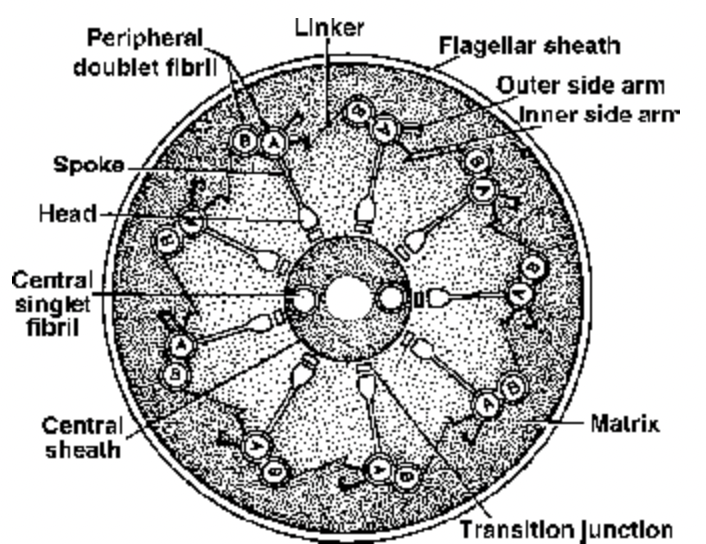
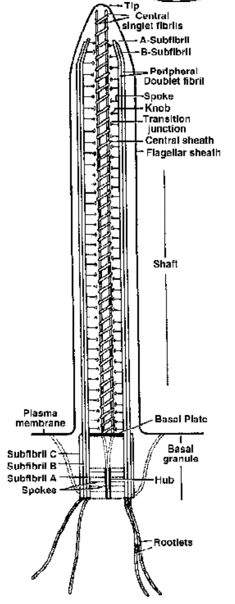
Fig. Ultrastructure of flagellum in cross-section
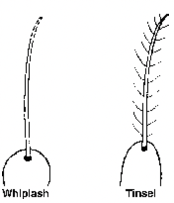
Fig. Types of flagella
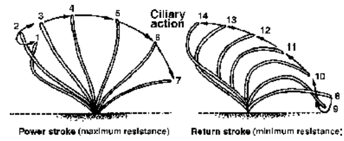
Fig. Movement in Cilia Fig. L.S. of a flagellum (Diagrammatic)
(i) Shaft/axoneme: consists of nine pairs of microtubule doublets (9 + 2 pattern). Each doublet also has pairs of arms projecting from subtubule A towards a neighbouring doublet. A radial spoke extends from subtubule A towards the internal pair of microtubules with their central sheath. These microtubules are similar to those found in the cytoplasm. Each is made up of two types of tubulin subunits, that resemble the contractile protein actin in their composition.
(ii) Basal plate - Area of high density which lies above the basal body at the level of plasma membrane.
In this region one subfibre of each peripheral fibril disappears. The central fibrils develop in this area.
(iii) Basal body / kinetosome / basal granule / blepharoplast: lies in the cytoplasm at the base of each cilium or flagellum. It is short cylinder with nine microtubule triplets around its periphery (9+0 pattern)
and is separated from the rest of the organelles by a basal plate. The basal body directs the construction of these organelles.
(iv) Rootlets Striated fibrillar outgrowths which develop from the outer lower part of body and are meant for providing support to the basal body. They are made of bundles of microfilaments. Cilia and flagella appear to grow through the addition of preformed microtubule subunits at their tips. Cilia and flagella bend because adjacent microtubule doublets slide along one another while maintaining their individual lengths. The doublet arms, 15 nm long, are made up of the protein dynein. Adenosine triphosphate (ATP) powers the movement of cilia and flagella and isolated dynein hydrolyses ATP. It appears that dynein arms interact with the subtubule B of adjacent doublets to cause the sliding motion.
Cilia and flagella beat at a rate of about 10-40 strokes or waves per second and propel the microorganisms rapidly. Such speeds are equivalent to or are much faster than those seen in higher animals.
