
Chromosomes
Structure of Cell of Class 11
Observed by Nageli (1842) and named by Waldeyer (1888).
The morphology of the chromosomes is best studied during metaphase and anaphase, which are the periods of maximal contraction.
Chromosomes are classified into four types according to their shape, which is determined by the position of the centromere (point of attachment to the mitotic spindle).
(i) Telocentric chromosomes have the centromere located on one end.
(ii) Acrocentric chromosomes have a very small or even imperceptible short arm.
(iii) Submetacentric chromosomes have arms of unequal length.
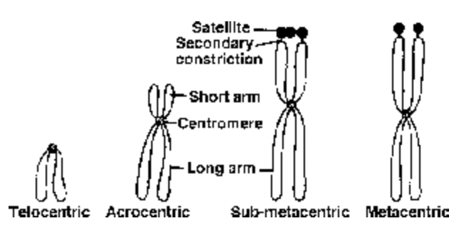
Fig. Types of chromosomes
(iv) Metacentric chromosomes have equal or almost equal arms. During anaphase movements, the chromosomes bend at the centromere, so that metacentric chromosomes are V-shaped and acrocentric chromosomes are rod-shaped.
Chromatid : At metaphase of mitosis each chromosome consists of two symmetrical structures, the chromatids, each one of which contains a single DNA molecule. The chromatids are attached to each other
only by the centromere and become separated at the start of anaphase, when the sister chromatids migrate to opposite poles. Therefore, anaphase chromosomes have only one chromatid, while metaphase chromosomes have two.
Chromonema (ta) : During prophase (and sometimes during interphase) the chromosomal material becomes visible as very thin filaments, which are called chromonemata and which represent chromatids in early stages of condensation “chromatid” and “chromonema”, therefore, are two names for the same structure: a single linear DNA molecule with its associated proteins.
Chromomeres : These components are bead-like accumulations of chromatin material that are sometimes visible along interphase chromosomes. Chromomeres are especially obvious in polytene chromosomes, where they become aligned side by side, constituting the chromosome bands. These tightly folded regions of DNA may correspond to the units of genetic function in the chromosome. At
metaphase the chromosome is tightly coiled and the chromomeres are no longer visible.
Centromere or Kinetochore: This is the region of the chromosome that becomes attached to the mitotic spindle. The centromere lies within a thinner segment of the chromosome, the primary constriction.
Centromeres contain specific DNA sequences with special proteins bound to them, forming a disc-shaped structure to which microtubules bind, called as kinetochore. Between 4 to 40 microtubules become attached to the kinetochore and provide the force for chromosomal movement during mitosis.
The function of the kinetochore is to provide a centre of assembly for microtubules.
The vast majority of chromosomes have only one kinetochore (monocentric chromosomes). Some species have diffuse kinetochores, with microtubules attached along the length of the chromosome (holocentric chromosomes).
In some chromosomal abnormalities, chromosomes may break and fuse with other ones, producing chromosomes without kinetochores (acentric) or with two kinetochores (dicentric).
Telomere : The term applies to the tips of the chromosomes.
Secondary Constrictions / Nucleolar Organizers : Morphological chracteristic of chromosomes, constant in their position and extent, these constrictions are useful in identifying particular chromosomes in a set.
Nucleolar Organizers are certain secondary constrictions that contain the genes coding for 18S and 28S ribosomal RNA and that induce the formation of nucleoli.
In man, the nucleolar organizers are located in the secondary constrictions of chromosomes 13, 14, 15, 21 and 22, all of which are acrocentrics and have satellites.
Satellites : Morphological element present in certain chromosomes. This is a rounded body separated from the rest of the chromosomes by a secondary constriction. The satellite and the constriction are constant
in shape and size for each particular chromosome.
Chromosome satellites are a morphological entity and should not be confused with the satellite DNAs, which are highly repeated DNA sequences.
Karyotype
It is the name given to the whole group of characteristics that allow the identification of a particular chromosomal set, i.e. the number of chromosomes, relative size, position of the centromere, length of the arms, secondary constrictions, and satellites.
The karyotype is characterististic of an individual, species, genus, or larger grouping and may be represented by a diagram in which the pairs of homologues are ordered in a series of decreasing size.
Some species may have special characteristics; for example, the mouse has acrocentric chromosomes, many amphibia have only metacentric chromosomes, and plants frequently have heterochromatic regions at the telomeres.
Techniques for Karyotyping
(i) Banding technique : Very useful for preparing karyotype in which the linear differentiation due to distinct banding patterns, allow identification of chromosomes that are morphologically similar. It also helps
in detecting the regions with repetitive DNA. Variety of bands — Q, C, G or R have been used in animal karyotyping. C and N bands have been used for plant karyotyping.
(ii) Fluorescence in situ hybridization (FISH) and multicolor fluorescence in situ hybridization (McFISH) have been extensively used in plants and animals. DNA probes, labelled with radioactive or non-
radioactive molecules are used to locate the position of specific DNA sequence on chromosomes.
In Mc FISH, labelling with fluorochromes allow use of one or more colours to locate the position of one or more DNA sequences simultaneously on same chromosome.
(iii) Flow cytometry : A suspension of thousands of chromosomes is made and stained with a DNA binding fluorochrome. These chromosome are then passed through cytometer, where fluorescence of each
chromosome is measured and the result is represented as histogram. Each peak in this histogram represents one chromosome or a group of chromosomes of same size.
This technique allows the detection of differences as small as 1.5 to 4.0 Mbp (mega base pairs).
It helps in detection of aneuploidy, duplication or deletion.
Idiogram : The arrangement of chromosomes as per in order of their length, size, morphological features and diagrammatic representation of the haploid set of a species is called idiogram.
Special Types of Chromosomes
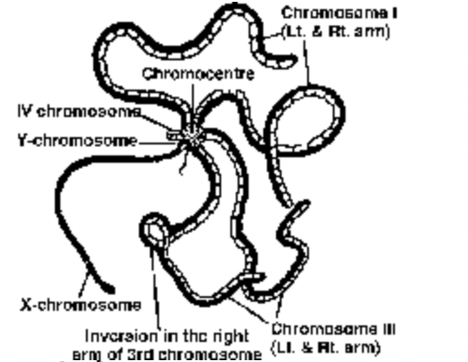
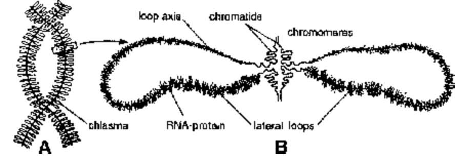
Fig. Salivary Gland Chromosome Fig. Lampbrush chromosome
1. Polytene Chromosomes First seen by Balbiani (1881) in salivary glands of larva of Chironomus insect (midges — belongs to order Diptera).
Term ‘Polytene’ by Koller (1888). Later on such chromosomes were observed in several other insects like Drosophila, mosquitoes.
Polytene chromosomes are composed of many chromonema (512 to several thousands).
Polyteny of giant chromosomes is achieved by replication of chromosomal DNA several times without nuclear division (endore duplication) and the resulting daughter chromatids do not separate but remain aligned side by side. These cells are unable to undergo mitosis and are destined to die during metamorphosis.
In stained preparation, they appear striped with alternate dark and light bands. Dark bands have high amount of histone. Several puffy and swollen areas are seen on polytene chromosomes called as Balbiani
rings or puffs. These are site of rapid m-RNA synthesis. Ecdysone hormone (moulting hormone) of insects stimulates formation of balbiani rings.
Polytene chromosomes are concerned with metamorphosis in insects.
2. Lampbrush Chromosomes – Ruckert (1892). Seen in Oocytes of animals like shark, amphibians, reptiles and birds (most distinct in Diplotene stage). Also found in spermatocytes of several species, giant nucleus of Acetabularia and even in plants.
Giant size of these chromosomes is due to increase in size of chromonema (not due to number). Chromonema grow in size to form lateral loops where synthesis of m-RNA occurs. Lampbrush Chromosomes are concerned with vitellogenesis (Yolk formation).
