
Epithelial Tissue
Animal Tissues of Class 11
The term ‘epithelium’ was coined by Dutch anatomist Ruysch (1638-1731)
Epithelium means ‘epi’-upon; thelio-grows i.e. tissues that grow upon other tissues.
Epithelium consists of one or a few layers of compactly arranged cells overlying a non-cellular basement membrane.
Occurs on external and internal exposed surfaces of the body parts and forms protective covering.
Epithelium is formed first in the embryo and it arises from all the three primary germ layers: ectoderm, mesoderm and endoderm, of the embryo.
The epithelium lining heart, blood vessels, lymphatic vessels and serous cavities is called endothelium.
Epithelium that lines pleural, pericardial and peritoneal cavities is called mesothelium derived from mesoderm.
Epithelium lining major part of alimentary tract (except buccal cavity and anal canal), respiratory tract and distal part of urinogenital tract develops from endoderm.
Epithelium with its supporting connective tissue is regarded as single unit, also called mucosa.
It is called serosa when covered by mesothelium.
There is no intercellular space or very narrow space. Adjacent cells are held together by various types of cell junctions (junctional complexes).
Vascular supply is absent, nutrition being provided by diffusion from capillaries of underlying connective tissue.
Cells are typically polygonal. Shape depends on cytoplasmic contents and pressure from surrounding tissue.
Epithelia regenerate very quickly.
Cells may remain soft or superficial layers may become keratinised.
Non-cellular layer basement membrane lies beneath the epithelium. It is synthesized partly by epithelium and partly by underlying connective tissue. It has two parts:
(a) Basal lamina (outer)
(b) Reticular lamina (inner)
Functions of basement membrane :
(a) Anchors epithelial tissue to underlying connective tissue.
(b) Constitutes a selectively permeable barrier.
(c) Acts as a pathway for diffusion of materials between epithelial tissue and vascular supply.
(d) Determines polarity, metabolism, cell division, repair and movement of other tissues.
Epithelium is subjected to wear, tear and injury; its cells divide and produce new cells.
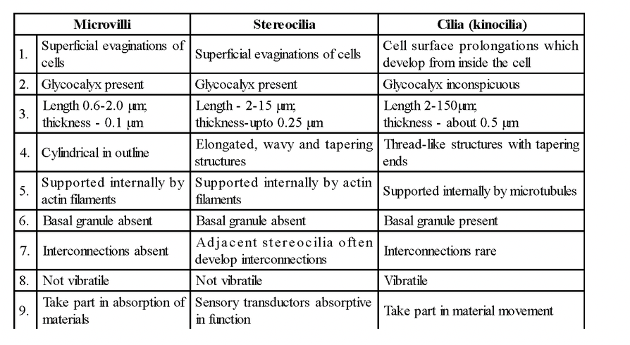
Epithelial cells may have microvilli, stereocilia or kinocilia.
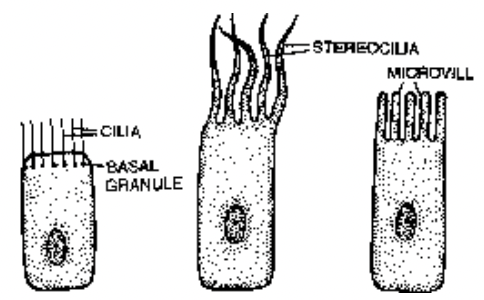
Fig. Epithelial cells showing projections in the form of cilia, stereocilia and microvilli
Table : Differences amongst Microvilli, Stereocilia and Cilia
Junctions:
Epithelial cells develop contacts, which are of two types
(a) Specialized
(b) Unspecialized
Unspecialized contacts are established by trans- membrane proteins called cadherins. Contacts between cells and extracellular material is maintained through integrins (glycoprotein).
Specialized contacts occur by means of intercellular bridges, interdigitations, tight junctions, desmosomes, terminal bars and gap junctions.
(i) Intercellular bridges
- Projections from adjacent cells into common intercellular space. Aid in quick transfer of stimuli.
(ii) Interdigitations
- Adjacent cells show outgrowths which fit into each other. Enhance adherence and provide large areas for mutual exchange.
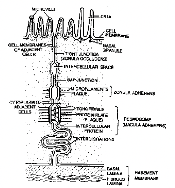
Fig. Microvilli, cilia, cell junctions and
basement membrane
(iii) Tight junctions :
(Occluding junctions, Zonula occludens)
- Plasma membrane of adjacent cells are fused at intervals.
- They create diffusion barrier; prevent leakage of toxic substances.
(iv) Desmosomes (Macula adherens)
- Disc-like junctions thicker and stronger than zonula adherens.
- They have intercellular protein. They form plaque-like structures which are much thicker.
- Microfilaments extend from plaque-like structure and these microfilaments are called tonofibrils.
- Desmosomes serve anchoring functions.
- In hemidesmosomes, the single membrane is thick on one side.
- Hemidesmosomes join epithelial cells to basal lamina.
(v) Gap Junctions (Communicating junctions, Maculae communicans)
- Fine hydrophilic channels between adjacent cells.
- Formed with the help of cylindrical proteins called connexons. Each has six protein subunits.
- Opening, narrowing or closure of channels is influenced by pH and Ca2+ ion concentration.
Epithelia are of two types:
(A) Covering and lining epithelium
(B) Glandular epithelium
